More Related Content
Similar to Tech experience overview (20)
Tech experience overview
- 1. TECHNICAL EXPERIENCE OVERVIEW:
Project: Deciphering innervation dynamics during skeletal
muscle regeneration.
Confocal micrographs of whole-mount, 3rd-digit extensor
digitorum longus muscle isolated from Thy1:CFP transgenic
mouse. Thy1 fluorescence (cyan) allows for visualization of adult,
axonal processes that extend to innervate individual skeletal
muscle myofibers at neuromuscular junctions (NMJs) labeled via
alpha-bungarotoxin (red). Micrographs depict NMJ-associated
axonal growth cones extensions (left, DAPI in blue; star identifies
zoomed region, right).
Leica SPV Confocal - 20x and 63x z-stack respectively
FIJI (ImageJ) - post-acquisition processing
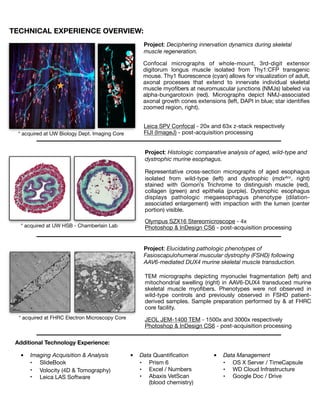
Project: Histologic comparative analysis of aged, wild-type and
dystrophic murine esophagus.
Representative cross-section micrographs of aged esophagus
isolated from wild-type (left) and dystrophic (mdx4cv, right)
stained with Gomori’s Trichrome to distinguish muscle (red),
collagen (green) and epithelia (purple). Dystrophic esophagus
displays pathologic megaesophagus phenotype (dilation-
associated enlargement) with impaction with the lumen (center
portion) visible.
Olympus SZX16 Stereomicroscope - 4x
Photoshop & InDesign CS6 - post-acquisition processing
Project: Elucidating pathologic phenotypes of
Fasioscapulohumeral muscular dystrophy (FSHD) following
AAV6-mediated DUX4 murine skeletal muscle transduction.
TEM micrographs depicting myonuclei fragmentation (left) and
mitochondrial swelling (right) in AAV6-DUX4 transduced murine
skeletal muscle myofibers. Phenotypes were not observed in
wild-type controls and previously observed in FSHD patient-
derived samples. Sample preparation performed by & at FHRC
core facility.
JEOL JEM-1400 TEM - 1500x and 3000x respectively
Photoshop & InDesign CS6 - post-acquisition processing
* acquired at UW Biology Dept. Imaging Core
* acquired at UW HSB - Chamberlain Lab
* acquired at FHRC Electron Microscopy Core
Additional Technology Experience:
• Imaging Acquisition & Analysis
• SlideBook
• Volocity (4D & Tomography)
• Leica LAS Software
• Data Quantification
• Prism 6
• Excel / Numbers
• Abaxis VetScan
(blood chemistry)
• Data Management
• OS X Server / TimeCapsule
• WD Cloud Infrastructure
• Google Doc / Drive