1. BRIEF REPORT
MDMA Increases Glutamate Release and Reduces
Parvalbumin-Positive GABAergic Cells in the Dorsal
Hippocampus of the Rat: Role of Cyclooxygenase
John H. Anneken & Jacobi I. Cunningham & Stuart A. Collins &
Bryan K. Yamamoto & Gary A. Gudelsky
Received: 29 August 2012 /Accepted: 5 November 2012 /Published online: 18 November 2012
# Springer Science+Business Media New York 2012
Abstract 3,4-Methylenedioxymethamphetamine (MDMA;
Ecstasy) is a popular drug of abuse with well-documented
acute effects on serotonergic, dopaminergic, and cholinergic
transmitter systems, as well as evidence of long-term dis-
ruption of serotoninergic systems in the rat brain. Recently,
it was demonstrated that MDMA evokes a delayed and
sustained increase in glutamate release in the hippocampus.
The purpose of the present study was to determine the role
of inflammatory mediators in the MDMA-induced increase
in glutamate release, as well as the contribution of inflam-
matory pathways in the persistent neurochemical toxicity
associated with repeated MDMA treatment. Treatment with
the non-selective cyclooxygenase (COX) inhibitor ketopro-
fen and the COX-2 selective inhibitor nimesulide attenuated
the increase in extracellular glutamate in the hippocampus
evoked by repeated MDMA exposure (10 mg/kg, i.p., every
2 h); no attenuation was observed in rats treated with the
COX-1 selective inhibitor piroxicam. Reverse dialysis of a
major product of COX activity, prostaglandin E2, also resulted
in a significant increase in extracellular glutamate in the hip-
pocampus . Repeated exposure to MDMA diminished the
number of parvalbumin-positive GABA interneurons in the
dentate gyrus of the hippocampus, an effect that was attenuated
by ketoprofen treatment. However, COX inhibition with keto-
profen did not prevent the long-term depletion of 5-HT in the
hippocampus evoked by MDMA treatment. These data are
supportive of the view that cyclooxygenase activity contributes
to the mechanism underlying both the increased release of
glutamate and decreased number of GABA interneurons in
the rat hippocampus produced by repeated MDMA exposure.
Keywords MDMA . Glutamate . Cyclooxygenase . GABA
Introduction
3,4-Methylenedioxymethamphetamine (MDMA, Ecstasy)
has generally been viewed as selectively neurotoxic for 5-
HT axon terminals in view of the well documented reduc-
tions in brain 5-HT concentrations, 5-HT uptake sites, 5-HT
transporter immunoreactivity and reductions in 5-HT immu-
noreactive fibers following repeated exposure to MDMA
(c.f., Green et al. 2003; Gudelsky and Yamamoto 2003).
Moreover, MDMA-induced 5-HT neurotoxicity has been
considered to resemble a distal axotomy affecting 5-HT
axon terminals (Xie et al. 2006).
However, there are several reports that treatment of rats
with MDMA produces neuronal degeneration in several
brain regions, including the hippocampus (Kermanian et
al. 2012; Riezzo et al. 2010; Schmued 2003; Wang et al.
2009; Warren et al. 2007). Investigators from these and
other studies have concluded that MDMA promotes apopto-
tic cell death on the basis of increases in caspase-3, TUNEL
staining and altered Bcl-2 family gene expression produced
by MDMA (Meyer et al. 2004; Asi et al. 2012; Riezzo et al.
2010; Wang et al. 2009). Consistent with these data, Capela
et al. (2007, 2012) have reported that MDMA promotes
apoptotic cell death in cultured cortical and hippocampal
neurons in vitro. Although these data support the view that
MDMA neurotoxicity extends beyond 5-HT axon terminals
to neuronal cell bodies themselves within the hippocampus,
the identity of neurons potentially damaged by MDMA has
not been established.
J. H. Anneken :G. A. Gudelsky (*)
James Winkle College of Pharmacy, University of Cincinnati,
3225 Eden Ave,
Cincinnati, OH 45267, USA
e-mail: Gary.Gudelsky@uc.edu
J. I. Cunningham :S. A. Collins :B. K. Yamamoto
Department of Neurosciences, University of Toledo
College of Medicine, Toledo, OH, USA
J Neuroimmune Pharmacol (2013) 8:58–65
DOI 10.1007/s11481-012-9420-x
2. A recent report from our lab documented the finding that
MDMA produces a delayed and sustained increase in the
extracellular concentration of glutamate in the hippocampus
(Anneken and Gudelsky 2012). The MDMA-induced in-
crease in hippocampal glutamate release was suppressed
by fluoxetine and the 5-HT2 antagonist ketanserin, but
was still evident in the presence of tetrodotoxin. Anneken
and Gudelsky (2012) concluded that 5-HT, released by
MDMA, activates 5-HT2A/C receptors, thereby promoting
the release of glutamate, presumably from astrocytes, in
the hippocampus.
Although the consequences of an increased release of hip-
pocampal glutamate by MDMA are unknown, several studies
suggest that hippocampal neurons are vulnerable to excitotoxic
effects of elevated glutamate through the activation of gluta-
mate receptors. For example, parvalbumin-positive GABA
neurons in the hippocampus express Group 1 metabotropic
glutamate receptors (Kerner et al. 1997), as well as AMPA
receptors containing GluR1 and GluR3 subunits, but not GluR2
subunits (Moga et al. 2003). In support of this vulnerability,
parvalbumin-positive GABAergic neurons are selectively vul-
nerable to the toxic effects of kainic acid, an effect blocked by
glutamate antagonists (Sanon et al. 2005).
Given the aforementioned reports that 1) MDMA produ-
ces neuronal damage in the hippocampus, 2) MDMA
increases glutamate release in the hippocampus and 3)
parvalbumin-positive GABA neurons in the hippocampus
are sensitive to glutamate-mediated neurotoxicity, the pres-
ent study was undertaken to evaluate the effects of repeated
exposure to MDMA on parvalbumin-positive GABA neu-
rons in the dorsal hippocampus. In view of the findings that
MDMA-induced glutamate release is dependent upon 5-
HT2 receptor activation and that 5-HT2 receptor activation
appears to increase the activity of cyclooxygenase (COX),
we also sought to ascertain the role of COX in the effects of
MDMA on hippocampal glutamate release and PV-positive
GABAergic neurons.
Materials and methods
Animals and drug treatments
Adult male Sprague–Dawley rats (250–350 g) (Harlan Labo-
ratories, Indianapolis, IN) were used in this study. Animals
were given free access to food and water in a temperature and
humidity controlled room. The animals were singly housed
following cannula implantation until the day of the experi-
ment. All procedures were performed in adherence to the
National Institutes of Health guidelines and were approved
by the institutional animal care and use committee.
MDMA was generously provided by the National Insti-
tute on Drug Abuse (Bethesda). Ketoprofen and piroxicam
were obtained from Sigma-Aldrich (St. Louis, MO), while
nimesulide and prostaglandin E2 were obtained from Tocris
Bioscience (Ellisville, MO). MDMA was dissolved in
0.15 M NaCl and injected i.p. at a dose of 10 mg/kg at 2 h
intervals for a total of 2 or 3 injections. Ketoprofen was
dissolved in a 30 % Transcutol solution and administered in
3 injections at a dose of 5 mg/kg, s.c., 1 h prior to and 1 h
and 3 h following the first MDMA injection. Both pirox-
icam and nimesulide were delivered in a 2 % polyvinylpyr-
rolidone suspension. Piroxicam was administered as 3
injections at a dose of 3 mg/kg, i.p., in the same time course
as ketoprofen. Nimesulide was administered as 3 injections
at a dose of 7.5 mg/kg, i.p., also on the same time course.
Prostaglandin E2 (PGE2) was dissolved in the dialysis buffer
and was administered by reverse dialysis through the probe
at a concentration of 30 μM for 1.5 h and then at 100 μM for
an additional 1.5 h. Doses of COX inhibitors were based on
those used in previous studies (Asanuma et al. 2003;
Candelario-Jalil et al. 2004; Terao et al. 1998).
Microdialysis
Rats were implanted with a stainless steel guide cannula
under ketamine/xylazine (70/6 mg/kg, i.p.) anesthesia 48–
72 h prior to the insertion of the dialysis probe. On the
evening prior to the experiment, a concentric style dialysis
probe was inserted through the guide cannula into the dorsal
hippocampus; the coordinates for the tip of the probe were:
A/P, -3.6 mm, L, 2.0 mm, and D/V −4.0 mm. The active
portion of the membrane for the probes was 2.0 mm. The
probes were connected to an infusion pump set to deliver
modified Dulbecco's phosphate buffered saline containing
1.2 mM CaCl2 and 5 mM glucose at a flow rate of 1 μl/min
overnight. On the morning of the experiment, the flow rate
was increased to 2 μl/min and the probes were allowed to
equilibrate for 1.5 h. Three collections were then taken at
30 min intervals to establish values; thereafter, samples were
collected every hour for the duration of the experiment. Data
were calculated as a percentage of the baseline value for
glutamate which was obtained by averaging the three base-
line samples.
HPLC glutamate analysis
Glutamate was derivitized according to the method described
by Donzanti and Yamamoto (1988) and quantified by HPLC
with electrochemical detection, as described previously
(Anneken and Gudelsky 2012).
Analysis of tissue serotonin (5-HT)
All tissue samples were homogenized in 0.2 N perchloric
acid. Concentrations of 5-HT were determined via HPLC in
J Neuroimmune Pharmacol (2013) 8:58–65 59
3. 20μL aliquots of supernatants from tissue homogenates, as
described previously (Shankaran et al. 2001).
Tissue preparation for GABA neuron counts
Rats were deeply anesthetized with ketamine hydrochlo-
ride (70 mg/kg, ip) and xylazine (6 mg/kg, ip) and were
transcardially perfused with 0.1 M PBS (100 mL) prior to
the perfusion with 4 % paraformaldehyde (400 mL).
Brains were removed and post-fixed for 2 h in 4 % parafor-
maldehyde before cryoprotection in a series of glycerol sol-
utions; 10 % glycerol with 2 % dimethylsufide (DMS0) in
0.1 M PBS overnight, followed by 20 % glycerol with 2 %
DMS0 in 0.1 M PBS overnight. The following day, brains
were flash frozen in 2-methylbutane for 30 min. Coronal
sectioning was at a thickness of 50 μm through the dorsal
extent of the hippocampus (approximately −3.30 mm to −4.80
from bregma, (Paxinos and Watson 1998) as described
previously (Muller et al. 2001). Each section was collected in
four uninterrupted series with a 200 μm section interval and
stored in 15 % glycerol in 0.1 M PBS at −80 °C until
immunostaining.
Parvalbumin immunostaining
Free-floating brain sections were stained for parvalbumin.
Sections were washed in 0.1 M PBS and then treated with
1 % H2O2 for 20 min room at temperature (RT). After
several washes, the sections were blocked for 1 h at RT with
3 % normal goat serum (NGS; Invitrogen, Carlsbad, CA,
USA) in 0.1 M PBS containing 0.5 % Triton-X 100 and
Avidin block (Vector Laboratories, Burlingame, CA, USA).
Sections were then incubated for 36 h at 4 °C with a
mouse monoclonal parvalbumin antibody (1:3000; Swant,
Bellinzona, Switzerland) in 0.1 M PBS containing 0.5 %
Triton-X 100, 1 % NGS and Biotin block (Vector Laborato-
ries). Sections were washed and incubated in goat anti-mouse
secondary antibody (Millipore, Billerica, MA, USA) for 2 h
at RT followed by incubation in avidin-biotin-horseradish
peroxidase (Vectastain Elite ABC Kit; Vector Laboratories)
for 2 h at RT. Sections were washed and developed in
diaminobenzidine.
Quantitative cell counts
Stereological estimations of the total number of parvalbumin-
immunoreactive (PV-ir) interneurons were assessed using a
BX51 Olympus microscope equipped with a DVC camera
interfaced with StereoInvestigator 8.21 software (MBF Bio-
science, Williston, VT, USA). A modified optical fractionator
technique (Gundersen et al. 1999; West et al. 1991) was used
to quantify neurons throughout the right dorsal hippocampus.
Pilot studies were used for sampling procedures to ensure that
the coefficient of error (Gundersen et al. 1999) was less than
1 % for all counts. All slides were coded and the code not
broken until the end of quantitative analysis. Every fourth
section was systematically sampled and all subfields (dentate
gyrus [DG] and cornu ammonis 1 and 3 [CA1 & CA3]) were
outlined using a 5X objective. The DG included the hilus and
subgranular zone, but not the principle cell layer of the CA3.
Counting was performed using a 60X oil objective (NA
0.16). Due to the limited number of PV-ir interneurons in
the DG, exhaustive sampling was performed with grid
dimensions of 100 μm x 100 μm and a sampling area of
100 μm x 100 μm. For both the CA1 and CA3 subfield,
the grid dimensions were 200 μm x 300 μm with a
sampling area of 300 μm x 300 μm. Guard zones above
and below the dissector were not used due to the massive
tissue shrinkage during processing, as previously described
(Czeh et al. 2005; Lister et al. 2006).
Statistical analysis
All microdialysis data were analyzed using two-way repeat-
ed measures ANOVA, and multiple pairwise comparisons
were performed using post-hoc analysis with the Student-
Newman-Keuls test. Tissue 5-HT data were analyzed using
a two-way ANOVA, as were the stereological neuron
counts. Treatment differences were considered statistically
significant at p<0.05.
Results
Ketoprofen and nimesulide, but not piroxicam, attenuate the
MDMA-induced increase in extracellular glutamate concen-
trations in the rat hippocampus.
The involvement of cyclooxygenase (COX) activity in
the MDMA-induced increase in extracellular glutamate in
the rat hippocampus was investigated using the non-
selective COX inhibitor ketoprofen. Rats received injec-
tions of vehicle or ketoprofen (5 mg/kg, s.c.) 1 h prior to
and 1 h and 3 h following the first injection of MDMA
(10 mg/kg, i.p.). As shown in Fig. 1, ketoprofen signif-
icantly attenuated (p<0.05) the increase in the extracel-
lular concentration of glutamate in the hippocampus
evoked by 2 injections of MDMA. A two-way repeated
measures ANOVA revealed a significant effect of treatment
(F(3,286)03.27; p<0.05).
The relative contribution of each isoform of COX to the
increased efflux of hippocampal glutamate elicited by
MDMA was assessed using the relatively specific inhibitors
piroxicam (COX-1) and nimesulide (COX-2) (Fig. 1, inset).
Piroxicam (3 mg/kg, i.p.), nimesulide (7.5 mg/kg, i.p.), or
vehicle was administered 1 h prior to and 1 h and 3 h
following the initiation of MDMA treatment (10 mg/kg,
60 J Neuroimmune Pharmacol (2013) 8:58–65
4. i.p.). A two-way repeated measures ANOVA revealed a
significant treatment effect (F(3,169)012.00; p<0.001) as well
as a significant effect of time (F(10,169)02.86; p<0.01) and a
significant treatment x time interaction (F(30,169)02.44; p<
0.001). Results of the ANOVA revealed no significant
difference in the glutamate reponses of animals treated with
piroxicam + MDMA and those receiving MDMA alone
(p>0.05). In contrast, nimesulide significantly diminished
(p<0.05) the MDMA-induced increase in the extracellular
concentration of glutamate. Treatment with the COX inhib-
itors alone did not significantly alter glutamate concentrations
(data not shown).
Reverse dialysis of prostaglandin E2 increases extracellular
glutamate concentration in the rat hippocampus
The local effect of inflammatory mediators on hippocampal
glutamate was assessed using reverse dialysis of prostaglan-
din E2 (PGE2), the most biologically active product of COX
activity. PGE2 was delivered in the dialysis buffer at a
concentration of 30 μM for 1.5 h and was continued at
100 μM for another 1.5 h. As shown in Fig. 2, PGE2
produced a significant elevation (p<0.05) in the extracellu-
lar concentration of glutamate in the hippocampus when
compared to control animals; this was evident at the end
of the 30 μM perfusion and during the 100 μM perfusion.
Results of the ANOVA revealed a significant main effect of
drug treatment (F(1,87)015.18; p<0.01).
Ketoprofen prevents the MDMA-induced reduction
in parvalbumin-positive GABA neurons in the dentate gyrus
The effects of MDMA on a subpopulation of GABAergic
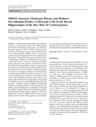
neurons in the hippocampus was determined. Figure 3a,b illus-
trates the effect of MDMA on parvalbumin-immunoreactive
GABA neurons in the dentate gyrus. Repeated treatment with
MDMA (Panel B) resulted in a significant (p<0.05) reduction
of approximately 36 % in pavralbumin-positive GABA neu-
rons in the dentate gyrus compared to control animals (Panel
A). There was no significant effect of MDMA on these cells in
the CA1 or CA3 regions (data not shown). Notably, the
MDMA-induced reduction in parvalbumin-immunoreactive
GABA neurons was not evident in rats treated with ketoprofen
(Fig. 3c). The number of parvalbumin-positive neurons in the
dentate gyrus of rats treated with ketoprofen + MDMA was
significantly (p<0.05) greater than that in rats treated with
vehicle + MDMA.
Ketoprofen does not prevent the MDMA-induced depletion
of 5-HT in the rat hippocampus
In order to ascertain whether inhibition of COX activity also
would afford protection against the long-term reductions in
biochemical markers of 5-HT axon terminals produced by
MDMA, rats were treated with the previously described reg-
imen of ketoprofen or vehicle, and concomitantly adminis-
tered MDMA (2×10 mg/kg, i.p.) or vehicle. Concentrations
of 5-HT in the hippocampus were determined 7 days follow-
ing MDMA treatment. The hippocampal concentration of 5-
HT was depleted by approximately 25 % (p<0.05) in rats
Fig. 1 Effect of the COX 1/2 inhibitor ketoprofen. COX-1 inhibitor
piroxicam, and COX-2 inhibitor nimesulide on the MDMA-induced
efflux of glutamate in the rat hippocampus. Rats received ketoprofen
(5 mg/kg, s.c.), piroxicam (3 mg/kg, i.p.) (inset), nimesulide (7.5 mg/
kg, i.p.) (inset) or vehicle 1 h prior to and 1 h and 3 h following the first
injection of MDMA (10 mg/kg, i.p.) or vehicle. (n04–14 per group).
Average basal glutamate for VEH-MDMA group was 2.54±0.34 ng/
20μL (uncorrected for recovery). Arrows indicate injections with MDMA
or Veh. * Indicates values that differ significantly (p<0.05) from
Veh-MDMA animals
Fig. 2 Extracellular glutamate concentrations in the hippocampus
following reverse dialysis of PGE2. PGE2 (30 μM) or vehicle was
infused via the dialysis buffer into the hippocampus for a duration of
90 min after three 30 min baseline samples were taken. The concen-
tration of PGE2 was then increased to 100 μM for an additional 90 min.
(n05–8 per group) *Indicates values that differ significantly (p<0.05)
from those animals that received vehicle
J Neuroimmune Pharmacol (2013) 8:58–65 61
5. treated with MDMA, and there was no significant difference
in the depletion of hippocampal 5-HT produced by MDMA in
vehicle- and ketoprofen-treated animals (p>0.05). The values
(ng/mg tissue) for hippocampal 5-HT were: vehicle-vehicle,
0.33±0.02; ketoprofen-vehicle, 0.34±0.02; vehicle-MDMA,
0.21±0.02; ketoprofen-MDMA, 0.21±0.01. (n06–12)
Discussion
The key findings of the present study include the demon-
stration that 1) the MDMA-induced increase in the extra-
cellular concentration of glutamate in the hippocampus is
attenuated in rats treated with ketoprofen and nimesulide,
but not piroxicam, 2) infusion of PGE2 produces a signif-
icant increase in the extracellular concentration of gluta-
mate, 3) MDMA reduces the number of parvalbumin-
positive neurons in the dentate gyrus of the hippocampus,
an effect attenuated by ketoprofen and 4) ketoprofen does
not attenuate the depletion of 5-HT in the hippocampus
produced by MDMA.
We have previously reported that MDMA produces a
delayed and sustained increase in the extracellular concentra-
tion of glutamate in the hippocampus, but not in the striatum
or prefrontal cortex (Anneken and Gudelsky 2012). Moreover,
fluoxetine and ketanserin, a 5-HT2A/C antagonist, were
found to suppress the glutamate response to MDMA, and it
was suggested that MDMA increases glutamate release in the
hippocampus subsequent to an increased release of 5-HT and
increased activation of 5-HT2A/C receptors (Anneken and
Gudelsky 2012). The present study extends the previous find-
ings to further investigate additional mechanisms and conse-
quences of the increased release of hippocampal glutamate
produced by MDMA.
In the present study, the MDMA-evoked increase in
extracellular glutamate was suppressed by ketoprofen and
nimesulide, but not piroxicam, which is supportive of the
involvement of COX-2, rather than COX-1, in the mecha-
nism of MDMA-induced glutamate release. Although the
doses of COX inhibitors used in this study were based on
those used previously by other investigators (Asanuma et al.
2003; Terao et al. 1998), we cannot exclude the possibility
that these dosage regimens did not selectively and/or effec-
tively inhibit the respective COX isoforms. Nevertheless,
the present results are consistent with previous findings that
an increase in COX-2 activity in hippocampal tissue results
in an increased extracellular concentration of glutamate and
exacerbation of glutamate-associated excitotoxicity (Bezzi
et al. 1998; Kelley et al. 1999; Sang et al. 2011). Moreover,
reverse dialysis of a major product of COX activity, PGE2,
also produced a significant increase in extracellular gluta-
mate in the hippocampus. To our knowledge, this is the first
in vivo demonstration of glutamate release evoked by PGE2
exposure. This finding is in accord with data from in vitro
Fig. 3 Effect of ketoprofen on
the MDMA-induced reduction of
parvalbumin-reactive GABAer-
gic neurons in the rat hippocam-
pus. Panel A is a representative
photomicrograph depicting
parvalbumin-immunoreactive
cells in the dentate gyrus in con-
trol animals, as highlighted by the
arrows, while panel B is repre-
sentative of the dentate gyrus of
animals given repeated doses of
MDMA. Panel C depicts the
quantitative assessment of PV-ir
neurons in the dentate gyrus of
vehicle- and MDMA-treated rats.
Rats received ketoprofen (5 mg/
kg, s.c.) or vehicle 1 h prior to
and 1 h and 3 h following the first
of 4 injections of MDMA
(10 mg/kg, i.p.) or vehicle. (n04–
8 per group) * indicates values
that differ significantly (p<0.05)
from those for animals that
received VEH-VEH. # indicates
values that differ significantly
(p<0.05) from those for animals
that received VEH-MDMA
62 J Neuroimmune Pharmacol (2013) 8:58–65
6. studies (Bezzi et al. 1998; Sanzgiri et al. 1999) wherein it
was demonstrated that multiple prostaglandins, including
PGE2, evoke calcium-dependent glutamate release from
astrocytes in brain tissue culture.
There are multiple signaling pathways through which
MDMA may increase COX activity and the subsequent pro-
duction of prostaglandins. MDMA may directly or indirectly
activate 5-HT2A/C receptors to increase the release of arach-
idonic acid, thereby increasing COX activity and prostanoid
formation. Indeed, activation of 5-HT2 receptors on glia has
been shown to result in arachidonic acid release, presumably
through the activation of phospholipase A2 (Garcia and Kim
1997; Mackowiak et al. 2002). Furthermore, 5-HT2 receptor
stimulation has been shown to increase glutamate release from
astrocytes in a calcium dependent manner (Bezzi et al. 1998;
Meller et al. 2002). These findings together with our report
that MDMA-induced glutamate release is dependent upon 5-
HT2 receptor activation and is independent of neuronal activ-
ity (Anneken and Gudelsky 2012) support the view that the
MDMA-induced increase in extracellular glutamate is
the result of: 1) increased 5-HT release and a resulting 5-
HT2A/C receptor-dependent activation of COX, 2) an in-
creased formation of prostanoids and 3) a prostanoid-induced
release of glutamate from astrocytes.
Until recently, there has been little evidence that MDMA
produces persistent deficits in neurotransmitter systems be-
yond 5-HT axon terminals. Perrine et al. (2010) has demon-
strated that repeated treatment with MDMA reduces the
hippocampal concentration of GABA. Armstrong and
Noguchi (2004) reported that chronic MDMA administration
reduced the binding of [3
H]-flunitrazapam in the hippocampus
and concluded that GABAergic terminals or interneurons may
be damaged by MDMA. In the present study, MDMA treat-
ment resulted in a reduction in parvalbumin-positive GABA
neurons in the dentate gyrus. No effect of MDMAwas evident
in the CA1 or CA3 regions of the hippocampus. Thus, it
appears that repeated treatment with MDMA results in a
persistent reduction in markers of GABAergic neurons. Future
studies are needed to examine the effects of MDMA on other
subpopulations of GABAergic cells such as calbindin- and
calretinin-positive neurons.
Previous studies have indicated that MDMA produces
neuronal degeneration in the hippocampus under in vivo and
in vitro conditions (Meyer et al. 2004; Riezzo et al. 2010;
Wang et al. 2009). MDMA treatment increases caspase-3 and
TUNEL staining and promotes apoptotic cell death in cultured
hippocampal neurons (Capela et al. 2012). On the basis of the
results of the present study, it is tempting to propose that
MDMA-induced cell death occurs in GABAergic neurons
within the hippocampus.
In the present study, ketoprofen suppressed not only the
MDMA-induced increase in glutamate release but also the
MDMA-induced reduction in parvalbumin-positive GABA
neurons in the hippocampus. Thus, COX activity and neuro-
inflammatory products also appear to contribute to the mech-
anism of MDMA-induced deficits in GABA neurons. In view
of the inhibitory effect of ketoprofen on MDMA-stimulated
glutamate release and the sensitivity of parvalbumin-positive
GABA neurons to glutamate-mediated damage (Sanon et al.
2005), it is suggested that MDMA-induced damage to GABA
neurons involves glutamate-mediated excitotoxicity. Addi-
tional studies are necessary to establish a causal relationship
between these two phenomena.
Although inhibition of COX activity with ketoprofen
greatly attenuated the MDMA-induced release of glutamate,
it did not prevent the long term depletion of 5-HT in the
hippocampus following repeated MDMA treatment. Thus, it
appears that neither neuroinflammatory mediators nor glu-
tamate excitotoxicity is a contributing factor in MDMA-
induced 5-HT depletion. This finding is in agreement with
earlier work by Farfel and Seiden (1995) who reported
MDMA-induced 5-HT depletion to be insensitive to NMDA
receptor antagonists under hyperthermic conditions.
Repeated exposure of rats to MDMA has been to shown to
produce deficits in reference memory (Able et al. 2006; Asi et
al. 2011; Cunningham et al. 2009; Kay et al. 2011; Skelton et
al. 2006), a function subserved by the hippocampus, and in
particular the dentate gyrus (Morris et al. 2012; Okada and
Okaichi 2009). Moreover, human abusers of MDMA exhibit
deficits in verbal memory (Burgess et al. 2011; de Sola Llopis
et al. 2008; Raj et al. 2010), and altered function of GABA
interneurons in the hippocampus (Jacobsen et al. 2004). Fur-
ther studies are warranted to investigate the hypothesis that
MDMA-induced impairment of GABA interneurons in the
hippocampus underlies the cognitive impairments associated
with repeated exposure to MDMA.
In summary, the data are consistent with the hypothesis
that MDMA produces damage to GABA neurons within the
hippocampus through a mechanism involving enhanced for-
mation of neuroinflammatory mediators and a subsequent
increase in glutamate release.
Acknowledgments This work was supported by awards from the
National Institute on Drug Abuse DA 07427 (GG) and DA016886 (BY).
Conflict of Interest The authors declare no conflicts of interest.
References
Able JA, Gudelsky GA, Vorhees CV, Williams MT (2006) 3,4-meth-
ylenedioxymethamphetamine in adult rats produces deficits in
path integration and spatial reference memory. Biol Psychiatry
59:1219–1226
Anneken JH, Gudelsky GA (2012) MDMA produces a delayed and
sustained increase in the extracellular concentration of glutamate
in the rat hippocampus. Neuropharmacology 63(6):1022–1027
J Neuroimmune Pharmacol (2013) 8:58–65 63
7. Armstrong BD, Noguchi KK (2004) The neurotoxic effects of 3,4-meth-
ylenedioxymethamphetamine (MDMA) and methamphetamine on
serotonin, dopamine, and GABA-ergic terminals: an in-vitro auto-
radiographic study in rats. Neurotoxicology 25:905–914
Asanuma M, Tsuji T, Miyazaki I, Miyoshi K, Ogawa N (2003)
Methamphetamine-induced neurotoxicity in mouse brain is atten-
uated by ketoprofen, a non-steroidal anti-inflammatory drug.
Neurosci Lett 352:13–16
Asi SS, Farhadi HM, Naghdi N, Choopani S, Samzadeh-Kermani A,
Mehdizadeh M (2011) Non-acute effects of different doses of 3,4-
methylenedioxymethamphetamine on spatial memory in the morris
water maze in Sprague–Dawley male rats. Neural Regen Res 6:1715
Asi SS, Farhadi HM, Mousavizadeh K, Samadikuchaksaraei A,
Soleimani M, Jameie SB, Joghataei MT, Samzadeh-Kermani
A, Hashemi-Nasl H, Mehdizadeh, M (2012) Evaluation of
Bcl-2 Family Gene Expression in Hippocampus of 3,4-meth-
ylenedioxymethamphetamine Treated Rats. Cell J Vol 13, No 4,
Winter 2012
Bezzi P, Carmignoto G, Pasti L, Vesce S, Rossi D, Rizzini BL, Pozzan
T, Volterra A (1998) Prostaglandins stimulate calcium-dependent
glutamate release in astrocytes. Nature 391:281–285
Burgess AP, Venables L, Jones H, Edwards R, Parrott AC (2011) Event
related potential (ERP) evidence for selective impairment of ver-
bal recollection in abstinent recreational methylenedioxymetham-
phetamine (“ecstasy”)/polydrug users. Psychopharmacol (Berl)
216:545–556
Candelario-Jalil E, Gonzalez-Falcon A, Garcia-Cabrera M, Leon OS,
Fiebich BL (2004) Wide therapeutic time window for nimesulide
neuroprotection in a model of transient focal cerebral ischemia in
the rat. Brain Res 1007:98–108
Capela JP, Fernandes E, Remiao F, Bastos ML, Meisel A, Carvalho F
(2007) Ecstasy induces apoptosis via 5-HT(2A)-receptor stimula-
tion in cortical neurons. Neurotoxicology 28:868–875
Capela JP, da Costa Araujo S, Costa VM, Ruscher K, Fernandes E, Bastos
MD, Dirnagl U, Meisel A, Carvalho F (2012) The neurotoxicity of
hallucinogenic amphetamines in primary cultures of hippocampal
neurons. Neurotoxicology. doi:10.1016/j.neuro.2012.09.005
Cunningham JI, Raudensky J, Tonkiss J, Yamamoto BK (2009)
MDMA pretreatment leads to mild chronic unpredictable stress-
induced impairments in spatial learning. Behav Neurosci 123:1076–
1084
Czeh B, Simon M, van der Hart MG, Schmelting B, Hesselink MB, Fuchs
E (2005) Chronic stress decreases the number of parvalbumin-
immunoreactive interneurons in the hippocampus: Prevention by
treatment with a substance P receptor (NK1) antagonist. Neuropsy-
chopharmacology 30:67–79
de Sola Llopis S, Miguelez-Pan M, Pena-Casanova J, Poudevida S,
Farre M, Pacifici R, Bohm P, Abanades S, Verdejo Garcia A,
Langohr K, Zuccaro P, de la Torre R (2008) Cognitive performance
in recreational ecstasy polydrug users: a two-year follow-up study. J
Psychopharmacol 22:498–510
Donzanti BA, Yamamoto BK (1988) An improved and rapid HPLC-
EC method for the isocratic separation of amino acid neurotrans-
mitters from brain tissue and microdialysis perfusates. Life Sci
43:913–922
Farfel GM, Seiden LS (1995) Role of hypothermia in the mechanism
of protection against serotonergic toxicity. I. experiments using
3,4-methylenedioxymethamphetamine, dizocilpine, CGS 19755
and NBQX. J Pharmacol Exp Ther 272:860–867
Garcia MC, Kim HY (1997) Mobilization of arachidonate and doco-
sahexaenoate by stimulation of the 5-HT2A receptor in rat C6
glioma cells. Brain Res 768:43–48
Green AR, Mechan AO, Elliott JM, O'Shea E, Colado MI (2003) The
pharmacology and clinical pharmacology of 3,4-methylenediox-
ymethamphetamine (MDMA, “ecstasy”). Pharmacol Rev 55:463–
508
Gudelsky GA, Yamamoto BK (2003) Neuropharmacology and neuro-
toxicity of 3,4-methylenedioxymethamphetamine. Methods Mol
Med 79:55–73
Gundersen HJ, Jensen EB, Kieu K, Nielsen J (1999) The efficiency of
systematic sampling in stereology–reconsidered. J Microsc
193:199–211
Jacobsen LK, Mencl WE, Pugh KR, Skudlarski P, Krystal JH (2004)
Preliminary evidence of hippocampal dysfunction in adolescent
MDMA ("ecstasy") users: Possible relationship to neurotoxic
effects. Psychopharmacol (Berl) 173:383–390
Kay C, Harper DN, Hunt M (2011) The effects of binge MDMA on
acquisition and reversal learning in a radial-arm maze task. Neu-
robiol Learn Mem 95:473–483
Kelley KA, Ho L, Winger D, Freire-Moar J, Borelli CB, Aisen PS,
Pasinetti GM (1999) Potentiation of excitotoxicity in transgenic
mice overexpressing neuronal cyclooxygenase-2. Am J Pathol
155:995–1004
Kermanian F, Mehdizadeh M, Soleimani M, Ebrahimzadeh Bideskan
AR, Asadi-Shekaari M, Kheradmand H, Haghir H (2012) The
role of adenosine receptor agonist and antagonist on hippocampal
MDMA detrimental effects; a structural and behavioral study.
Metab Brain Dis
Kerner JA, Standaert DG, Penney JB Jr, Young AB, Landwehrmeyer
GB (1997) Expression of group one metabotropic glutamate re-
ceptor subunit mRNAs in neurochemically identified neurons in
the rat neostriatum, neocortex, and hippocampus. Brain Res Mol
Brain Res 48:259–269
Lister JP, Tonkiss J, Blatt GJ, Kemper TL, DeBassio WA, Galler JR,
Rosene DL (2006) Asymmetry of neuron numbers in the hippocam-
pal formation of prenatally malnourished and normally nourished
rats: a stereological investigation. Hippocampus 16:946–958
Mackowiak M, Chocyk A, Sanak M, Czyrak A, Fijal K, Wedzony K
(2002) DOI, an agonist of 5-HT2A/2C serotonin receptor, alters
the expression of cyclooxygenase-2 in the rat parietal cortex. J
Physiol Pharmacol 53:395–407
Meller R, Harrison PJ, Elliott JM, Sharp T (2002) In vitro evidence that
5-hydroxytryptamine increases efflux of glial glutamate via 5-HT
(2A) receptor activation. J Neurosci Res 67:399–405
Meyer JS, Grande M, Johnson K, Ali SF (2004) Neurotoxic effects of
MDMA ("ecstasy") administration to neonatal rats. Int J Dev
Neurosci 22:261–271
Moga DE, Janssen WG, Vissavajjhala P, Czelusniak SM, Moran TM,
Hof PR, Morrison JH (2003) Glutamate receptor subunit 3
(GluR3) immunoreactivity delineates a subpopulation of
parvalbumin-containing interneurons in the rat hippocampus. J
Comp Neurol 462:15–28
Morris AM, Churchwell JC, Kesner RP, Gilbert PE (2012) Selective
lesions of the dentate gyrus produce disruptions in place learning
for adjacent spatial locations. Neurobiol Learn Mem 97:326–331
Muller GJ, Moller A, Johansen FF (2001) Stereological cell counts of
GABAergic neurons in rat dentate hilus following transient cere-
bral ischemia. Exp Brain Res 141:380–388
Okada K, Okaichi H (2009) Functional differentiation and cooperation
among the hippocampal subregions in rats to effect spatial mem-
ory processes. Behav Brain Res 200:181–191
Paxinos G, Watson C (1998) The rat brain in stereotaxic coordinates,
4th edn. Academic, San Diego
Perrine SA, Ghoddoussi F, Michaels MS, Hyde EM, Kuhn DM,
Galloway MP (2010) MDMA administration decreases serotonin
but not N-acetylaspartate in the rat brain. Neurotoxicology
31:654–661
Raj V, Liang HC, Woodward ND, Bauernfeind AL, Lee J, Dietrich
MS, Park S, Cowan RL (2010) MDMA (ecstasy) use is associated
with reduced BOLD signal change during semantic recognition in
abstinent human polydrug users: a preliminary fMRI study. J
Psychopharmacol 24:187–201
64 J Neuroimmune Pharmacol (2013) 8:58–65
8. Riezzo I, Cerretani D, Fiore C, Bello S, Centini F, D'Errico S, Fiaschi
AI, Giorgi G, Neri M, Pomara C, Turillazzi E, Fineschi V (2010)
Enzymatic-nonenzymatic cellular antioxidant defense systems re-
sponse and immunohistochemical detection of MDMA, VMAT2,
HSP70, and apoptosis as biomarkers for MDMA (ecstasy) neuro-
toxicity. J Neurosci Res 88:905–916
Sang N, Yun Y, Yao GY, Li HY, Guo L, Li GK (2011) SO(2)-induced
neurotoxicity is mediated by cyclooxygenases-2-derived prosta-
glandin E(2) and its downstream signaling pathway in rat hippo-
campal neurons. Toxicol Sci 124:400–413
Sanon N, Carmant L, Emond M, Congar P, Lacaille JC (2005) Short-
term effects of kainic acid on CA1 hippocampal interneurons
differentially vulnerable to excitotoxicity. Epilepsia 46:837–848
Sanzgiri RP, Araque A, Haydon PG (1999) Prostaglandin E(2) stim-
ulates glutamate receptor-dependent astrocyte neuromodulation in
cultured hippocampal cells. J Neurobiol 41:221–229
Schmued LC (2003) Demonstration and localization of neuronal de-
generation in the rat forebrain following a single exposure to
MDMA. Brain Res 974:127–133
Shankaran M, Yamamoto BK, Gudelsky GA (2001) Ascorbic acid
prevents 3,4-methylenedioxymethamphetamine (MDMA)-in-
duced hydroxyl radical formation and the behavioral and neuro-
chemical consequences of the depletion of brain 5-HT. Synapse
40:55–64
Skelton MR, Williams MT, Vorhees CV (2006) Treatment with
MDMA from P11-20 disrupts spatial learning and path integration
learning in adolescent rats but only spatial learning in older rats.
Psychopharmacol (Berl) 189:307–318
Terao A, Matsumura H, Saito M (1998) Interleukin-1 induces slow-
wave sleep at the prostaglandin D2-sensitive sleep-promoting
zone in the rat brain. J Neurosci 18:6599–6607
Wang X, Zhu SP, Kuang WH, Li J, Sun X, Huang MS, Sun XL
(2009) Neuron apoptosis induced by 3,4-methylenedioxy
methamphetamine and expression of apoptosis-related factors
in rat brain. Sichuan Da Xue Xue Bao Yi Xue Ban 40(1000–
2):1037
Warren MW, Larner SF, Kobeissy FH, Brezing CA, Jeung JA, Hayes
RL, Gold MS, Wang KK (2007) Calpain and caspase proteolytic
markers co-localize with rat cortical neurons after exposure to
methamphetamine and MDMA. Acta Neuropathol 114:277–286
West MJ, Slomianka L, Gundersen HJ (1991) Unbiased stereological
estimation of the total number of neurons in thesubdivisions of the
rat hippocampus using the optical fractionator. Anat Rec 231:482–
497
Xie T, Tong L, McLane MW, Hatzidimitriou G, Yuan J, McCann U,
Ricaurte G (2006) Loss of serotonin transporter protein after
MDMA and other ring-substituted amphetamines. Neuropsycho-
pharmacology 31:2639–2651
J Neuroimmune Pharmacol (2013) 8:58–65 65