
Recommended
More Related Content
Similar to Cranial and Peripheral Nerves - Martin.pdf
Similar to Cranial and Peripheral Nerves - Martin.pdf (20)
Recently uploaded
Recently uploaded (20)
Cranial and Peripheral Nerves - Martin.pdf
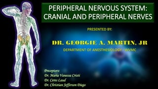
- 1. PERIPHERAL NERVOUS SYSTEM: CRANIAL AND PERIPHERAL NERVES Preceptors: Dr. Maria Vanessa Cristi Dr. Ceres Laud Dr. Christian Jefferson Diego
- 2. PERIPHERAL NERVOUS SYSTEM • Nerves and ganglia outside the brain and spinal cord • Connects the CNS to limbs and organs • Somatic Nervous System – voluntary movements • Autonomic Nervous System – involuntary movements • Sympathetic Nervous System • Parasympathetic Nervous System
- 4. CRANIAL NERVES ➢ 12 pairs ➢ Exits skull through the foramina ➢ Mainly innervate the head and neck ➢ 3 groups: ➢ Sensory ➢ Motor ➢ Mixed
- 5. CRANIAL NERVES: OLFACTORY NERVE (I) ➢ First and shortest cranial nerve ➢ Transmits information relating to SMELL ➢ Special visceral afferent nerve ➢ Fibers of the olfactory nerve are the central processes of the olfactory cells
- 6. CRANIAL NERVES: OLFACTORY NERVE (I) ➢ Sense smell detected by olfactory receptors at the nasal epithelium ➢ Signal is sent through the filia olfactoria ➢ Synapse with the Mitral Cells ➢ Pass on the second order neurons to the primary olfactory cortex
- 7. CRANIAL NERVES: OPTIC NERVE (II) ➢ Nerve of Vision ➢ Developed from the optic vesicle, an out pocketing of the fore brain ➢ Devoid of neurilemmal sheath ➢ Surrounded by the extension of the meninges and subarachnoid space
- 8. CRANIAL NERVES: OPTIC NERVE (II) ➢ Carries afferent impulses for vison ➢ Convergence of axons from the retinal ganglion ➢ Enter the optic canal and converge at the optic chiasm ➢ Left optic tract – contains fibers from left temporal (lateral) retina and right nasal (medial) retina ➢ Right optic tract – contains fibers from right temporal (lateral) retina and left nasal (medial) retina
- 9. CRANIAL NERVES: OCULOMOTOR (III) ➢ Motor ➢ Extraocular muscles of the eye (levator palpebrae superioris, superior rectus, inferior rectus, medial rectus and inferior oblique) ➢ Parasympathetic ➢ Sphincter pupillae and the ciliary muscles of the eye
- 10. CRANIAL NERVES: OCULOMOTOR (III) ➢ Originates from the oculomotor nucleus ➢ Emerge at the anterior aspect of the midbrain ➢ Pierces the dura mater and enters the cavernous sinus ➢ Leaves the cranial cavity via superior orbital fissure ➢ Superior branch ➢ Inferior branch
- 11. CRANIAL NERVES: TROCHLEAR (IV) ➢ Smallest cranial nerve but with the longest intracranial course ➢ Purely a motor ➢ supplies the superior oblique ➢ Arise from the trochlear nucleus and emerged on the posterior surface of the midbrain ➢ Passes through the cranial middle fossa ➢ Enters the orbit to superior orbital fissure
- 12. CRANIAL NERVES: TROCHLEAR (IV) ➢ Motor innervation to the superior oblique ➢ Moves eye downward and laterally ➢ A lesion of the trochlear nerve results in paralysis of the superior oblique muscle with the result that diplopia occurs when the patient attempts to turn the eye downwards and laterally.
- 13. CRANIAL NERVES: TRIGEMINAL (V) ➢ Largest cranial nerve ➢ Principal sensory nerve of the face, orbit, nose and mouth, and because its branches are eminently suitable for accurate anesthetic blockade
- 14. CRANIAL NERVES: TRIGEMINAL (V) ➢ Leaves the anterior aspect of the pons as a small motor root and a large sensory root ➢ Passes forward out of the posterior cranial fossa and rest on the apex of the middle cranial fossa ➢ In the middle cranial fossa ➢ Sensory root expands into 3 trigeminal ganglion supplying 3 facial division ➢ Motor root supplies the mandibular division
- 15. CRANIAL NERVES: TRIGEMINAL (V) V1 OPHTHALMIC NERVE ➢ First division of the CN V and is entirely sensory ➢ Innervates skin and mucous membrane of: ➢ Forehead, scalp, frontal and ethmoid sinus, upper eyelid and its conjunctiva, cornea and dorsum of the nose ➢ Provides parasympathetic supply to: ➢ Lacrimal gland
- 16. CRANIAL NERVES: TRIGEMINAL (V) V2 MAXILLARY NERVE ➢ Second division of the trigeminal nerve, entirely sensory ➢ Innervates the skin, mucous membrane and sinuses of: ➢ Lower eyelid and its conjunctiva, cheeks and maxillary sinus nasal cavity and lateral nose, upper lip upper molar, incisor and canine and associated gingiva, superior palate ➢ Provides parasympathetic supply to: ➢ Lacrimal gland and nasal gland
- 17. CRANIAL NERVES: TRIGEMINAL(V) V3 MANDIBULAR NERVE ➢ Sensory supply: mucous membrane and floor of the oral cavity, external ear, lower lip, chin, anterior 2/3 of tongue, lower molar, incisor and canine teeth and the associated ganglia ➢ Motor supply: muscle of mastication (medial pterygoid, masseter, temporalis), Anterior belly of the digastric muscle and the mylohyoid muscle (these are suprahyoid muscle), Tensor veli palatini, Tensor tympani ➢ Parasympathetic supply: submandibular, sublingual and parotid
- 18. CRANIAL NERVES: TRIGEMINAL (V) TRIGEMINAL NERVE BLOCK Indication: Trigeminal neuralgia, intractable facial cancer, cluster headache Target: Depending on the site of pain ➢ Gasserian Ganglion Block ➢ Maxillary Nerve Block ➢ Mandibular Nerve Block ➢ Inferior Alveolar Nerve Block
- 19. CRANIAL NERVES: TRIGEMINAL (V) SEMILUNAR (GASSERIAN) NERVE BLOCK ➢ Most comprehensive blockade of the trigeminal nerve ➢ Performed under fluoroscopic guidance ➢ Injection site: posterior 1/3 of the zygomatic bone, opposite the second upper molar tooth
- 20. CRANIAL NERVES: TRIGEMINAL (V) MAXILLARY NERVE BLOCK ➢ Maxillary nerve block is performed for acute or chronic herpetic neuralgia, trigeminal neuralgia and cancer pain. ➢ Injection site: pterygopalatine fossa after emerging from the foramen rotundum. ➢ It is reached by inserting a needle through the mid- point of the coronoid notch beneath the zygomatic arch.
- 21. CRANIAL NERVES: TRIGEMINAL (V) ➢ MANDIBULAR NERVE BLOCK ➢ Indication: dental and maxillary surgery, for inferior dental pain, trigeminal neuralgia on the third branch, Temporomandibular junction dysfunction ➢ Needle inserted midpoint of the coronoid process beneath the zygomatic arch ➢ Target: muscles of mastication, lower jaw, the side of the tongue and the skin overlying the mandible
- 22. CRANIAL NERVES: TRIGEMINAL (V) ➢ INFERIOR ALVEOLAR NERVE BLOCK ➢ Injection site: medial to the anterior border of the mandibular ramus ➢ Anaesthesia of the lower teeth, the skin and mucosa of the lower lip ➢ Loss of sensation of the side of the tongue owing to involvement of the lingual nerve
- 23. CRANIAL NERVES: ABDUCENS (VI) ➢ Purely somatic motor nerve supplying the Lateral Rectus ➢ Arises from the abducens nucleus in the pons ➢ Exits at the junction of the pons and medulla ➢ Passes through the cavernous sinus, lying below and lateral to the internal carotid artery ➢ Enters the orbit through the superior orbital fissure ➢ When damaged, it gives rise to diplopia and a convergent squint
- 24. CRANIAL NERVES: FACIAL NERVE (VII) ➢ The facial nerve supplies the muscles of facial expression, conveys secretomotor fibers to the lacrimal gland, and to the submandibular and sublingual salivary glands, and transmits taste fibers from the anterior two-thirds of the tongue.
- 25. CRANIAL NERVES: FACIAL NERVE (VII) ➢ Damage to the facial nerve or its central pathway results in facial palsy ➢ Both nuclear and infranuclear palsies – complete facial paralysis involving all facial muscles on one side ➢ Supranuclear palsies – no involvement of the muscles above the palpebral fissure
- 26. CRANIAL NERVES: VESTIBULOCOCHLEAR NERVE (VIII) ➢ COCHLEAR FIBERS – concerned with hearing ➢ Arises from the ventral and dorsal cochlear nuclei in the inferior cerebellar peduncle ➢ VESTIBULAR FIBER – concerned with balance ➢ Arises from the vestibular nuclei complex in the pons and medulla
- 27. CRANIAL NERVES: VESTIBULOCOCHLEAR NERVE (VIII) ➢ Lesions of the vestibular division of the labyrinth or of the vestibulocerebellar pathway result in vertigo, ataxia and nystagmus ➢ Lesions of the cochlear division result in deafness that may or may not be accompanied by tinnitus ➢ Temporal lobe tumors may give rise to auditory hallucinations if they encroach upon the auditory radiation or superior temporal gyrus.
- 28. CRANIAL NERVES: GLOSSOPHARYNGEAL NERVE (IX) ➢ Leaves the anterolateral surface of the upper part of the medulla oblongata as several rootlets ➢ Leaves the cranial cavity via the jugular foramen ➢ Superior and inferior glossopharyngeal sensory ganglia ➢ Give rise to carotid sinus nerve
- 29. CRANIAL NERVES: GLOSSOPHARYNGEAL NERVE (IX) ➢ Sensory: Innervates the oropharynx, carotid body and sinus, posterior 1/3 of the tongue, middle ear cavity and Eustachian tube. ➢ Special sensory: Provides taste sensation to the posterior 1/3 of the tongue. ➢ Parasympathetic: Provides parasympathetic innervation to the parotid gland. ➢ Motor: Innervates the stylopharyngeus muscle of the pharynx.
- 30. CRANIAL NERVES: GLOSSOPHARYNGEAL NERVE (IX) GLOSSOPHARYNGEAL NERVE BLOCK ➢ Indication: glossopharyngeal neuralgia, palliative care for patients with head and neck malignancy ➢ Injection site: midpoint through mastoid process and angle of mandible ➢ Targets the nerve as it emerges the jugular foramen ➢ Anesthesia may spread to CN X (Vagus), XI (Accessory) and XII (Hypoglossal)
- 31. CRANIAL NERVES: VAGUS NERVE (X) ➢ Largest and most widely distributed of the cranial nerves ➢ Originates from the medulla of the brainstem ➢ Exits through the jugular foramen after giving off the auricular branch
- 32. CRANIAL NERVES: VAGUS NERVE (X) ➢ Sensory: skin of the external acoustic meatus, internal surfaces of the laryngopharynx and larynx, visceral sensation to the heart and abdominal viscera ➢ Special Sensory: taste sensation to the epiglottis and root of the tongue ➢ Motor: majority of the muscles of the pharynx, soft palate and larynx ➢ Parasympathetic: smooth muscle of the trachea, bronchi and gastro-intestinal tract and regulates heart rhythm
- 33. CRANIAL NERVES: ACCESSORY NERVE (XI) ➢ Purely somatic nerve that provides motor innervation to the sternocleidomastoid and trapezius muscle ➢ Cranial Component – arise from the lateral aspect of the medulla oblongata ➢ Exits through the jugular foramen ➢ Combines with the Vagus Nerve
- 34. ➢ Spinal Component – arise from the neurons of the upper spinal cord (C1 – C5/6) ➢ Runs superiorly and enter the foramen magnum ➢ Exits through the jugular foramen ➢ Descend along the internal carotid artery to the sternocleidomastoid and moves to the trapezius CRANIAL NERVES: ACCESSORY NERVE (XI)
- 35. CRANIAL NERVES: HYPOGLOSSAL NERVE (XII) ➢ Supplies all intrinsic and extrinsic muscle of the tongue except the palatoglossus ➢ Arise from the hypoglossal nucleus of the medulla oblongata of the brainstem ➢ Exits the cranium via the hypoglossal canal ➢ Travels along with branches from the cervical plexus, C1/C2 spinal nerve root ➢ Pass inferiorly to the angle of the mandible and moves to the direction of the tongue
- 36. ➢ Division of the hypoglossal nerve, or lesions involving its nucleus, result in ipsilateral paralysis and wasting of the muscles of the tongue. ➢ This is detected clinically by deviation of the protruded tongue to the affected side. CRANIAL NERVES: HYPOGLOSSAL NERVE (XII)
- 38. SPINAL NERVES
- 39. SPINAL NERVES ➢ 31 Pairs ➢ 8 Cervical ➢ 12 Thoracic ➢ 5 Lumbar ➢ 5 Sacral ➢ 1 Coccygeal ➢ Pass through the Intervetebral Foramina ➢ Divides into Branches or Rami ➢ Posterior Ramus ➢ Anterior Ramus
- 40. SPINAL NERVES ➢ Dorsal (Posterior) Root ➢ Afferent (Sensory) Fibers ➢ Ventral (Anterior) Root ➢ Efferent (Motor) Fibers ➢ Mixed Spinal Nerve ➢ Anterior Ramus ➢ Posterior Ramus Afferent fibers Arrive - SENSORY Efferent fibers Exit - MOTOR
- 41. SPINAL NERVES ➢ Paravertebral Space ➢ A potential space along the outside of the vertebral canal ➢ Filled with loose fat and areolar tissue ➢ Contains the nerve roots issuing from the spinal cord and blood vessels ➢ Allows access for blocking the nerve without invading the epidural or subarachnoid space ➢ Provide unilateral nerve blocks extending two to four segments
- 42. SPINAL NERVES ➢ Nerve Plexus ➢ A network of intersecting nerve serving the same part of the body ➢ Composed of afferent and efferent fibers that arise from the merging of anterior rami of spinal nerves ➢ Nerve Plexuses except in the thoracic region ➢ Cervical Plexus ➢ Brachial Plexus ➢ Lumbar Plexus ➢ Sacral and Coccygeal Plexus
- 43. ➢ Myotome ➢ Specific muscle/s supplied by motor innervation ➢ Dermatome ➢ Specific area of the skin supplied by sensory innervation SPINAL NERVES
- 44. SPINAL NERVES: CERVICAL PLEXUS ➢ Formed by the upper four Cervical Nerves, C1 – C4 ➢ Provides innervation to skin and muscles of the neck and the diaphragm ➢ Located on the posterior triangle of the neck ➢ Divided into two groups: ➢ Deep Muscular Branch ➢ Sensory Superficial Branch
- 45. SPINAL NERVES: CERVICAL PLEXUS ➢ Deep Muscular Branch ➢ Nerve Root: C1 ➢ Nerves to geniohyoid and thyrohyoid ➢ Travels along with the hypoglossal nerve ➢ Ansa Cervicalis (C1 – C3) ➢ Give off four branches to the infrahyoid muscles ➢ Phrenic Nerve (C3 – C5) ➢ Motor innervation to the diaphragm
- 46. SPINAL NERVES: CERVICAL PLEXUS ➢ Sensory Branches ➢ Greater Auricular Nerve (C2 – C3) ➢ External ear, skin over the parotid gland ➢ Largest ascending plexus branch ➢ Transverse Cervical Nerve(C2 – C3) ➢ Sensation to the anterior neck ➢ Lesser Occipital Nerve (C2, contribution from C3) ➢ Posterosuperior scalp ➢ Supraclavicular Nerve (C3 – C4) ➢ Skin overlying the supraclavicular fossa, upper thoracic region, sternoclavicular joint
- 47. SPINAL NERVES: CERVICAL PLEXUS ➢ SUPERFICIAL AND DEEP CERVICAL NERVE BLOCK ➢ Indication: provides regional anesthesia for neck surgery (e.g. carotid endarectomy, thyroidectomy, and cervical lymph node dissection) ➢ Superficial plexus: behind the posterior border of the middle portion of the sternocleidomastoid ➢ Deep Plexus: transverse process of the 2nd, 3rd and 4th cervical vertebrae
- 48. SPINAL NERVES: CERVICAL PLEXUS ➢ OCCIPITAL NERVE BLOCK ➢ Indication: Diagnostic step in evaluating head and neck pain ➢ Complications: rare, intravascular injection
- 49. SPINAL NERVES: BRACHIAL PLEXUS ➢ Provides motor innervation and nearly all sensory supply of the upper limb
- 50. SPINAL NERVES: BRACHIAL PLEXUS ➢ Pass through the anterior and medial scalene muscles ➢ Covered sheaths of fibrous tissue ➢ Upper Trunk – root of C5 and C6 ➢ Middle trunk – C7 ➢ Lower Trunk – root of C8 and T1
- 51. SPINAL NERVES: BRACHIAL PLEXUS ➢ Lateral border of the first rib ➢ 3 Anterior Division ➢ 3 Posterior Division ➢ Leave through the posterior triangle and pass into the axilla
- 52. SPINAL NERVES: BRACHIAL PLEXUS ➢ Enter the apex of the axilla ➢ Grouped together: ➢ Medial Cords ➢ Lateral Cords ➢ Posterior Cords
- 53. BRACHIAL PLEXUS : MAJOR BRANCHES MUSCULOCUTANEOUS NERVE • Roots: C5, C6, C7. • Motor Functions: Innervates the brachialis, biceps brachii and coracobrachialis muscles. • Sensory Functions: Gives off the lateral cutaneous branch of the forearm, which innervates the lateral half of the anterior forearm, and a small lateral portion of the posterior forearm.
- 54. BRACHIAL PLEXUS : MAJOR BRANCHES MUSCULOCUTANEOUS NERVE • Roots: C5, C6, C7. • Motor Functions: Innervates the brachialis, biceps brachii and coracobrachialis muscles. • Sensory Functions: Gives off the lateral cutaneous branch of the forearm, which innervates the lateral half of the anterior forearm, and a small lateral portion of the posterior forearm.
- 55. BRACHIAL PLEXUS : MAJOR BRANCHES MUSCULOCUTANEOUS NERVE • Roots: C5, C6, C7. • Motor Functions: Innervates the brachialis, biceps brachii and coracobrachialis muscles. • Sensory Functions: Gives off the lateral cutaneous branch of the forearm, which innervates the lateral half of the anterior forearm, and a small lateral portion of the posterior forearm.
- 56. BRACHIAL PLEXUS : MAJOR BRANCHES AXILLARY NERVE • Roots: C5 and C6. • Motor Functions: Innervates the teres minor and deltoid muscles. • Sensory Functions: Gives off the superior lateral cutaneous nerve of arm, which innervates the inferior region of the deltoid (“regimental badge area”).
- 57. BRACHIAL PLEXUS : MAJOR BRANCHES AXILLARY NERVE • Roots: C5 and C6. • Motor Functions: Innervates the teres minor and deltoid muscles. • Sensory Functions: Gives off the superior lateral cutaneous nerve of arm, which innervates the inferior region of the deltoid (“regimental badge area”).
- 58. BRACHIAL PLEXUS : MAJOR BRANCHES AXILLARY NERVE • Roots: C5 and C6. • Motor Functions: Innervates the teres minor and deltoid muscles. • Sensory Functions: Gives off the superior lateral cutaneous nerve of arm, which innervates the inferior region of the deltoid (“regimental badge area”).
- 59. BRACHIAL PLEXUS : MAJOR BRANCHES RADIAL NERVE • Nerve roots – C5-T1. • Sensory – Innervates most of the skin of the posterior forearm, the lateral aspect of the dorsum of the hand, and the dorsal surface of the lateral three and a half digits. • Motor – Innervates the triceps brachii and the extensor muscles in the forearm.
- 60. BRACHIAL PLEXUS : MAJOR BRANCHES RADIAL NERVE • Nerve roots – C5-T1. • Sensory – Innervates most of the skin of the posterior forearm, the lateral aspect of the dorsum of the hand, and the dorsal surface of the lateral three and a half digits. • Motor – Innervates the triceps brachii and the extensor muscles in the forearm.
- 61. BRACHIAL PLEXUS : MAJOR BRANCHES RADIAL NERVE • Nerve roots – C5-T1. • Sensory – Innervates most of the skin of the posterior forearm, the lateral aspect of the dorsum of the hand, and the dorsal surface of the lateral three and a half digits. • Motor – Innervates the triceps brachii and the extensor muscles in the forearm.
- 62. BRACHIAL PLEXUS : MAJOR BRANCHES MEDIAN NERVE • Nerve roots: C6 – T1 (may contain C5 fibers) • Motor functions: Innervates the flexor and pronator muscles; thenar muscles and lateral two lumbricals in the hand • Sensory functions: palmar cutaneous branch – lateral aspect of the palm digital cutaneous branch – lateral three and a half fingers on the anterior (palmar) surface of the hand.
- 63. BRACHIAL PLEXUS : MAJOR BRANCHES MEDIAN NERVE • Nerve roots: C6 – T1 (may contain C5 fibers) • Motor functions: Innervates the flexor and pronator muscles; thenar muscles and lateral two lumbricals in the hand • Sensory functions: palmar cutaneous branch – lateral aspect of the palm digital cutaneous branch – lateral three and a half fingers on the anterior (palmar) surface of the hand.
- 64. BRACHIAL PLEXUS : MAJOR BRANCHES MEDIAN NERVE • Nerve roots: C6 – T1 (may contain C5 fibers) • Motor functions: Innervates the flexor and pronator muscles; thenar muscles and lateral two lumbricals in the hand • Sensory functions: palmar cutaneous branch – lateral aspect of the palm digital cutaneous branch – lateral three and a half fingers on the anterior (palmar) surface of the hand.
- 65. BRACHIAL PLEXUS : MAJOR BRANCHES • ULNAR NERVE • Spinal roots: C8-T1. • Motor functions: • Two muscles of the anterior forearm – flexor carpi ulnaris and medial half of flexor digitorum profundus • Intrinsic muscles of the hand (apart from the thenar muscles and two lateral lumbricals) • Sensory functions: Medial one and half fingers and the associated palm area.
- 66. BRACHIAL PLEXUS : MAJOR BRANCHES • ULNAR NERVE • Spinal roots: C8-T1. • Motor functions: • Two muscles of the anterior forearm – flexor carpi ulnaris and medial half of flexor digitorum profundus • Intrinsic muscles of the hand (apart from the thenar muscles and two lateral lumbricals) • Sensory functions: Medial one and half fingers and the associated palm area.
- 67. BRACHIAL PLEXUS : MAJOR BRANCHES • ULNAR NERVE • Spinal roots: C8-T1. • Motor functions: • Two muscles of the anterior forearm – flexor carpi ulnaris and medial half of flexor digitorum profundus • Intrinsic muscles of the hand (apart from the thenar muscles and two lateral lumbricals) • Sensory functions: Medial one and half fingers and the associated palm area.
- 68. SPINAL NERVES: BRACHIAL PLEXUS BRACHIAL PLEXUS NERVE BLOCK INTERSCALENE NERVE BLOCK ➢ procedure of upper arm and shoulder. SUBCLAVIAN NERVE BLOCK ➢ Procedure at the distal elbow INFRACLAVICULAR NERVE BLOCK ➢ Procedure at the distal elbow AXILLARY NERVE BLOCK ➢ Procedure at elbow, forearm and hand
- 69. SPINAL NERVES: BRACHIAL PLEXUS BRACHIAL PLEXUS NERVE BLOCK AXILLARY BRACHIAL PLEXUS BLOCK ➢Indication: for procedures of the elbow, forearm and hand ➢ Targets the Ulnar, Radial, and Median Nerve
- 70. SPINAL NERVES: BRACHIAL PLEXUS BRACHIAL PLEXUS NERVE BLOCK MEDIAN NERVE BLOCK • Indication: for injuries requiring procedure on the radial side of the palm, palmar surface and tip of the thumb, index, middle and ring finger
- 71. BRACHIAL PLEXUS : MAJOR BRANCHES ULNAR BLOCK • Indication: for rescue analgesia or surgical anesthesia for surgery on the fifth digit
- 72. SPINAL NERVES: THORACIC NERVE ➢12 pairs of thoracic anterior primary rami ➢11 intercostal nerve ➢1 subcostal nerve ➢Nerve Root: T1 – T12 ➢Motor: muscles of the intercostal space, anterior abdominal wall ➢Sensory: skin of the medial aspect of the upper arm, anterior and lateral aspects of the trunk from the level of the angle of Louis to just above the groin
- 73. SPINAL NERVES: THORACIC NERVE ➢Intercostal nerve travels together with the ribs on the inferior aspect ➢Between intercostal muscle and innermost intercostal muscle ➢Sensory innervation to the parietal pleura
- 74. SPINAL NERVES: THORACIC NERVE ➢Motor and sensory functions - determined by its vertebral level ➢T1 and T2 - top of the chest, arm and hand ➢T3, T4, and T5 - chest wall and aid in breathing ➢T6, T7, and T8 - chest and/or down into the abdomen ➢T9, T10, T11, and T12 - abdomen and/or lower in the back
- 75. SPINAL NERVES: THORACIC NERVE INTERCOSTAL NERVE BLOCK ➢Indication: for relief of pain associated with rib fractures, herpes zoster, and cancer ➢ Targeted to the various vertebral level that correspond to the area of the body wall to be anesthetized ➢Complication: intravascular local injection, pneumothorax.
- 76. SPINAL NERVES: CERVICOTHORACIC BLOCK ➢ STELLATE GANGLION BLOCK ➢ Combined structure of the inferior cervical sympathetic and the first thoracic ganglion ➢ Indications: ➢ Pain syndrome (refractory angina pectoris, phantom limb, etc) ➢ Vascular insufficiency in the arm ➢ Adequate block develops Horner’s Syndrome
- 77. SPINAL NERVES: LUMBAR PLEXUS ➢Nerve Root: L1, L2, L3, L4 (contribution from L4) ➢Divide into several cords to form six major peripheral branches: 1. Iliohypogastric 2. Ilioinguinal 3. Genitofemoral 4. Dorsal division: 1. Lateral cutaneous nerve 2. Femoral nerve 5. Ventral Division 1. obturator nerve 2. accessory obturator nerve
- 78. LUMBAR PLEXUS: MAJOR BRANCHES ILIOHYPOGASTRIC NERVE • Roots: L1 (with contributions from T12) • Motor Functions: Innervates the internal oblique and transversus abdominis • Sensory functions: innervates the posterolateral gluteal skin in the pubic region
- 79. LUMBAR PLEXUS: MAJOR BRANCHES ILIOINGUINAL NERVE • Roots: L1 • Motor Functions: Innervates the internal oblique and transversus abdominis. • Sensory Functions: Innervates the skin on the superior antero-medial thigh. • Males - over the root of the penis and anterior scrotum • Females - skin over mons pubis and labia majora
- 80. LUMBAR PLEXUS: MAJOR BRANCHES GENITOFEMORAL NERVE • Roots: L1, L2 • Motor Functions: The genital branch innervates the cremasteric muscle. • Sensory Functions: • genital branch innervates the skin of the anterior scrotum (in males) or the skin over mons pubis and labia majora (in females) • femoral branch innervates the skin on the upper anterior thigh.
- 81. LUMBAR PLEXUS: MAJOR BRANCHES LATERAL CUTANEOUS NERVE • Roots: L2, L3 • Sensory Functions: Innervates the anterior and lateral thigh down to the level of the knee. • Anterior branch – skin over the anterolateral aspect of the thigh down to the knee • Posterior branch – lateral aspect of the leg from the greater trochanter to the mid-thigh
- 82. LUMBAR PLEXUS: MAJOR BRANCHES FEMORAL NERVE • Roots: L2, L3, L4. • Motor Functions: Innervates the muscles of the anterior thigh – the illiacus, pectineus, sartorius and quadriceps femoris. • Sensory Functions: Innervates the skin on the anterior thigh and the medial leg.
- 83. LUMBAR PLEXUS: MAJOR BRANCHES
- 84. LUMBAR PLEXUS: MAJOR BRANCHES OBTURATOR NERVE • Roots: L2, L3, L4 • Motor Functions: Innervates the muscles of the medial thigh – the obturator externus, adductor longus, adductor brevis, adductor magnus and gracilis. • Sensory Functions: Innervates the skin over the medial thigh.
- 85. LUMBAR PLEXUS: MAJOR BRANCHES OBTURATOR NERVE • Roots: L2, L3, L4 • Motor Functions: Innervates the muscles of the medial thigh – the obturator externus, adductor longus, adductor brevis, adductor magnus and gracilis. • Sensory Functions: Innervates the skin over the medial thigh.
- 86. LUMBAR PLEXUS: MAJOR BRANCHES OBTURATOR NERVE • Roots: L2, L3, L4 • Motor Functions: Innervates the muscles of the medial thigh – the obturator externus, adductor longus, adductor brevis, adductor magnus and gracilis. • Sensory Functions: Innervates the skin over the medial thigh.
- 87. SPINAL NERVES: LUMBAR PLEXUS LUMBAR PLEXUS BLOCK LUMBAR PLEXUS NERVE BLOCK ➢ Indication: hip surgery and for intermittent or continuous analgesia in selected patients ➢ Blocks the femoral, obturator and lateral cutaneous nerve of the thigh
- 88. SPINAL NERVES: LUMBAR PLEXUS LUMBAR PLEXUS BLOCK FEMORAL NERVE BLOCK ➢ Indication: provide postoperative analgesia for hip, thigh, knee, and ankle (for the saphenous nerve) procedures ➢ Blocks the entire anterior thigh and most of the femur and knee joint, skin on the medial aspect of the leg below the knee joint
- 89. SPINAL NERVES: LUMBAR PLEXUS LUMBAR PLEXUS BLOCK OBTURATOR NERVE BLOCK ➢ Indication: hip joint pain, relief of adductor muscle spasm associated with hemi- or paraplegia ➢ Anesthesia of the adductor muscles and knee
- 90. SACRAL AND COCCYGEAL PLEXUSES SACRAL PLEXUS ➢Nerve Root: L5 – S3, contributions from L4 and S4 COCCYGEAL PLEXUS ➢Nerve Root: S4, S5 and anterior primary ramus of the coccygeal nerve
- 91. SACRAL PLEXUS: MAJOR BRANCHES SUPERIOR GLUTEAL NERVE • Roots: L4, L5, S1 • Motor Functions: Innervates the gluteus minimus, gluteus medius and tensor fascia lata.
- 92. SACRAL PLEXUS: MAJOR BRANCHES INFERIOR GLUTEAL NERVE • Roots: L5, S1, S2. • Motor Functions: Innervates gluteus maximus
- 93. SACRAL PLEXUS: MAJOR BRANCHES POSTERIOR FEMORAL CUTANEOUS (POSTERIOR CUTANEOUS NERVE OF THE THIGH) • Roots: Roots: S1, S2, S3 • Sensory Functions: Innervates the skin on the posterior surface of the thigh and leg. Also innervates the skin of the perineum.
- 94. SACRAL PLEXUS: MAJOR BRANCHES PUDENDAL NERVE • Roots: S2, S3, S4 • Motor Functions: Innervates the skeletal muscles in the perineum, the external urethral sphincter, the external anal sphincter, levator ani. • Sensory Functions: Innervates the penis and the clitoris and most of the skin of the perineum.
- 95. SACRAL PLEXUS: MAJOR BRANCHES SCIATIC NERVE • Roots: L4, L5, S1, S2, S3 • Largest peripheral nerve of the body • Made up of two nerves: the tibial nerve and the peroneal nerve
- 96. SACRAL PLEXUS: MAJOR BRANCHES SCIATIC NERVE The branches of the sciatic nerve can be grouped into the following: 1. Muscular – semitendinous, semimembranous, adductor magnus, biceps femoris 2. Articular to the hip joint 3. Terminal – common peroneal and tibial nerve
- 97. SACRAL PLEXUS: MAJOR BRANCHES SCIATIC NERVE • Motor Functions: • Tibial portion – muscles in the posterior compartment of the thigh, leg and sole of the feet • Common fibular portion – Short head of biceps femoris, all muscles in the anterior and lateral compartments of the leg and extensor digitorum brevis.
- 98. SACRAL PLEXUS: MAJOR BRANCHES SCIATIC NERVE • Sensory Functions: • Tibial portion: supplies the skin of the posterolateral leg, lateral foot and the sole of the foot. • Common Peroneal portion: supplies the skin of the lateral leg and the dorsum of the foot.
- 99. SACRAL PLEXUS: MAJOR BRANCHES BRANCHES OF SCIATIC NERVE: TIBIAL NERVE • Nerve roots: L4-S3 • Sensory: Innervates the skin of the posterolateral leg, lateral foot and the sole of the foot. • Motor: Innervates the posterior compartment of the leg and the majority of the intrinsic foot muscles.
- 100. SACRAL PLEXUS: MAJOR BRANCHES BRANCHES OF SCIATIC NERVE: TIBIAL NERVE • Nerve roots: L4-S3 • Sensory: Innervates the skin of the posterolateral leg, lateral foot and the sole of the foot. • Motor: Innervates the posterior compartment of the leg and the majority of the intrinsic foot muscles.
- 101. SACRAL PLEXUS: MAJOR BRANCHES BRANCHES OF SCIATIC NERVE: COMMON PERONEAL NERVE • Nerve roots: L4 – S2 • Motor: Innervates the short head of the biceps femoris directly. Also supplies (via branches) the muscles in the lateral and anterior compartments of the leg. • Sensory: Innervates the skin of the lateral leg and the dorsum of the foot.
- 102. SACRAL PLEXUS: MAJOR BRANCHES BRANCHES OF SCIATIC NERVE: COMMON PERONEAL NERVE • Superficial Peroneal nerve: Innervates the muscles of the lateral compartment of the leg; fibularis longus and brevis. • Deep Peroneal nerve: Innervates the muscles of the anterior compartment of the leg; tibialis anterior, extensor digitorum longus and extensor hallucis longus.
- 103. SCIATIC NERVE BLOCK ➢ Indication: for surgical procedures involving the hip, thigh, knee, lower leg, and foot ➢ Posterior approach – most popular and reliable SPINAL NERVE: SACRAL PLEXUS BLOCK
- 104. PUDENDAL NERVE BLOCK ➢ Indication: for evaluating patients with perineal somatosensory pain ➢ Numbness of the genitalia SPINAL NERVE: SACRAL PLEXUS BLOCK
- 105. NERVE BLOCKS TO THE ANKLE ➢ Five nerves pass the malleoli at the ankle: ➢ posterior tibial nerve, ➢ sural nerve ➢ deep peroneal nerve ➢ superficial peroneal nerve ➢ saphenous nerve SPINAL NERVE: SACRAL PLEXUS BLOCK
- 106. SPINAL NERVES: COCCYGEAL PLEXUS • Nerve Root: part of S4, S5 and Co. 1 • Forms a single trunk – Anococcygeal Nerve • Sensory Function: supply the skin over the coccyx
- 107. References: • Barash Clinical Anesthesia 8th ed • Anatomy for Anaesthetists 9th ed • Snell’s Clinical Neuroanatomy 8th ed
- 108. END. . . THANK YOU!