SlideShare a Scribd company logo
1 of 259
PRESENTED BY:-
DR.POORVI YADAV
MDS 1ST YR
ORAL MEDICINE AND RADIOLOGY
GUIDED BY:-
DR.SANJAY NB
DR.SHWETA HEGDE
DR.SALONA KALRA
 INTRODUCTION
 DEVELOPMENT OF CRANIAL NERVES
 CRANIAL NERVE FUNCTIONAL COMPONENETS
 OLFACTORY NERVE[I]
 OPTIC NERVE[II]
 OCULOMOTOR NERVE[III]
 TROCLEAR NERVR[IV]
 TRIGEMINAL NERVE[V]
 ABDUCENT NERVE[VI]
 FACIAL NERVE[VII]
 VESTIBULOCOCHLEAR NERVE[VIII]
 GLOSSPHYRYNGEAL NERVE[IX]
 VAGUS NERVE[X]
 ACCESORY NERVE[XI]
 HYPOGLOSSAL NERVE[XII]
 VASCULARIZATION OF CRANIAL NERVE
 EXAMINATION OF CRANIAL NERVE
 APPLIED ANATOMY
 The 12 pairs of cranial nerves are part of the peripheral
nervous system (PNS) and pass through foramina or
fissures in the cranial cavity.
 All nerves except one, the accessory nerve[XI],originate
from the brain.
 Having to similar somatic and visceral components as
spinal nerves, some cranial nerve special sensory and
motor components.
 The special sensory components are associated with
hearing, seeing , smelling , blanching , and tasting.
 Special motor components include those that innervate
skeletal muscles derived embryologically from the
pharyngeal arches and not from somites.
 In human embryology , six pharyngeal arches are
designated, but 5th pharyngeal arch never develops.
 Cranial nerves carry efferent fibers that innervate the
musculature derived from the pharyngeal arch.
 Innervation of the musculature derived from the five
pharyngeal arches that do develop is as follows:-
1-first arch – trigeminal nerve
2-second arch - facial nerve
3-third arch – glossopharyngeal nerve
4-fourth arch – superior laryngeal branch of
the vagus nerve
5- sixth arch – recurrent laryngeal branch of
the vagus nerve
 The Olfactory nerve [I] carries special afferent (SA)
fibers for the sense of smell.
 Its sensory neurons have:-
- peripheral processes that act as
receptors in the nasal mucosa.
- central processes that return information
to the brain.
 The receptors are in the roof and upper parts of nasal
cavity and the central processes , after joining into small
bundles , enter the cranial cavity by passing through the
cribriform plate of the ethmoid bone.
 They terminate by synpasing with secondary neurons in
the olfactory bulbs.
 OLFACTORY NERVE
 There are a number of disorder of the sense of smell,
which can result:-
 hyposomia
 hypersomia
 anosomia
 parosmia
 PERIPHERAL LESION:-
 LOCOREGIONAL:-
 Six can be mentioned:-
 nasal polyps
 Acute viral or bacterial rhinitis
 Nonallergic eosinophillic rhinitis
 Sinusitis
 Allergic rhinitis
 Tumor involvement
 VIRAL:-
 Herpes
 Viral hepatitis
TOXIC:-
Tobacco
Cement
ammonia
Cocaine
 POST TRAUAMTIC
 Fracture of the anterior floor of the base of the skull ith
involvement of the CRIBRIFORM PLATE.
 Usually caused by the occipital shock.
 in order of frequency :- olfactroy filaments ,nasal and
sinus cavities,cerebral centers
 Immediate or delayed anosmia
 Recovery is rare.
 POSTOPERATIVE , IATROGENIC
Seen after surgical traction on the frontal lobe
 CENTRAL LESION:-
 Frontal or occipital trauma
 Neurotoxic medications
 EXPANSIVE PROCESSES
 OTHER CAUSE:-
 ENDOCRINE:-
 Cushing syndrome
 Hypothyroidism
 Diabetes mellitus
 NEUROLOGIC:-
Alzheimer diseasse
 Parkinson disease
 Down syndrome
 Multiple sclerosis
 Epilepsy
 KALLMANN DE MORSIER SYNDROME
 Hypogonadotropic hypogonadism
 Delayed puberty
 Often transmitted by X chromosome
 Congenital anosmia
 Atresia of choana
 Various
 Paget
 Renal failure
 Hepatic failure
 Alcoholic cirrhosis
 AIDS
IDOPATHIC
Usually advanced age
 The optic nerve carries SA fibers for vision.
 These fibers return information to the brain from
photoreceptors in the retina.
 Neuronal processes leave the retinal receptors, join ,into
small bundles, and are carried by the optic nerves to
other components of the visual system in the brain.
 The optic nerves enter the cranial cavity through the
optic canals.
 The occulomotor nerve carries two types of fibers :-
- general soamtic efferent (GSE) fibers
innervate most of the extra – ocular muscles.
- general visceral efferent (GVE) fibers are
part of the parasympathetic parts of the autonomic
division of the PNS.
 Motor: Innervates a number of the extraocular muscles.
 Parasympathetic: Supplies the sphincter pupillae and
the ciliary muscles of the eye.
 Sympathetic: No direct function, but sympathetic fibres
run with the oculomotor nerve to innervate the superior
tarsal muscle (helps to raise the eyelid).
Paralysis of occulomotor nerve causes:-
ptosis
external strabismus
inability to move the occular globe
upward , downward ,or medially.
Intrinsic lesion produces Mydriasis ,that does not react to
light.
 Paralysis of the Trochlear nerve results Diplopia.
 Paralysis of Abdunce nerve results CONVERGENT
STRABISMUS in paralyzed eye.
ABDUNCE NERVE
PALSY
 ETIOLOGY:-
 Deficiency of thiamine(B1)
 Characterized by clinical triad:-
 Mental status change
 Ataxia
 Eye sign( nystagmus , opthalmoplagia)
 The trochlear nerve is a cranial nerve that carries GSE
fibers to innervate the superior oblique muscle , an
extra- ocular muscle in the orbit.
 The trigeminal nerve is the largest cranial nerve.
 It carries general somatic afferent (GSA) and branchial
efferent (BE) fibers.
 The trigeminal nerve is the major general sensory nerve
of the head , and also innervates muscles that move the
lower jaw.
 Trigeminal nerve has to roots:-
(a) sensory
(b) motor
. Sensory root has 3 division:-
(a) V1 – Ophthalmic
(b) V2 – Maxillary
(c) V3 – Mandibular
 both motor and sensory root are attached ventrally to
junction of pons and middle ceberal peduncle with motor
root lying ventromedially to the sensory root.
 Passes anteriorly in middle cranial fossa to lie below
tentorium cerebelli in cavum trigeminale, here motor root
lies to sensory root.
 It has both efferent component for the muscle of
mastication as well as some other cranial
muscles, and an afferent component for teeth
tongue and oral cavity , as well as most of the
skin of face and head.
 Trigeminal nerve has no preganglionic
parasympathetic fibers, postganglionic
parasymapthetic fibers travel along with its
branch.
 The first division of the sensory root of the trigeminal
nerve is the OPTHALMIC NERVE.
 This smallest branch serve as an afferent nerve for the
conjunctiva , cornea , eyeball ,orbit , forehead , and
ethmoid and frontal sinuses, plus a portion of the dura
mater.
 The FRONTAL NERVE is an afferent nerve located in
the Orbit and is composed of merger of the
SUPRAORBITAL NERVE from the forehead and
anterior scalp and the SUPRATROCHLEAR NERVE
from the bridge of the nose and medial portions of the
upper eyelid and forehead.
 The nerve courses along the roof of the orbit toward the
superior orbital fissure of the sphenoid bone , where it is
joined by the lacrimal and nasociliary nerves to form V1.
 The lacrimal nerve serve as a afferent nerve for the
lateral portion of the eyelid, conjunctiva , and lacrimal
gland.
 These nerves also responsible for the production of
lacrimal fluid or tears.
 The nerve runs posteriorly along the lateral roof of the
orbit and then joins the frontal and nasociliary nerves
near superior orbital fissure of the sphenoid bone to form
V1.
 Several afferent nerve branches converge to form the
NASOCILIARY NERVE.
 These branches include the INFRATROCHLEAR
NERVE from the skin of the medial portion of the eyelid
and the side of the nose, CILIARY NERVE to and from
the eyeball and ANTERIOR ETHMOIDAL NERVE from
nasal cavity and paranasal sinuses.
 The nasociliary nerve is an afferent nerve that runs
within the orbit , superior to the second cranial nerve to
join the frontal and lacrimal nerves near superior orbital
fissure of the sphenoid bone to form V1.
 The second division V2 from the sensory root of
trigeminal nerve is the MAXILLARY NERVE.
 The afferent nerve branches of the maxillary nerve carry
sensory information for the maxilla and overlying skin ,
maxillary sinuses , nasal cavity , palate ,and
nasopharynx and a portion of the dura mater.
 The maxillary is a nerve trunk formed in the
ptrygopalatine fossa by the convergence of many
nerves.
 The largest contributor is the infraorbital nerve.
 The tributaries of the maxillary nerve trunk include:-
 1-zygomatic
 2-anterior
 3-middle
 4-posterior
 5-superior alveolar
 6-greater and lesser palatine
 7-nasopalatine nerves
 The zygomatic is an afferent nerve composed of merger
of the zygomaticofacial nerve and the
zygomaticotemporal nerve in the orbit.
 The zygomatic nerve courses posteriorly along the
lateral orbit floor , enters the pterygopalatine fossa
through the inferior orbital fissure , between the
sphenoid bone and maxilla, and finally joins V2.
 The infraorbital nerve is afferent nerve formed from the
merger of cutaneous branches from the upper lip ,
medial portion of the cheek , lower eyelid and side of
nose.
 The infraorbital nerve then passes into the infraorbital
foramen of the maxilla and travels posteriorly through
infraorbital canal along with the infraorbital blood
vessels ,where it is joined by the anterior superior
alveolar nerve.
 From the infraorbital canal and groove the infraorbital
nerve passes into the ptrygopatatine fossa through the
inferior orbital fissure.
 After it leaves the infraorbital groove and within the
pterygopalatine fossa , the infraorbital nerve receives
the posterior superior alveolar nerve.
 The ASA serves as an afferent nerve of sensation
including pain for the maxillary central incisors , lateral
incisors and canine as well as their associated tissues.
 The ASA nerve originates from dental branches in the
pulp tissue of these teeth that exit through the apical
foramina.
 The ASA nerve also innervates the overlying facial
gingiva.
 The ASA nerve then ascends along the anterior wall of
the maxillary sinus to join the IO in the infraorbital canal.
 The MIDDLE SUPERIOR ALVEOLAR NERVE serves as
an afferent nerve of sensation (including pain), typically
for the maxillary premolar teeth and the mesiobuccal
root of the maxillary first molar and their associated
periodontium and overlying buccal gingiva.
 The MSA originates from dental branches in the pulp
tissue that exit the teeth through the apical foramina , as
well as interdental and interradicular branches from
periodontium.
 MSA nerve like PSA and ASA forms the dental plexus or
nerve network in the maxilla.
 The MSA nerve then ascends to join the IO nerve by
running in the lateral wall of maxillary sinus.
 The PSA joins the IO nerve in the pterygopalatine fossa.
 The PSA nerve serve as an afferent nerve of sensation
(including pain)for most potions of the maxillary molar
teeth and their periodontium and buccal gingiva as well
as the maxillary sinus.
 Some branches of PSA nerve remain external to the
posterior surface of the maxilla. These external branches
provide afferent innervation for the buccal gingiva that
overlies the maxillary molars.
 Other afferent nerve branches of the PSA nerve
originate from dental branches in the pulp tissue of the
each of the maxillary molar teeth that exit the teeth by
way of apical foramina.
 These dental branches are then joined by interdental
branches and interradicular branches from the
periodontium forming a dental plexus or a nerve network
in the maxilla for the region.
 All these internal branches of PSA nerve exit from
several posterior superior alveolar foramina on the
maxillary tuberosity of the maxilla.
 Both the external and internal branches of the PSA
nerve then ascend together along the maxillary
tuberosity , which forms the posterolateral wall of the
maxillary sinus to join either the IO nerve or maxillary
nerve.
 The PSA typically provides afferent innervation for the
maxillary second and third molars and the palatal and
distal buccal root of the maxillary first molar , as well as
the mucous membranes of the maxillary sinus.
 The MSA nerve serves as an afferent nerve of sensation
including pain typically for the maxillary premolar teeth
and mesiobuccal root of the maxillary first molar and
their associated periodontium and overlying bucaal
gingiva.
 MSA nerve originate from dental branches in pulp tissue
that exit the teeth through apical foramina,as well as
interdental and imterradicular branches from the
periodontium.
 MSA also form dental plexus in maxilla.
 MSA nerve then ascends to join INFERIOR ALVEOLAR
NERVE by running in the lateral wall of maxillary sinus.
 Posterior superior alveolar nerve or PSA joins the
INFERIOR ALVEOLAR NERVE in the pterygopalatine
fossa.
 The nerve serve as an afferent nerve of sensation
(including pain) for most portions of the maxillary molar
teeth and their periodontium and buccal gingiva as well
as the maxillary sinus.
 Some branches of the PSA nerve remain external to the
posterior surface of maxilla.
 These external branches provide afferent innervation for
the buccal gingiva that overlies the maxillary molars.
 Other afferent nerve branches of PSA nerve originate
from dental branches in the pulp tissue of each of
maxillary teeth and exit from the apical foramina of the
teeth.
 These dental branch are then joined by the
interdental branches and interradicular branches
from the periodontium and form the nerve
plexus in the maxilla for the region.
 All these internal branches of PSA nerve exit
from several POSTERIOR SUPERIOR
FORAMINA on the maxillary tuberosity.
 Both the external and internal branches of PSA then
ascend together along the maxillary sinus which form
the posterolateral wall of the maxillary sinus.
 Then it join the either IO or Maxillary nerve.
 The PSA nerve typically provides afferent innervation for
the maxillary 2nd & 3rd molars and palatal and distal
buccal root of the 1st molar as well as mucous
membranes of the maxillary sinus.
 Both palatine nerves join the maxillary nerve from the
palate.
 GREATER PALATINE NERVE
 It is also known as Anterior palatine nerve.
 It is located between the periosteum and bone of the
anterior hard palate.
 Nerve serves as an afferent nerve for the posterior hard
palate and posterior palatal gingiva.
 Posteriorly,the GP nerve enters the greater palatine
foramen in the palatine bone near the maxillary 2nd or 3rd
molar to travel in the pterygopalatine canal along with
greater palatine blood vessels.
Lesser Palatine Nerve
 It is also called Posterior Palatine nerve.
 Serve as an afferent nerve for the soft palate and
tonsillar tissues.
 The lesser palatine nerve enters the lesser palatine
foramen in the palatine bone near its junction with
pterygoid process of sphenoid bone,along ith lesser
palatine blood vessels.
 The lesser palatine nerve join the greater palatine nerve
in the pterygopalatine canal.
 Both palatine nerves ascend through the
pterygopalatine canal ,towards the maxillary nerve in the
pterygopalatine fossa.
 On the way , palatine nerves are joined by the lateral
nasal branches, which are afferent nerves from the
posterior nasal cavity.
 The Nasopalatine nerve or NP originate from the
mucosa of the anterior hard palate, palatal to the anterior
maxillary teeth.
 The right and left NP nerves enter the incisive canal by
way of the incisive foramen , beneath the incisive papilla
, thus exiting the oral cavity.
 The NP serve as an afferent nerve for the anterior hard
palate , and the palatal gingiva of the maxillary anterior
teeth as well as the nasal septal tissues.
 The third division (V3) of the trigeminal nerve is the
mandibular nerve.
 Which is a short main trunk formed by the merger of a
smaller anterior trunk and a larger posterior trunk in the
infraorbital fossa,before the nerve passes through the
foramen ovale of the sphenoid bone.
 The mandibular nerve then joins with the opthalmic and
maxillary nerve to form the TRIGEMINAL GANGLION of
the TRIGEMINAL NERVE.
 Few branches arises from the V3 trunk before its
separation to Anterior and Posterior trunks.
 These branches from the undivided mandibular nerve
include the MENINGEAL BRANCHES, which are
afferent nerves for portions of the Dura matter.
 also from the undivided branch from the mandibular
nerve are MASCULAR NERVE.
 They are efferent nerves for the
 medial pterygoid
 tensor tympani
 tensor veli palatine
 Anterior trunk formed by the merger of the:-
 Buccal nerve
 additional muscular nerve branches
 Posterior trunk formed by the merger of the :-
 auriculotemporal nerve
 lingual nerve
 inferior alveolar nerve
 The buccal nerve also known as long buccal nerve serve
as an afferent nerve for skin of cheek , buccal mucous
membranes and buccal gingiva of the mandibular
posterior teeth.
 The nerve is located on the surface of the buccinator
muscle.
 The buccal nerve then travels posteriorly in the cheek,
deep to the masseter muscle.
 at the level of the occlusal plane of the last manibular
molar , the nerve crosses in front the anterior border of
the ramus of the mandible then goes between the two
heads of the lateral pterygoid muscle to join the anterior
trunk of the V3.
 They arises from the motor root of the trigeminal nerve.
 Muscular branches are:-
 1- deep temporal nerve
 2- masseteric nerve
 3- lateral pterygoid muscle
 1- DEEP TEMPORAL NERVE
 Usually 2 in number
 Anterior and posterior,efferent nerves
 They passes between the sphenoid bone and superior border
of the lateral ptrygoid muscle then turn around the
infratemporal crest of the sphenoid bone to end in the deep
surface of the temporal muscle that they innervate.
 2- MASSETRIC NERVE
 It is also an efferent nerve.
 It is passes between sphenoid bone and superior border
of the lateral pterygoid muscle.
 The nerve then accompanies the masseteric blood
vessels through the mandibular notch to innervate the
massetric muscle.
 3- LATERAL PTERYGOID MUSCLE
 A small sensory branch goes to the temporalmandibular
joint,after the short course it enters the deep surface of
the lateral pterygoid muscle.
 Between the muscle’s two heads of origin and serve as
an efferent nerve for the muscle.
 It travels with superficial temporal artery and vein serve
as an afferent nerve for the scalp and external ear.
 It also carries the postganglionic parasympathetic fibers
to the parotid salivary gland.
 The parasympathetic fibers arises from the lesser
petrosal branch of the IXth cranial nerve.
 The nerve runs deep to the lateral pterygoid muscle and
the neck of the mandible ,then splits to encircle the
middle meningeal artery and finally join the posterior
trunk of the V3.
 It is formed by the afferent branches of the body of the
tongue that travels along the lateral surface of the
tongue.
 Then it passes posteriorly, passing from the medial to
the lateral surface of the duct of the sub mandibular
galnd by going under the duct.
 The lingual nerve communicate with the submandibualr
ganglion located superior to the deep lobe of the
submandibular gland.
 At the base of the tongue, the lingual nerve ascends and
runs between the medial pterygoid muscle and the
mandible, anterior and slightly medial to the inferior
alveolar nerve.
 The lingual nerve then continues to travel upward to join
the posterior trunk of V3.
 It is serve as an afferent nerve for the general sensation
for the body of the tongue, floor of the mouth and lingual
gingiva of the mandibular teeth.
 It is a main branch of the lower jaw.
 It is an afferent nerve formed by the merger of the
MENTAL NERVE & INCISIVE NERVE.
 After forming inferior alveolar nerve travel posterior
through the mandibular canal along with the inferior
alveolar artery and vein.
 Join by the interdental and interradicualr branches of the
peridontium and form the nerve plexus in the region.
 then it exit the mandible through mandibular foramen ,
where it is joined by the mylohyoid nerve.
 Then travels lateral to the pterygoid muscle and
between the ramus of the mandible and
sphenomandibular ligamentwwithin the
pterygomandibualr space.
 Then it join the posterior trunk of the V3.
 It is composed of the external branches that serve as an
afferent nerve for the chin ,lower lip ,labial mucosa of
premolars and anterior teeth.
 The nerve then enters the mental foramen on the
anterolateral surface of the mandible, usually between
the apices of the mandibular premoalrs.
 Then it merge with the incisive nerve to form the inferior
alveolar nerve in the mandibular canal.
 It is an afferent nerve composed of dental branches from
the mandibular premolars and anterior teeth that
originate in the pulp tissue exit the teeth through the
apical foramina , and then join with interdental branches
from the surrounding periodontium forming the dental
plexus in the region.
 It is merge to the mental nerve just posterior to the
mental foramen to form the inferior alveolar nerve in the
mandibular canal.
 It is serve as an afferent nerve for the mandibular
premolars and anterior teeth.
 MYLOHYOID NERVE
 After the inferior alveolar nerve exits the mandibular
foramen, small branches occur called mylohyoid nerve.
 It pierces the sphenomandibular ligament and runs
inferiorly and anteriorly in the mylohyoid groove then
onto the inferior surface of the mylohyoid muscle.
 It serves as an efferent nerve to the mylohyoid muscle
and anterior belly of digestric muscle.
 It is distinct neurological syndrome of deficits that can
arise due to the closeness of the cerebellopalatine angle
to specific cranial nerve.
 ETIOLOGIES:-
 Schwannoma
 Meningioma
 Tumor of the petrous bone
CLINICAL FEATURES:-
Unilateral sensorineural hearing loss
Unilateral tinnitus
Disequilibrium (vertigo) rare
NERVE AFFECTED:-
cranial nerve V,VII,VIII,sometimes IX
 Gradenigo syndrome first describe by GUISEPPE
GRADENIGO in 1904.
 ETIOLOGIES:-
 petrositis
 tumor of the apex of the petrous bone
 CLINICAL FEATURES
 Periorbital unilateral pain related to trigeminal
nerveinvolvement
 Diplopia due to VI nerve paralysis
 Persistent otorrhea
 LOCALIZATION:-
 Apex of petrosal
 NERVE INVOLVED:-
 V
 VI
 It is rare disorder.
 CLINICAL FEATURE:-
 Unilateral headache
 Orbital pain
 Weakness &paralysis of extraocular muscle
 LOCALZIATION :-
 Lateral wall of cavernous sinus
 ETIOLOGIES:-
 Aneurysm /thrombosis of cavernous sinus
 NERVED INVOLVED:-
 III,IV, VI, V1 ,V2
 ETIOLOGIES:-
 Aneurysm /thrombosis of cavernous sinus
 NERVED INVOLVED:-
 III,IV, VI, V1 ,V2
 LOCALIZATION :-
 Superior orbital fissure
 ETIOLOGIES:-
 Invasive tumor of the sphenoid sinus
 Aneruysm
 Inflammatory granuloma
 Invasive sellar tumor
 NERVES INVOLVED:-
 III
 IV
 VI
 V1
 It is very rare pathological condition.
 Resulting from neuralgia of nasocilliary nerve.
 It is also known as nasal nerve syndrome.
 CLINICAL FEATURE:-
 Severe unilateral pain , generated by irritation of the
ciliary ganglion.
 Affected areas:-
 Above and outside of nose
 Above the inner canthus
 Inferior angle of medial tarsal ligament
 ASSOCIATED SYMPTOMS:-
 Unilateral vasomotor obstruction of the nasal fossa
 Sneezing
 Hydrorrhoea
 Cutaneous hyperasthesia
 Conjectival hypermia
 Keratitis
 Corneal ulcer
 There might be:-
 slight inflammatory swelling of upper eyelid
 Photophobia
 Ciliary & conjectival infection
 DIFFERENTIAL DIAGNOSIS
 cluster headache
 Trigeminal neuralgia
 Demyelinating disease
 It is also known as shingles.
 It is secondary manifestation of varicella zoster infection.
 CLINICAL PRESENTATION:-
 Form of multiple vesicles along the course of one of the
three division.
 Prodrome fever
 Headache
 Rash
 Malaise
 nausea
 Rash
 Malaise
 Nausea
 Hutchinson’s sign
 ORAL MANIFESTATION
 Vesicles affecting the palate , uvula ,tonsils , tongue
Buccal mucosa floor of the mouth
-devitalized teeth
- internal resorption
- Pulpal necrosis
- Severe periodontitis
 It also known as STYLOHYOD SYNDROME,STYLOID
SYNDROME , STYLOID –STYLOHYOID SYNDROME
,STYLOID CAROTID ARTERY SYNDROME.
 It is first described by Watt Weems eagle in 1937.
 It is characterized by elongated styloid process , or
calcification of stylohyoid ligament.
 Classical eagle syndrome is ipsilateral.
 CLINICAL FEATURE:-
 Sharp , shooting pain in the jaw ,back of the throat ,
base of tongue , ears ,neck or face.
 Difficulty swallowing
 Pain from chweing , swallowing , turning the neck or
touching the back of the throat.
 tinnitus
 NERVE INVOLVED:-
 Glossophyrangeal
 Facial nerve
 Hypoglossal nerve
 Vegus nerve
 V3
 DIIFERENTIAL DIAGNOSIS
 Pulpitis
 Idiopathic trigeminal neuralgia
 TMJ disorder
 The abducent nerve carries GSE fibers to innervate the
lateral rectus muscle in the orbit.
 Nerve exits the skull through the superior orbital fissure
of sphenoid bone on its way to the orbit.
 Runs through the sinus , close to the internal carotid
artery.
 It contains both afferent and efferent components.
 Nerve carries the Efferent component for the:-
 muscle of facial expression
 preganglionic parasympathetic fiber of the lacrimal
gland , submandibular gland and sublingual gland.
 Afferent component serves:-
 tiny patch skin behind the ear
 taste sensation
 body of the tongue
 Nerve leaves the cranial cavity by passing through the
internal acoustic meatus in the petrous region of the
temporal bone.
 Within the bone the nerve gives off small efferent branch
to the muscle in the middle ear and two large branches
GREATER PETROSAL AND CHORDA TYMAPNI.
 Both of which carry parasympathetic fibers.
 The main trunk emerges from the skull through the
stylomastoid foramen of the temporal bone and gives off
two branches POSTERIOR AURICULAR NERVE and
NERVE TO POSTERIOR BELLY OF DIGESTRIC
MUSCLE &STYLOID MUSCLE.
 Then passes into the parotid salivary gland divides into
several branches &innervate muscles of facial
expression.
 It carries the efferent fibers preganglionic
parasymapthrtic fibers to ptrygopalatine ganglion in
pterygopalatine fossa.
 Also carries the afferent fibers for taste sensation in the
palate.
 It arises before the facial nerve exit from the skull, join
with the branches of the maxillary nerve of the trigeminal
nerve,to be carried to the lacrimal gland , nasal cavity
minor salivary gland of the hard and soft palate.
 It also carries afferent nerve fibers for taste sensation in
the palate.
 It is the small branch of the facial nerve.
 Parasympathetic efferent nerve for the submandibular
and sublingual galnd.
 Afferent nerve for the taste sensation.
 It is branching off the facial nerve within the petrous
portion of the temporal bone.
 Crosses the medial surface of the tympanic membrane.
 Exit the skull by petrotymapni fissure,travel with the
lingual nerve along the floor of the mouth.
 It is arises opposite the pyramid of the middle ear , and
supplies the stapedius muscle.
 The muscle dampens excessive vibrations of the stapes
caused by high - pitched sounds.
 Paralysis of stapedius muscle caused Hyperacusis.
 It arises just below the stylomastoid foramen.
 It ascends between the mastoid process and the
external acoustic meatus.
 It supplies :-
 auricularis posterior
 occipitalis
 intrinsic muscles on the back of the auricle.
 It arises close to the stylomastoid foramen.
 It is short and supplies to posterior belly of digestric
muscle.
 It arises with the digestric branch.
 It is long and supplies the stylohyoid muscle.
 THE TEMPORAL BRANCH
 It crosses the zygomatic arch and supply:-
 auricularis anterior
 Auricularis superior
 Intrinsic muscles on the lateral side of the ear
 Frontalis
 Orbicularis oris
 Corrugator supercilii
 It runs across the zygomatic bone and supplies the orbicularis
oculi.
 THE BUCCAL BRANCHES
 It is two in number.
 Upper buccal nerve:- it runs above the parotid duct.
 Lower buccal branch:-it runs below the parotid duct.
 Innervates :-
 They supply muscles in that vicinity especially the
buccinator.
 It runs below the angle of the mandible deep to
platysma.
 It crosses the body of the mandible and supplies muscle
of lower lip and chin.
 THE CERVICAL BRANCH
 It emerges from the apex of the parotid gland and runs
downwards and forwards in the neck.
 It supply the platysma.
 For effective coordination between the movements of the
muscles of the first , second , and the third branchial
arches , the motor nerves of the three arches
communicate with each other.
 The facial nerve also communicates with sensory nerves
distributed over its motor territory.
 PERIPHERAL FACIAL PARALYSIS
 CLINICAL PRESENTATION:-
 Unilateral facial weakness
 Loss of taste
 Hyperacusis
 Decreased salivation
 Decreased tear secretion
 It is also known as BOGOARD’S SYNDROME.
 It is rare complication.
 ETIOLOGY:-
 Aberent regeneration after Trauma
 CLINICAL PRESENTATION
 Lacrimation during eating
 Involvement of geniculate ganglia by HERPES ZOSTER.
 CLINICAL PRESENTATION:-
 Hyperacusis
 Loss of lacrimation
 Loss of sensation of taste in anterior two third of tongue
 Bell’s palsy
 Lack of salivation
 Vesicle on auricle
 It is rare condition.
 It is serious autoimmune disorder in which the immune
system attacks healthy nerve cells in peripheral nervous
syndrome.
 CLINCAL PRESENTATION:-
 Tingling sensation in toes , feet , and legs .
 Muscle weakness
 Difficulty moving eyes face chewing or swallowing
 Severe lower back ache
 Rapid heart rate
 Difficulty in breathing
 paralysis
 It is rare neurological disorder
 Characterized by paralysis of multiple cranial nerve ,
most often the VI and VII cranial nerve.
 CLINICAL PRESENTATION:-
 Micrognathia
 Microstomia
 Cleft palate
 Dental abnoramalties
 Strabismus
 Ptosis
 Hypotonia
 Hypoacusis
 Mask like face
 Incompetent lips
 Ulceration
 It is also known as BAILLARGER’S SYNDROME ,
DUPUY’S SYNDROME , AURICULOTEMPORAL
SYNDROME , FREY-BAILLARGER SYNDROME.
 It is rare postoperative phenomenon following salivary
gland surgery , facelift procedures, trauma.
 It is characterized by gustatory sweating and flushing in
the preauricular area in response to mastication or
salivary stimulation.
 It was first described by Lucie Frey in 1923 and was
termed auriculotemporal syndrome.
 CLINICAL PRESENTATION:-
 flushing
 Sweating
 Burning
 Itching
 It is serve as an afferent nerve for hearing and balance.
 Nerve enters the cranial nerve through the internal
acoustic meatus of the temporal bone.
 LARGE VESTIBULAR AQUEDUCT SYNDROME:-
 It is congenital malformation of cochlea and semicircular
canal.
 Hypoacusis
 It is an illusion rotatory movement due to disturbed
orientation of the body in space.
 It is due to disease of vestibular nerve.
 ETIOLOGIES:-
 Meniere’s disease
 Vestibular neuritis
 Head or neck injury
 Tumor or stroke
 Migraine headache
 CLINICAL PRESENTATION:-
 Dizziness
 Nausea
 Nystagmus
 Sweating
 Hearing loss in one ear
 tinnitus
 It is very frequent disorder.
 It consist of perception of a sensation of sound localized
in one or both ear.
 TYPE OF TINNITUS
 Subejctive
 objective
 Carries the effernt component for the phyryngeal
muscle, stylopharygeus muscle and preganglionic
parasympathetic innervation for the parotid galnd.
 Afferent component for the:-
 pharynx
 taste
 general sensation of the base of the tongue
 Pharyngeal branch – combines with fibres of the
vagus nerve to form the pharyngeal plexus. It innervates
the mucosa of the oropharynx.
 Lingual branch – provides the posterior 1/3 of the
tongue with general and taste sensation
 Tonsillar branch – forms a network of nerves,
known as the tonsillar plexus, which innervates the
palatine tonsils.
 Lesion of glossophyrangeal nerve causes:-
 Absence of taste from posterior one third of tongue
 Absence of secretion of parotid gland
 Loss of pain sensations from tongue , tonsil , pharynx ,
and soft palate
 Gag reflex is absent
 It is sharp ,shooting severe attack of pain affecting
posterior part of pharynx.
 JUGULAR FORAMEN SYNDROME
 It is also known as VERNET’S SYNDROME.
 It involved usually IX , X , XI cranial nerve
 CLINICAL PRESENTATION:-
 Dysphonia
 Soft palate dropping
 Deviation of uvula towards the normal side
 Dysphagia
 Loss of sensory function from the posterior 1/3rd of
tongue
 Sternocleidomastoid and trapezius muscle paralysis
 ETIOLOGIES:-
 1-Tumors:-
 Glommus jugulare tumors
 Meningioma
 2-Inflammation:-
 Meningitis
 Malignant otitis externa
 Sarcoidosis
 3- Trauma
 4- narrowing of jugular foramen
 It is rare disease.
 It involved the IX X XI XII cranial nerve.
 Characterized by:-
 Constricted pupil(miosis)
 Drooping of upper eyelid(ptosis)
 Absence of sweating (anhidrosis)
 enopthalmous
 NERVE INVOLVED:-
 Facial nerve
 Occulomotor nerve
 It is also known as Pnumogastric nerve.
 Large efferent component for the muscles of the:-
 soft palate
 larynx
 Parasympathetic fibers for the:-
 thorax
 heart
 stomach
 thymus gland
 Carries small afferent fibers for :-
 small amount of skin around ear
 taste sensation
 epiglottis
 In the Head :-
 Within the cranium, the auricular branch arises.
This supplies sensation to the posterior part of the
external auditory canal and external ear.
 In the Neck
 In the neck, the vagus nerve passes into the carotid
sheath, travelling inferiorly with the internal jugular vein
and common carotid artery. At the base of the neck, the
right and left nerves have differing pathways:
 The right vagus nerve passes anterior to the
subclavian artery and posterior to the sternoclavicular
joint, entering the thorax.
 The left vagus nerve passes inferiorly between the left
common carotid and left subclavian arteries, posterior to
the sternoclavicular joint, entering the thorax.
 Several branches arise in the neck:
 Pharyngeal branches – Provides motor innervation
to the majority of the muscles of the pharynx and
soft palate.
 Superior laryngeal nerve – Splits into internal and
external branches. The external laryngeal nerve
innervates the cricothyroid muscle of the larynx. The
internal laryngeal provides sensory innervation to
the laryngopharynx and superior part of the larynx.
 Recurrent laryngeal nerve (right side only) – Hooks
underneath the right subclavian artery, then ascends
towards to the larynx. It innervates the majority of the
intrinsic muscles of the larynx
 In the Thorax
 In the thorax, the right vagus nerve forms the posterior
vagal trunk, and the left forms the anterior vagal
trunk. Branches from the vagal trunks contribute to the
formation of the oesophageal plexus, which innervates
the smooth muscle of the oesophagus.
 Two other branches arise in the thorax:
 Left recurrent laryngeal nerve – it hooks under the
arch of the aorta, ascending to innervate the majority of
the intrinsic muscles of the larynx.
 Cardiac branches – these innervate regulate heart rate
and provide visceral sensation to the organ.
 The vagal trunks enter the abdomen via the
oesophageal hiatus, an opening in the diaphragm.
 In the Abdomen
 In the abdomen, the vagal trunks terminate by dividing
into branches that supply the oesophagus, stomach and
the small and large bowel (up to the splenic flexure)
 It carries the efferent component for the TRAPEZIUS
AND STERNOCLEIDOMASTOID muscle as well as for
muscle of soft palate and pharynx.
 It passes through the skull by the way of jugular foramen
between occipital and temporal bone.
 It function as efferent nerve for:-
 intrinsic muscle
 extrinsic muscle of the tongue
 It exit the skull through the hypoglossal canal In the
occipital bone.
TAPIA’S SYNDROME:
-it invoved the X , XII cranial nerve.
CLINICAL PRESENTATION:-
Dysphonia
Deviation of tongue
Dysphagia
 A bulbar palsy refers to disease affecting the
glossopharyngeal, vagus, accessory and hypoglossal
nerves and is due to lower motor neuron pathology.
 CLINICAL PRESENTATION:-
 dysphagia,
 dysarthria,
 flaccid pareses,
 atrophy and fasciculation of muscles supplied by those
cranial nerves and fibrilliation of the tongue
 weakness of the palate
 reduced or absent gag reflex,
 dribbling of saliva and
 nasal speech.
 The inferolateral trunk of carotid siphon vascualrize the:-
 V1
 III
 IV
 VI
 The ascending pharyngeal artery vacularize :-
 IX
 X
 XI
 XII
 The middle meningeal and accessory arteries ,the
inferolateral trunk of the carotid siphon and ascending
pharyngeal artery have abundant anastomoses.
 Textbook of anatomy ;gray’s 39th edition
 Textbook of antomy of the head and neck-
margaret j. fehrenbach, susan . Herring
 Textbook of cranial nerve- Dominique doyon
,Kathlyn marsot – dupuch ,Jean –paul Francke
 Textbook of human antomy-B D Chaurasia vol 3 6th
edition
 Textbookof oral medicine – Burket’s 10th edition
 Journal of medical case reports ISSN :1752-147
 Erol FS,Kaplan M, Ozveren MF.Jugular foramen
syndrome caused by choleastatoma.Clin Neurol
Neurosurg.2005jun;107(4):342-6. PMID:15885397
 Chandrashekhar S , PeterfreundRA.Horner’s
syndrome following very low concentration
Bupivacaine infusion for labor epidural
analgesia .J Clin Anesth. 2003;15:217-19
 Malik U,Sunil MK ,Gupta C , Kumari
M.Trigeminal HerpesZoster:Early Recognition
and Treatment-A Case Report.J Indian Acad
Oral Radiol 2017;2;350-3.
 Singh MP,Mukherji A , Vats Ak. Arare case of
Charlin’s syndrome.J Indian Acad Oral Med
Radiol 2017;29;129-31
 Raina D , Gothi R, Rajan S .J Radiol
Imaginig.2009 May;19(2):107-108
 Motz M K , Kim J Y,Otolaryngeal Clin North
Am.2016 Apr;49(2):501-501
 Adams et al., Principles of Neurology, 6th ed,
p1091; Brain 1992 Dec;115(Pt 6):1889-1900)
 seminar on cranial nerve

More Related Content

What's hot (20)

Facial nerve anatomy
Facial nerve anatomyFacial nerve anatomy
Facial nerve anatomy
 
Trigeminal nerve
Trigeminal nerveTrigeminal nerve
Trigeminal nerve
 
FACIAL NERVE ANATOMY
FACIAL NERVE ANATOMYFACIAL NERVE ANATOMY
FACIAL NERVE ANATOMY
 
Cranial nerves
Cranial nervesCranial nerves
Cranial nerves
 
Anatomy of the cranial nerves
Anatomy of the cranial nervesAnatomy of the cranial nerves
Anatomy of the cranial nerves
 
Facial nerve
Facial nerveFacial nerve
Facial nerve
 
Clinical anatomy of facial nerve and facial nerve palsy
Clinical anatomy of facial nerve and facial nerve palsy Clinical anatomy of facial nerve and facial nerve palsy
Clinical anatomy of facial nerve and facial nerve palsy
 
Trigeminal nerve
Trigeminal nerveTrigeminal nerve
Trigeminal nerve
 
Cranial nerves
Cranial nerves Cranial nerves
Cranial nerves
 
trigeminal nerve
trigeminal  nerve trigeminal  nerve
trigeminal nerve
 
Surgical anatomy of nose
Surgical anatomy of noseSurgical anatomy of nose
Surgical anatomy of nose
 
Vagus nerve
Vagus nerveVagus nerve
Vagus nerve
 
Trigeminal nerve
Trigeminal nerveTrigeminal nerve
Trigeminal nerve
 
Cranial nerves
Cranial nerves Cranial nerves
Cranial nerves
 
Head And Neck
Head And NeckHead And Neck
Head And Neck
 
MICROSURGICAL ANATOMY OF CRANIAL NERVES
MICROSURGICAL ANATOMY OF CRANIAL NERVESMICROSURGICAL ANATOMY OF CRANIAL NERVES
MICROSURGICAL ANATOMY OF CRANIAL NERVES
 
Facial nerve
Facial nerveFacial nerve
Facial nerve
 
Ansa cervicalis
Ansa cervicalisAnsa cervicalis
Ansa cervicalis
 
External carotid artery
External carotid arteryExternal carotid artery
External carotid artery
 
FACIAL NERVE
FACIAL NERVEFACIAL NERVE
FACIAL NERVE
 

Similar to seminar on cranial nerve

CRANIAL NERVES 1.pptx
CRANIAL NERVES 1.pptxCRANIAL NERVES 1.pptx
CRANIAL NERVES 1.pptxMeghna490298
 
Cranial Nerves - Origin, Course and Innervation
Cranial Nerves - Origin, Course and InnervationCranial Nerves - Origin, Course and Innervation
Cranial Nerves - Origin, Course and InnervationNelson Ekechukwu
 
Neuroanatomy seminar with special reference to trigeminal,facial and glossoph...
Neuroanatomy seminar with special reference to trigeminal,facial and glossoph...Neuroanatomy seminar with special reference to trigeminal,facial and glossoph...
Neuroanatomy seminar with special reference to trigeminal,facial and glossoph...Karishma Sirimulla
 
Cranial nerves i by prof dr nader el;nemr
Cranial nerves i by prof dr nader el;nemrCranial nerves i by prof dr nader el;nemr
Cranial nerves i by prof dr nader el;nemrsallamahmed1
 
Facial nerve ppt roger original
Facial nerve ppt  roger originalFacial nerve ppt  roger original
Facial nerve ppt roger originalRoger Paul
 
Ns2. The Nervous System And Some Developmental Problems. Compressed File
Ns2. The Nervous System And Some Developmental Problems. Compressed FileNs2. The Nervous System And Some Developmental Problems. Compressed File
Ns2. The Nervous System And Some Developmental Problems. Compressed Filemedical
 
Facial nerve and its prosthodontic implications
Facial nerve and its prosthodontic implicationsFacial nerve and its prosthodontic implications
Facial nerve and its prosthodontic implicationsRajvi Nahar
 
Cranial nerves for dental students
Cranial nerves for dental studentsCranial nerves for dental students
Cranial nerves for dental studentsDr. Avaneethram A R
 
FACIAL NERVE.pptx
FACIAL NERVE.pptxFACIAL NERVE.pptx
FACIAL NERVE.pptxmalti19
 
Trigeminal nerve and its importance in max-fac surgery
Trigeminal nerve and its importance in max-fac surgeryTrigeminal nerve and its importance in max-fac surgery
Trigeminal nerve and its importance in max-fac surgeryDr. Anindya Chakrabarty
 

Similar to seminar on cranial nerve (20)

1.facial nerve
1.facial nerve1.facial nerve
1.facial nerve
 
CRANIAL NERVES 1.pptx
CRANIAL NERVES 1.pptxCRANIAL NERVES 1.pptx
CRANIAL NERVES 1.pptx
 
Trigeminal nerve
Trigeminal nerveTrigeminal nerve
Trigeminal nerve
 
Cranial Nerves - Origin, Course and Innervation
Cranial Nerves - Origin, Course and InnervationCranial Nerves - Origin, Course and Innervation
Cranial Nerves - Origin, Course and Innervation
 
Trigeminal nerve
Trigeminal nerveTrigeminal nerve
Trigeminal nerve
 
Neuroanatomy seminar with special reference to trigeminal,facial and glossoph...
Neuroanatomy seminar with special reference to trigeminal,facial and glossoph...Neuroanatomy seminar with special reference to trigeminal,facial and glossoph...
Neuroanatomy seminar with special reference to trigeminal,facial and glossoph...
 
Cranial nerves 2
Cranial nerves  2 Cranial nerves  2
Cranial nerves 2
 
Cranial nerves i by prof dr nader el;nemr
Cranial nerves i by prof dr nader el;nemrCranial nerves i by prof dr nader el;nemr
Cranial nerves i by prof dr nader el;nemr
 
Facial nerve ppt roger original
Facial nerve ppt  roger originalFacial nerve ppt  roger original
Facial nerve ppt roger original
 
The trigeminal nerve
The trigeminal nerve The trigeminal nerve
The trigeminal nerve
 
Ns2. The Nervous System And Some Developmental Problems. Compressed File
Ns2. The Nervous System And Some Developmental Problems. Compressed FileNs2. The Nervous System And Some Developmental Problems. Compressed File
Ns2. The Nervous System And Some Developmental Problems. Compressed File
 
Cranial nerves x,xi & xii
Cranial nerves x,xi & xiiCranial nerves x,xi & xii
Cranial nerves x,xi & xii
 
Facial nerve and its prosthodontic implications
Facial nerve and its prosthodontic implicationsFacial nerve and its prosthodontic implications
Facial nerve and its prosthodontic implications
 
Cranial nerves for dental students
Cranial nerves for dental studentsCranial nerves for dental students
Cranial nerves for dental students
 
FACIAL NERVE.pptx
FACIAL NERVE.pptxFACIAL NERVE.pptx
FACIAL NERVE.pptx
 
Pns
PnsPns
Pns
 
Trigeminal nerve and its importance in max-fac surgery
Trigeminal nerve and its importance in max-fac surgeryTrigeminal nerve and its importance in max-fac surgery
Trigeminal nerve and its importance in max-fac surgery
 
Trigeminal Nerve
Trigeminal NerveTrigeminal Nerve
Trigeminal Nerve
 
Facial nerve
Facial nerveFacial nerve
Facial nerve
 
Facial nerve
Facial nerve Facial nerve
Facial nerve
 

Recently uploaded

Call Girls Hsr Layout Just Call 7001305949 Top Class Call Girl Service Available
Call Girls Hsr Layout Just Call 7001305949 Top Class Call Girl Service AvailableCall Girls Hsr Layout Just Call 7001305949 Top Class Call Girl Service Available
Call Girls Hsr Layout Just Call 7001305949 Top Class Call Girl Service Availablenarwatsonia7
 
Vip Call Girls Anna Salai Chennai 👉 8250192130 ❣️💯 Top Class Girls Available
Vip Call Girls Anna Salai Chennai 👉 8250192130 ❣️💯 Top Class Girls AvailableVip Call Girls Anna Salai Chennai 👉 8250192130 ❣️💯 Top Class Girls Available
Vip Call Girls Anna Salai Chennai 👉 8250192130 ❣️💯 Top Class Girls AvailableNehru place Escorts
 
Sonagachi Call Girls Services 9907093804 @24x7 High Class Babes Here Call Now
Sonagachi Call Girls Services 9907093804 @24x7 High Class Babes Here Call NowSonagachi Call Girls Services 9907093804 @24x7 High Class Babes Here Call Now
Sonagachi Call Girls Services 9907093804 @24x7 High Class Babes Here Call NowRiya Pathan
 
Housewife Call Girls Hoskote | 7001305949 At Low Cost Cash Payment Booking
Housewife Call Girls Hoskote | 7001305949 At Low Cost Cash Payment BookingHousewife Call Girls Hoskote | 7001305949 At Low Cost Cash Payment Booking
Housewife Call Girls Hoskote | 7001305949 At Low Cost Cash Payment Bookingnarwatsonia7
 
Call Girls Jayanagar Just Call 7001305949 Top Class Call Girl Service Available
Call Girls Jayanagar Just Call 7001305949 Top Class Call Girl Service AvailableCall Girls Jayanagar Just Call 7001305949 Top Class Call Girl Service Available
Call Girls Jayanagar Just Call 7001305949 Top Class Call Girl Service Availablenarwatsonia7
 
Low Rate Call Girls Pune Esha 9907093804 Short 1500 Night 6000 Best call girl...
Low Rate Call Girls Pune Esha 9907093804 Short 1500 Night 6000 Best call girl...Low Rate Call Girls Pune Esha 9907093804 Short 1500 Night 6000 Best call girl...
Low Rate Call Girls Pune Esha 9907093804 Short 1500 Night 6000 Best call girl...Miss joya
 
Book Call Girls in Kasavanahalli - 7001305949 with real photos and phone numbers
Book Call Girls in Kasavanahalli - 7001305949 with real photos and phone numbersBook Call Girls in Kasavanahalli - 7001305949 with real photos and phone numbers
Book Call Girls in Kasavanahalli - 7001305949 with real photos and phone numbersnarwatsonia7
 
Call Girls Whitefield Just Call 7001305949 Top Class Call Girl Service Available
Call Girls Whitefield Just Call 7001305949 Top Class Call Girl Service AvailableCall Girls Whitefield Just Call 7001305949 Top Class Call Girl Service Available
Call Girls Whitefield Just Call 7001305949 Top Class Call Girl Service Availablenarwatsonia7
 
Low Rate Call Girls Mumbai Suman 9910780858 Independent Escort Service Mumbai
Low Rate Call Girls Mumbai Suman 9910780858 Independent Escort Service MumbaiLow Rate Call Girls Mumbai Suman 9910780858 Independent Escort Service Mumbai
Low Rate Call Girls Mumbai Suman 9910780858 Independent Escort Service Mumbaisonalikaur4
 
Hemostasis Physiology and Clinical correlations by Dr Faiza.pdf
Hemostasis Physiology and Clinical correlations by Dr Faiza.pdfHemostasis Physiology and Clinical correlations by Dr Faiza.pdf
Hemostasis Physiology and Clinical correlations by Dr Faiza.pdfMedicoseAcademics
 
Call Girls Service Nandiambakkam | 7001305949 At Low Cost Cash Payment Booking
Call Girls Service Nandiambakkam | 7001305949 At Low Cost Cash Payment BookingCall Girls Service Nandiambakkam | 7001305949 At Low Cost Cash Payment Booking
Call Girls Service Nandiambakkam | 7001305949 At Low Cost Cash Payment BookingNehru place Escorts
 
Ahmedabad Call Girls CG Road 🔝9907093804 Short 1500 💋 Night 6000
Ahmedabad Call Girls CG Road 🔝9907093804  Short 1500  💋 Night 6000Ahmedabad Call Girls CG Road 🔝9907093804  Short 1500  💋 Night 6000
Ahmedabad Call Girls CG Road 🔝9907093804 Short 1500 💋 Night 6000aliya bhat
 
Asthma Review - GINA guidelines summary 2024
Asthma Review - GINA guidelines summary 2024Asthma Review - GINA guidelines summary 2024
Asthma Review - GINA guidelines summary 2024Gabriel Guevara MD
 
Russian Call Girls in Pune Riya 9907093804 Short 1500 Night 6000 Best call gi...
Russian Call Girls in Pune Riya 9907093804 Short 1500 Night 6000 Best call gi...Russian Call Girls in Pune Riya 9907093804 Short 1500 Night 6000 Best call gi...
Russian Call Girls in Pune Riya 9907093804 Short 1500 Night 6000 Best call gi...Miss joya
 
Call Girls Thane Just Call 9910780858 Get High Class Call Girls Service
Call Girls Thane Just Call 9910780858 Get High Class Call Girls ServiceCall Girls Thane Just Call 9910780858 Get High Class Call Girls Service
Call Girls Thane Just Call 9910780858 Get High Class Call Girls Servicesonalikaur4
 
Call Girl Koramangala | 7001305949 At Low Cost Cash Payment Booking
Call Girl Koramangala | 7001305949 At Low Cost Cash Payment BookingCall Girl Koramangala | 7001305949 At Low Cost Cash Payment Booking
Call Girl Koramangala | 7001305949 At Low Cost Cash Payment Bookingnarwatsonia7
 
Russian Call Girls Chickpet - 7001305949 Booking and charges genuine rate for...
Russian Call Girls Chickpet - 7001305949 Booking and charges genuine rate for...Russian Call Girls Chickpet - 7001305949 Booking and charges genuine rate for...
Russian Call Girls Chickpet - 7001305949 Booking and charges genuine rate for...narwatsonia7
 
Call Girls Hebbal Just Call 7001305949 Top Class Call Girl Service Available
Call Girls Hebbal Just Call 7001305949 Top Class Call Girl Service AvailableCall Girls Hebbal Just Call 7001305949 Top Class Call Girl Service Available
Call Girls Hebbal Just Call 7001305949 Top Class Call Girl Service Availablenarwatsonia7
 

Recently uploaded (20)

Call Girls Hsr Layout Just Call 7001305949 Top Class Call Girl Service Available
Call Girls Hsr Layout Just Call 7001305949 Top Class Call Girl Service AvailableCall Girls Hsr Layout Just Call 7001305949 Top Class Call Girl Service Available
Call Girls Hsr Layout Just Call 7001305949 Top Class Call Girl Service Available
 
Vip Call Girls Anna Salai Chennai 👉 8250192130 ❣️💯 Top Class Girls Available
Vip Call Girls Anna Salai Chennai 👉 8250192130 ❣️💯 Top Class Girls AvailableVip Call Girls Anna Salai Chennai 👉 8250192130 ❣️💯 Top Class Girls Available
Vip Call Girls Anna Salai Chennai 👉 8250192130 ❣️💯 Top Class Girls Available
 
Sonagachi Call Girls Services 9907093804 @24x7 High Class Babes Here Call Now
Sonagachi Call Girls Services 9907093804 @24x7 High Class Babes Here Call NowSonagachi Call Girls Services 9907093804 @24x7 High Class Babes Here Call Now
Sonagachi Call Girls Services 9907093804 @24x7 High Class Babes Here Call Now
 
sauth delhi call girls in Bhajanpura 🔝 9953056974 🔝 escort Service
sauth delhi call girls in Bhajanpura 🔝 9953056974 🔝 escort Servicesauth delhi call girls in Bhajanpura 🔝 9953056974 🔝 escort Service
sauth delhi call girls in Bhajanpura 🔝 9953056974 🔝 escort Service
 
Housewife Call Girls Hoskote | 7001305949 At Low Cost Cash Payment Booking
Housewife Call Girls Hoskote | 7001305949 At Low Cost Cash Payment BookingHousewife Call Girls Hoskote | 7001305949 At Low Cost Cash Payment Booking
Housewife Call Girls Hoskote | 7001305949 At Low Cost Cash Payment Booking
 
Escort Service Call Girls In Sarita Vihar,, 99530°56974 Delhi NCR
Escort Service Call Girls In Sarita Vihar,, 99530°56974 Delhi NCREscort Service Call Girls In Sarita Vihar,, 99530°56974 Delhi NCR
Escort Service Call Girls In Sarita Vihar,, 99530°56974 Delhi NCR
 
Call Girls Jayanagar Just Call 7001305949 Top Class Call Girl Service Available
Call Girls Jayanagar Just Call 7001305949 Top Class Call Girl Service AvailableCall Girls Jayanagar Just Call 7001305949 Top Class Call Girl Service Available
Call Girls Jayanagar Just Call 7001305949 Top Class Call Girl Service Available
 
Low Rate Call Girls Pune Esha 9907093804 Short 1500 Night 6000 Best call girl...
Low Rate Call Girls Pune Esha 9907093804 Short 1500 Night 6000 Best call girl...Low Rate Call Girls Pune Esha 9907093804 Short 1500 Night 6000 Best call girl...
Low Rate Call Girls Pune Esha 9907093804 Short 1500 Night 6000 Best call girl...
 
Book Call Girls in Kasavanahalli - 7001305949 with real photos and phone numbers
Book Call Girls in Kasavanahalli - 7001305949 with real photos and phone numbersBook Call Girls in Kasavanahalli - 7001305949 with real photos and phone numbers
Book Call Girls in Kasavanahalli - 7001305949 with real photos and phone numbers
 
Call Girls Whitefield Just Call 7001305949 Top Class Call Girl Service Available
Call Girls Whitefield Just Call 7001305949 Top Class Call Girl Service AvailableCall Girls Whitefield Just Call 7001305949 Top Class Call Girl Service Available
Call Girls Whitefield Just Call 7001305949 Top Class Call Girl Service Available
 
Low Rate Call Girls Mumbai Suman 9910780858 Independent Escort Service Mumbai
Low Rate Call Girls Mumbai Suman 9910780858 Independent Escort Service MumbaiLow Rate Call Girls Mumbai Suman 9910780858 Independent Escort Service Mumbai
Low Rate Call Girls Mumbai Suman 9910780858 Independent Escort Service Mumbai
 
Hemostasis Physiology and Clinical correlations by Dr Faiza.pdf
Hemostasis Physiology and Clinical correlations by Dr Faiza.pdfHemostasis Physiology and Clinical correlations by Dr Faiza.pdf
Hemostasis Physiology and Clinical correlations by Dr Faiza.pdf
 
Call Girls Service Nandiambakkam | 7001305949 At Low Cost Cash Payment Booking
Call Girls Service Nandiambakkam | 7001305949 At Low Cost Cash Payment BookingCall Girls Service Nandiambakkam | 7001305949 At Low Cost Cash Payment Booking
Call Girls Service Nandiambakkam | 7001305949 At Low Cost Cash Payment Booking
 
Ahmedabad Call Girls CG Road 🔝9907093804 Short 1500 💋 Night 6000
Ahmedabad Call Girls CG Road 🔝9907093804  Short 1500  💋 Night 6000Ahmedabad Call Girls CG Road 🔝9907093804  Short 1500  💋 Night 6000
Ahmedabad Call Girls CG Road 🔝9907093804 Short 1500 💋 Night 6000
 
Asthma Review - GINA guidelines summary 2024
Asthma Review - GINA guidelines summary 2024Asthma Review - GINA guidelines summary 2024
Asthma Review - GINA guidelines summary 2024
 
Russian Call Girls in Pune Riya 9907093804 Short 1500 Night 6000 Best call gi...
Russian Call Girls in Pune Riya 9907093804 Short 1500 Night 6000 Best call gi...Russian Call Girls in Pune Riya 9907093804 Short 1500 Night 6000 Best call gi...
Russian Call Girls in Pune Riya 9907093804 Short 1500 Night 6000 Best call gi...
 
Call Girls Thane Just Call 9910780858 Get High Class Call Girls Service
Call Girls Thane Just Call 9910780858 Get High Class Call Girls ServiceCall Girls Thane Just Call 9910780858 Get High Class Call Girls Service
Call Girls Thane Just Call 9910780858 Get High Class Call Girls Service
 
Call Girl Koramangala | 7001305949 At Low Cost Cash Payment Booking
Call Girl Koramangala | 7001305949 At Low Cost Cash Payment BookingCall Girl Koramangala | 7001305949 At Low Cost Cash Payment Booking
Call Girl Koramangala | 7001305949 At Low Cost Cash Payment Booking
 
Russian Call Girls Chickpet - 7001305949 Booking and charges genuine rate for...
Russian Call Girls Chickpet - 7001305949 Booking and charges genuine rate for...Russian Call Girls Chickpet - 7001305949 Booking and charges genuine rate for...
Russian Call Girls Chickpet - 7001305949 Booking and charges genuine rate for...
 
Call Girls Hebbal Just Call 7001305949 Top Class Call Girl Service Available
Call Girls Hebbal Just Call 7001305949 Top Class Call Girl Service AvailableCall Girls Hebbal Just Call 7001305949 Top Class Call Girl Service Available
Call Girls Hebbal Just Call 7001305949 Top Class Call Girl Service Available
 

seminar on cranial nerve

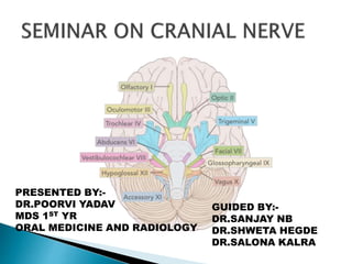
  • 1. PRESENTED BY:- DR.POORVI YADAV MDS 1ST YR ORAL MEDICINE AND RADIOLOGY GUIDED BY:- DR.SANJAY NB DR.SHWETA HEGDE DR.SALONA KALRA
  • 2.  INTRODUCTION  DEVELOPMENT OF CRANIAL NERVES  CRANIAL NERVE FUNCTIONAL COMPONENETS  OLFACTORY NERVE[I]  OPTIC NERVE[II]  OCULOMOTOR NERVE[III]  TROCLEAR NERVR[IV]  TRIGEMINAL NERVE[V]  ABDUCENT NERVE[VI]  FACIAL NERVE[VII]
  • 3.  VESTIBULOCOCHLEAR NERVE[VIII]  GLOSSPHYRYNGEAL NERVE[IX]  VAGUS NERVE[X]  ACCESORY NERVE[XI]  HYPOGLOSSAL NERVE[XII]  VASCULARIZATION OF CRANIAL NERVE  EXAMINATION OF CRANIAL NERVE  APPLIED ANATOMY
  • 4.
  • 5.  The 12 pairs of cranial nerves are part of the peripheral nervous system (PNS) and pass through foramina or fissures in the cranial cavity.  All nerves except one, the accessory nerve[XI],originate from the brain.  Having to similar somatic and visceral components as spinal nerves, some cranial nerve special sensory and motor components.
  • 6.  The special sensory components are associated with hearing, seeing , smelling , blanching , and tasting.  Special motor components include those that innervate skeletal muscles derived embryologically from the pharyngeal arches and not from somites.
  • 7.  In human embryology , six pharyngeal arches are designated, but 5th pharyngeal arch never develops.  Cranial nerves carry efferent fibers that innervate the musculature derived from the pharyngeal arch.
  • 8.  Innervation of the musculature derived from the five pharyngeal arches that do develop is as follows:- 1-first arch – trigeminal nerve 2-second arch - facial nerve 3-third arch – glossopharyngeal nerve 4-fourth arch – superior laryngeal branch of the vagus nerve 5- sixth arch – recurrent laryngeal branch of the vagus nerve
  • 9.
  • 10.
  • 11.
  • 12.
  • 13.
  • 14.
  • 15.
  • 16.  The Olfactory nerve [I] carries special afferent (SA) fibers for the sense of smell.  Its sensory neurons have:- - peripheral processes that act as receptors in the nasal mucosa. - central processes that return information to the brain.
  • 17.
  • 18.  The receptors are in the roof and upper parts of nasal cavity and the central processes , after joining into small bundles , enter the cranial cavity by passing through the cribriform plate of the ethmoid bone.  They terminate by synpasing with secondary neurons in the olfactory bulbs.
  • 20.
  • 21.
  • 22.
  • 23.
  • 24.  There are a number of disorder of the sense of smell, which can result:-  hyposomia  hypersomia  anosomia  parosmia
  • 25.  PERIPHERAL LESION:-  LOCOREGIONAL:-  Six can be mentioned:-  nasal polyps  Acute viral or bacterial rhinitis  Nonallergic eosinophillic rhinitis  Sinusitis  Allergic rhinitis  Tumor involvement
  • 26.  VIRAL:-  Herpes  Viral hepatitis TOXIC:- Tobacco Cement ammonia Cocaine
  • 27.  POST TRAUAMTIC  Fracture of the anterior floor of the base of the skull ith involvement of the CRIBRIFORM PLATE.  Usually caused by the occipital shock.  in order of frequency :- olfactroy filaments ,nasal and sinus cavities,cerebral centers  Immediate or delayed anosmia  Recovery is rare.
  • 28.  POSTOPERATIVE , IATROGENIC Seen after surgical traction on the frontal lobe  CENTRAL LESION:-  Frontal or occipital trauma  Neurotoxic medications
  • 30.  OTHER CAUSE:-  ENDOCRINE:-  Cushing syndrome  Hypothyroidism  Diabetes mellitus
  • 31.  NEUROLOGIC:- Alzheimer diseasse  Parkinson disease  Down syndrome  Multiple sclerosis  Epilepsy
  • 32.  KALLMANN DE MORSIER SYNDROME  Hypogonadotropic hypogonadism  Delayed puberty  Often transmitted by X chromosome  Congenital anosmia  Atresia of choana
  • 33.  Various  Paget  Renal failure  Hepatic failure  Alcoholic cirrhosis  AIDS IDOPATHIC Usually advanced age
  • 34.  The optic nerve carries SA fibers for vision.  These fibers return information to the brain from photoreceptors in the retina.  Neuronal processes leave the retinal receptors, join ,into small bundles, and are carried by the optic nerves to other components of the visual system in the brain.  The optic nerves enter the cranial cavity through the optic canals.
  • 35.
  • 36.
  • 37.
  • 38.
  • 39.
  • 40.
  • 41.
  • 42.
  • 43.
  • 44.
  • 45.  The occulomotor nerve carries two types of fibers :- - general soamtic efferent (GSE) fibers innervate most of the extra – ocular muscles. - general visceral efferent (GVE) fibers are part of the parasympathetic parts of the autonomic division of the PNS.
  • 46.
  • 47.  Motor: Innervates a number of the extraocular muscles.  Parasympathetic: Supplies the sphincter pupillae and the ciliary muscles of the eye.  Sympathetic: No direct function, but sympathetic fibres run with the oculomotor nerve to innervate the superior tarsal muscle (helps to raise the eyelid).
  • 48.
  • 49.
  • 50.
  • 51.
  • 52.
  • 53.
  • 54.
  • 55.
  • 56.
  • 57. Paralysis of occulomotor nerve causes:- ptosis external strabismus inability to move the occular globe upward , downward ,or medially. Intrinsic lesion produces Mydriasis ,that does not react to light.
  • 58.  Paralysis of the Trochlear nerve results Diplopia.  Paralysis of Abdunce nerve results CONVERGENT STRABISMUS in paralyzed eye. ABDUNCE NERVE PALSY
  • 59.
  • 60.  ETIOLOGY:-  Deficiency of thiamine(B1)  Characterized by clinical triad:-  Mental status change  Ataxia  Eye sign( nystagmus , opthalmoplagia)
  • 61.  The trochlear nerve is a cranial nerve that carries GSE fibers to innervate the superior oblique muscle , an extra- ocular muscle in the orbit.
  • 62.  The trigeminal nerve is the largest cranial nerve.  It carries general somatic afferent (GSA) and branchial efferent (BE) fibers.  The trigeminal nerve is the major general sensory nerve of the head , and also innervates muscles that move the lower jaw.
  • 63.
  • 64.  Trigeminal nerve has to roots:- (a) sensory (b) motor . Sensory root has 3 division:- (a) V1 – Ophthalmic (b) V2 – Maxillary (c) V3 – Mandibular
  • 65.  both motor and sensory root are attached ventrally to junction of pons and middle ceberal peduncle with motor root lying ventromedially to the sensory root.  Passes anteriorly in middle cranial fossa to lie below tentorium cerebelli in cavum trigeminale, here motor root lies to sensory root.
  • 66.
  • 67.  It has both efferent component for the muscle of mastication as well as some other cranial muscles, and an afferent component for teeth tongue and oral cavity , as well as most of the skin of face and head.  Trigeminal nerve has no preganglionic parasympathetic fibers, postganglionic parasymapthetic fibers travel along with its branch.
  • 68.
  • 69.  The first division of the sensory root of the trigeminal nerve is the OPTHALMIC NERVE.  This smallest branch serve as an afferent nerve for the conjunctiva , cornea , eyeball ,orbit , forehead , and ethmoid and frontal sinuses, plus a portion of the dura mater.
  • 70.
  • 71.  The FRONTAL NERVE is an afferent nerve located in the Orbit and is composed of merger of the SUPRAORBITAL NERVE from the forehead and anterior scalp and the SUPRATROCHLEAR NERVE from the bridge of the nose and medial portions of the upper eyelid and forehead.  The nerve courses along the roof of the orbit toward the superior orbital fissure of the sphenoid bone , where it is joined by the lacrimal and nasociliary nerves to form V1.
  • 72.
  • 73.  The lacrimal nerve serve as a afferent nerve for the lateral portion of the eyelid, conjunctiva , and lacrimal gland.  These nerves also responsible for the production of lacrimal fluid or tears.  The nerve runs posteriorly along the lateral roof of the orbit and then joins the frontal and nasociliary nerves near superior orbital fissure of the sphenoid bone to form V1.
  • 74.  Several afferent nerve branches converge to form the NASOCILIARY NERVE.  These branches include the INFRATROCHLEAR NERVE from the skin of the medial portion of the eyelid and the side of the nose, CILIARY NERVE to and from the eyeball and ANTERIOR ETHMOIDAL NERVE from nasal cavity and paranasal sinuses.  The nasociliary nerve is an afferent nerve that runs within the orbit , superior to the second cranial nerve to join the frontal and lacrimal nerves near superior orbital fissure of the sphenoid bone to form V1.
  • 75.
  • 76.
  • 77.  The second division V2 from the sensory root of trigeminal nerve is the MAXILLARY NERVE.  The afferent nerve branches of the maxillary nerve carry sensory information for the maxilla and overlying skin , maxillary sinuses , nasal cavity , palate ,and nasopharynx and a portion of the dura mater.  The maxillary is a nerve trunk formed in the ptrygopalatine fossa by the convergence of many nerves.
  • 78.  The largest contributor is the infraorbital nerve.  The tributaries of the maxillary nerve trunk include:-  1-zygomatic  2-anterior  3-middle  4-posterior  5-superior alveolar  6-greater and lesser palatine  7-nasopalatine nerves
  • 79.
  • 80.  The zygomatic is an afferent nerve composed of merger of the zygomaticofacial nerve and the zygomaticotemporal nerve in the orbit.  The zygomatic nerve courses posteriorly along the lateral orbit floor , enters the pterygopalatine fossa through the inferior orbital fissure , between the sphenoid bone and maxilla, and finally joins V2.
  • 81.  The infraorbital nerve is afferent nerve formed from the merger of cutaneous branches from the upper lip , medial portion of the cheek , lower eyelid and side of nose.  The infraorbital nerve then passes into the infraorbital foramen of the maxilla and travels posteriorly through infraorbital canal along with the infraorbital blood vessels ,where it is joined by the anterior superior alveolar nerve.
  • 82.  From the infraorbital canal and groove the infraorbital nerve passes into the ptrygopatatine fossa through the inferior orbital fissure.  After it leaves the infraorbital groove and within the pterygopalatine fossa , the infraorbital nerve receives the posterior superior alveolar nerve.
  • 83.  The ASA serves as an afferent nerve of sensation including pain for the maxillary central incisors , lateral incisors and canine as well as their associated tissues.  The ASA nerve originates from dental branches in the pulp tissue of these teeth that exit through the apical foramina.  The ASA nerve also innervates the overlying facial gingiva.
  • 84.  The ASA nerve then ascends along the anterior wall of the maxillary sinus to join the IO in the infraorbital canal.
  • 85.  The MIDDLE SUPERIOR ALVEOLAR NERVE serves as an afferent nerve of sensation (including pain), typically for the maxillary premolar teeth and the mesiobuccal root of the maxillary first molar and their associated periodontium and overlying buccal gingiva.  The MSA originates from dental branches in the pulp tissue that exit the teeth through the apical foramina , as well as interdental and interradicular branches from periodontium.
  • 86.  MSA nerve like PSA and ASA forms the dental plexus or nerve network in the maxilla.  The MSA nerve then ascends to join the IO nerve by running in the lateral wall of maxillary sinus.
  • 87.  The PSA joins the IO nerve in the pterygopalatine fossa.  The PSA nerve serve as an afferent nerve of sensation (including pain)for most potions of the maxillary molar teeth and their periodontium and buccal gingiva as well as the maxillary sinus.  Some branches of PSA nerve remain external to the posterior surface of the maxilla. These external branches provide afferent innervation for the buccal gingiva that overlies the maxillary molars.
  • 88.  Other afferent nerve branches of the PSA nerve originate from dental branches in the pulp tissue of the each of the maxillary molar teeth that exit the teeth by way of apical foramina.  These dental branches are then joined by interdental branches and interradicular branches from the periodontium forming a dental plexus or a nerve network in the maxilla for the region.
  • 89.  All these internal branches of PSA nerve exit from several posterior superior alveolar foramina on the maxillary tuberosity of the maxilla.  Both the external and internal branches of the PSA nerve then ascend together along the maxillary tuberosity , which forms the posterolateral wall of the maxillary sinus to join either the IO nerve or maxillary nerve.
  • 90.  The PSA typically provides afferent innervation for the maxillary second and third molars and the palatal and distal buccal root of the maxillary first molar , as well as the mucous membranes of the maxillary sinus.
  • 91.  The MSA nerve serves as an afferent nerve of sensation including pain typically for the maxillary premolar teeth and mesiobuccal root of the maxillary first molar and their associated periodontium and overlying bucaal gingiva.
  • 92.  MSA nerve originate from dental branches in pulp tissue that exit the teeth through apical foramina,as well as interdental and imterradicular branches from the periodontium.  MSA also form dental plexus in maxilla.  MSA nerve then ascends to join INFERIOR ALVEOLAR NERVE by running in the lateral wall of maxillary sinus.
  • 93.  Posterior superior alveolar nerve or PSA joins the INFERIOR ALVEOLAR NERVE in the pterygopalatine fossa.  The nerve serve as an afferent nerve of sensation (including pain) for most portions of the maxillary molar teeth and their periodontium and buccal gingiva as well as the maxillary sinus.
  • 94.  Some branches of the PSA nerve remain external to the posterior surface of maxilla.  These external branches provide afferent innervation for the buccal gingiva that overlies the maxillary molars.  Other afferent nerve branches of PSA nerve originate from dental branches in the pulp tissue of each of maxillary teeth and exit from the apical foramina of the teeth.
  • 95.  These dental branch are then joined by the interdental branches and interradicular branches from the periodontium and form the nerve plexus in the maxilla for the region.  All these internal branches of PSA nerve exit from several POSTERIOR SUPERIOR FORAMINA on the maxillary tuberosity.
  • 96.  Both the external and internal branches of PSA then ascend together along the maxillary sinus which form the posterolateral wall of the maxillary sinus.  Then it join the either IO or Maxillary nerve.  The PSA nerve typically provides afferent innervation for the maxillary 2nd & 3rd molars and palatal and distal buccal root of the 1st molar as well as mucous membranes of the maxillary sinus.
  • 97.  Both palatine nerves join the maxillary nerve from the palate.  GREATER PALATINE NERVE  It is also known as Anterior palatine nerve.  It is located between the periosteum and bone of the anterior hard palate.  Nerve serves as an afferent nerve for the posterior hard palate and posterior palatal gingiva.
  • 98.  Posteriorly,the GP nerve enters the greater palatine foramen in the palatine bone near the maxillary 2nd or 3rd molar to travel in the pterygopalatine canal along with greater palatine blood vessels. Lesser Palatine Nerve  It is also called Posterior Palatine nerve.  Serve as an afferent nerve for the soft palate and tonsillar tissues.
  • 99.  The lesser palatine nerve enters the lesser palatine foramen in the palatine bone near its junction with pterygoid process of sphenoid bone,along ith lesser palatine blood vessels.  The lesser palatine nerve join the greater palatine nerve in the pterygopalatine canal.
  • 100.  Both palatine nerves ascend through the pterygopalatine canal ,towards the maxillary nerve in the pterygopalatine fossa.  On the way , palatine nerves are joined by the lateral nasal branches, which are afferent nerves from the posterior nasal cavity.
  • 101.  The Nasopalatine nerve or NP originate from the mucosa of the anterior hard palate, palatal to the anterior maxillary teeth.  The right and left NP nerves enter the incisive canal by way of the incisive foramen , beneath the incisive papilla , thus exiting the oral cavity.  The NP serve as an afferent nerve for the anterior hard palate , and the palatal gingiva of the maxillary anterior teeth as well as the nasal septal tissues.
  • 102.  The third division (V3) of the trigeminal nerve is the mandibular nerve.  Which is a short main trunk formed by the merger of a smaller anterior trunk and a larger posterior trunk in the infraorbital fossa,before the nerve passes through the foramen ovale of the sphenoid bone.
  • 103.  The mandibular nerve then joins with the opthalmic and maxillary nerve to form the TRIGEMINAL GANGLION of the TRIGEMINAL NERVE.
  • 104.
  • 105.  Few branches arises from the V3 trunk before its separation to Anterior and Posterior trunks.  These branches from the undivided mandibular nerve include the MENINGEAL BRANCHES, which are afferent nerves for portions of the Dura matter.
  • 106.  also from the undivided branch from the mandibular nerve are MASCULAR NERVE.  They are efferent nerves for the  medial pterygoid  tensor tympani  tensor veli palatine
  • 107.  Anterior trunk formed by the merger of the:-  Buccal nerve  additional muscular nerve branches  Posterior trunk formed by the merger of the :-  auriculotemporal nerve  lingual nerve  inferior alveolar nerve
  • 108.
  • 109.  The buccal nerve also known as long buccal nerve serve as an afferent nerve for skin of cheek , buccal mucous membranes and buccal gingiva of the mandibular posterior teeth.  The nerve is located on the surface of the buccinator muscle.  The buccal nerve then travels posteriorly in the cheek, deep to the masseter muscle.
  • 110.  at the level of the occlusal plane of the last manibular molar , the nerve crosses in front the anterior border of the ramus of the mandible then goes between the two heads of the lateral pterygoid muscle to join the anterior trunk of the V3.
  • 111.  They arises from the motor root of the trigeminal nerve.  Muscular branches are:-  1- deep temporal nerve  2- masseteric nerve  3- lateral pterygoid muscle  1- DEEP TEMPORAL NERVE  Usually 2 in number  Anterior and posterior,efferent nerves  They passes between the sphenoid bone and superior border of the lateral ptrygoid muscle then turn around the infratemporal crest of the sphenoid bone to end in the deep surface of the temporal muscle that they innervate.
  • 112.  2- MASSETRIC NERVE  It is also an efferent nerve.  It is passes between sphenoid bone and superior border of the lateral pterygoid muscle.  The nerve then accompanies the masseteric blood vessels through the mandibular notch to innervate the massetric muscle.
  • 113.  3- LATERAL PTERYGOID MUSCLE  A small sensory branch goes to the temporalmandibular joint,after the short course it enters the deep surface of the lateral pterygoid muscle.  Between the muscle’s two heads of origin and serve as an efferent nerve for the muscle.
  • 114.
  • 115.  It travels with superficial temporal artery and vein serve as an afferent nerve for the scalp and external ear.  It also carries the postganglionic parasympathetic fibers to the parotid salivary gland.  The parasympathetic fibers arises from the lesser petrosal branch of the IXth cranial nerve.
  • 116.  The nerve runs deep to the lateral pterygoid muscle and the neck of the mandible ,then splits to encircle the middle meningeal artery and finally join the posterior trunk of the V3.
  • 117.  It is formed by the afferent branches of the body of the tongue that travels along the lateral surface of the tongue.  Then it passes posteriorly, passing from the medial to the lateral surface of the duct of the sub mandibular galnd by going under the duct.
  • 118.  The lingual nerve communicate with the submandibualr ganglion located superior to the deep lobe of the submandibular gland.  At the base of the tongue, the lingual nerve ascends and runs between the medial pterygoid muscle and the mandible, anterior and slightly medial to the inferior alveolar nerve.  The lingual nerve then continues to travel upward to join the posterior trunk of V3.
  • 119.  It is serve as an afferent nerve for the general sensation for the body of the tongue, floor of the mouth and lingual gingiva of the mandibular teeth.
  • 120.  It is a main branch of the lower jaw.  It is an afferent nerve formed by the merger of the MENTAL NERVE & INCISIVE NERVE.  After forming inferior alveolar nerve travel posterior through the mandibular canal along with the inferior alveolar artery and vein.  Join by the interdental and interradicualr branches of the peridontium and form the nerve plexus in the region.
  • 121.  then it exit the mandible through mandibular foramen , where it is joined by the mylohyoid nerve.  Then travels lateral to the pterygoid muscle and between the ramus of the mandible and sphenomandibular ligamentwwithin the pterygomandibualr space.  Then it join the posterior trunk of the V3.
  • 122.  It is composed of the external branches that serve as an afferent nerve for the chin ,lower lip ,labial mucosa of premolars and anterior teeth.  The nerve then enters the mental foramen on the anterolateral surface of the mandible, usually between the apices of the mandibular premoalrs.  Then it merge with the incisive nerve to form the inferior alveolar nerve in the mandibular canal.
  • 123.  It is an afferent nerve composed of dental branches from the mandibular premolars and anterior teeth that originate in the pulp tissue exit the teeth through the apical foramina , and then join with interdental branches from the surrounding periodontium forming the dental plexus in the region.  It is merge to the mental nerve just posterior to the mental foramen to form the inferior alveolar nerve in the mandibular canal.
  • 124.  It is serve as an afferent nerve for the mandibular premolars and anterior teeth.  MYLOHYOID NERVE  After the inferior alveolar nerve exits the mandibular foramen, small branches occur called mylohyoid nerve.  It pierces the sphenomandibular ligament and runs inferiorly and anteriorly in the mylohyoid groove then onto the inferior surface of the mylohyoid muscle.
  • 125.  It serves as an efferent nerve to the mylohyoid muscle and anterior belly of digestric muscle.
  • 126.
  • 127.
  • 128.
  • 129.
  • 130.
  • 131.
  • 132.
  • 133.  It is distinct neurological syndrome of deficits that can arise due to the closeness of the cerebellopalatine angle to specific cranial nerve.  ETIOLOGIES:-  Schwannoma  Meningioma  Tumor of the petrous bone
  • 134. CLINICAL FEATURES:- Unilateral sensorineural hearing loss Unilateral tinnitus Disequilibrium (vertigo) rare NERVE AFFECTED:- cranial nerve V,VII,VIII,sometimes IX
  • 135.  Gradenigo syndrome first describe by GUISEPPE GRADENIGO in 1904.  ETIOLOGIES:-  petrositis  tumor of the apex of the petrous bone  CLINICAL FEATURES  Periorbital unilateral pain related to trigeminal nerveinvolvement  Diplopia due to VI nerve paralysis  Persistent otorrhea
  • 136.  LOCALIZATION:-  Apex of petrosal  NERVE INVOLVED:-  V  VI
  • 137.  It is rare disorder.  CLINICAL FEATURE:-  Unilateral headache  Orbital pain  Weakness &paralysis of extraocular muscle  LOCALZIATION :-  Lateral wall of cavernous sinus  ETIOLOGIES:-  Aneurysm /thrombosis of cavernous sinus  NERVED INVOLVED:-  III,IV, VI, V1 ,V2
  • 138.  ETIOLOGIES:-  Aneurysm /thrombosis of cavernous sinus  NERVED INVOLVED:-  III,IV, VI, V1 ,V2
  • 139.  LOCALIZATION :-  Superior orbital fissure  ETIOLOGIES:-  Invasive tumor of the sphenoid sinus  Aneruysm  Inflammatory granuloma  Invasive sellar tumor
  • 140.  NERVES INVOLVED:-  III  IV  VI  V1
  • 141.  It is very rare pathological condition.  Resulting from neuralgia of nasocilliary nerve.  It is also known as nasal nerve syndrome.  CLINICAL FEATURE:-  Severe unilateral pain , generated by irritation of the ciliary ganglion.  Affected areas:-  Above and outside of nose  Above the inner canthus  Inferior angle of medial tarsal ligament
  • 142.  ASSOCIATED SYMPTOMS:-  Unilateral vasomotor obstruction of the nasal fossa  Sneezing  Hydrorrhoea  Cutaneous hyperasthesia  Conjectival hypermia  Keratitis  Corneal ulcer
  • 143.  There might be:-  slight inflammatory swelling of upper eyelid  Photophobia  Ciliary & conjectival infection
  • 144.  DIFFERENTIAL DIAGNOSIS  cluster headache  Trigeminal neuralgia  Demyelinating disease
  • 145.  It is also known as shingles.  It is secondary manifestation of varicella zoster infection.  CLINICAL PRESENTATION:-  Form of multiple vesicles along the course of one of the three division.  Prodrome fever  Headache  Rash  Malaise  nausea
  • 146.  Rash  Malaise  Nausea  Hutchinson’s sign
  • 147.  ORAL MANIFESTATION  Vesicles affecting the palate , uvula ,tonsils , tongue Buccal mucosa floor of the mouth -devitalized teeth - internal resorption - Pulpal necrosis - Severe periodontitis
  • 148.
  • 149.
  • 150.
  • 151.
  • 152.
  • 153.
  • 154.  It also known as STYLOHYOD SYNDROME,STYLOID SYNDROME , STYLOID –STYLOHYOID SYNDROME ,STYLOID CAROTID ARTERY SYNDROME.  It is first described by Watt Weems eagle in 1937.  It is characterized by elongated styloid process , or calcification of stylohyoid ligament.  Classical eagle syndrome is ipsilateral.
  • 155.  CLINICAL FEATURE:-  Sharp , shooting pain in the jaw ,back of the throat , base of tongue , ears ,neck or face.  Difficulty swallowing  Pain from chweing , swallowing , turning the neck or touching the back of the throat.  tinnitus
  • 156.
  • 157.  NERVE INVOLVED:-  Glossophyrangeal  Facial nerve  Hypoglossal nerve  Vegus nerve  V3
  • 158.  DIIFERENTIAL DIAGNOSIS  Pulpitis  Idiopathic trigeminal neuralgia  TMJ disorder
  • 159.  The abducent nerve carries GSE fibers to innervate the lateral rectus muscle in the orbit.
  • 160.  Nerve exits the skull through the superior orbital fissure of sphenoid bone on its way to the orbit.  Runs through the sinus , close to the internal carotid artery.
  • 161.
  • 162.  It contains both afferent and efferent components.  Nerve carries the Efferent component for the:-  muscle of facial expression  preganglionic parasympathetic fiber of the lacrimal gland , submandibular gland and sublingual gland.  Afferent component serves:-  tiny patch skin behind the ear  taste sensation  body of the tongue
  • 163.
  • 164.  Nerve leaves the cranial cavity by passing through the internal acoustic meatus in the petrous region of the temporal bone.  Within the bone the nerve gives off small efferent branch to the muscle in the middle ear and two large branches GREATER PETROSAL AND CHORDA TYMAPNI.  Both of which carry parasympathetic fibers.
  • 165.
  • 166.  The main trunk emerges from the skull through the stylomastoid foramen of the temporal bone and gives off two branches POSTERIOR AURICULAR NERVE and NERVE TO POSTERIOR BELLY OF DIGESTRIC MUSCLE &STYLOID MUSCLE.  Then passes into the parotid salivary gland divides into several branches &innervate muscles of facial expression.
  • 167.  It carries the efferent fibers preganglionic parasymapthrtic fibers to ptrygopalatine ganglion in pterygopalatine fossa.  Also carries the afferent fibers for taste sensation in the palate.
  • 168.
  • 169.  It arises before the facial nerve exit from the skull, join with the branches of the maxillary nerve of the trigeminal nerve,to be carried to the lacrimal gland , nasal cavity minor salivary gland of the hard and soft palate.  It also carries afferent nerve fibers for taste sensation in the palate.
  • 170.  It is the small branch of the facial nerve.  Parasympathetic efferent nerve for the submandibular and sublingual galnd.  Afferent nerve for the taste sensation.  It is branching off the facial nerve within the petrous portion of the temporal bone.
  • 171.
  • 172.  Crosses the medial surface of the tympanic membrane.  Exit the skull by petrotymapni fissure,travel with the lingual nerve along the floor of the mouth.
  • 173.
  • 174.  It is arises opposite the pyramid of the middle ear , and supplies the stapedius muscle.  The muscle dampens excessive vibrations of the stapes caused by high - pitched sounds.  Paralysis of stapedius muscle caused Hyperacusis.
  • 175.  It arises just below the stylomastoid foramen.  It ascends between the mastoid process and the external acoustic meatus.  It supplies :-  auricularis posterior  occipitalis  intrinsic muscles on the back of the auricle.
  • 176.
  • 177.  It arises close to the stylomastoid foramen.  It is short and supplies to posterior belly of digestric muscle.
  • 178.  It arises with the digestric branch.  It is long and supplies the stylohyoid muscle.  THE TEMPORAL BRANCH  It crosses the zygomatic arch and supply:-  auricularis anterior  Auricularis superior  Intrinsic muscles on the lateral side of the ear  Frontalis  Orbicularis oris  Corrugator supercilii
  • 179.  It runs across the zygomatic bone and supplies the orbicularis oculi.  THE BUCCAL BRANCHES  It is two in number.  Upper buccal nerve:- it runs above the parotid duct.  Lower buccal branch:-it runs below the parotid duct.  Innervates :-  They supply muscles in that vicinity especially the buccinator.
  • 180.  It runs below the angle of the mandible deep to platysma.  It crosses the body of the mandible and supplies muscle of lower lip and chin.  THE CERVICAL BRANCH  It emerges from the apex of the parotid gland and runs downwards and forwards in the neck.  It supply the platysma.
  • 181.  For effective coordination between the movements of the muscles of the first , second , and the third branchial arches , the motor nerves of the three arches communicate with each other.  The facial nerve also communicates with sensory nerves distributed over its motor territory.
  • 182.
  • 183.
  • 184.
  • 185.
  • 186.
  • 187.
  • 188.  PERIPHERAL FACIAL PARALYSIS  CLINICAL PRESENTATION:-  Unilateral facial weakness  Loss of taste  Hyperacusis  Decreased salivation  Decreased tear secretion
  • 189.
  • 190.
  • 191.  It is also known as BOGOARD’S SYNDROME.  It is rare complication.  ETIOLOGY:-  Aberent regeneration after Trauma  CLINICAL PRESENTATION  Lacrimation during eating
  • 192.  Involvement of geniculate ganglia by HERPES ZOSTER.  CLINICAL PRESENTATION:-  Hyperacusis  Loss of lacrimation  Loss of sensation of taste in anterior two third of tongue  Bell’s palsy  Lack of salivation  Vesicle on auricle
  • 193.
  • 194.  It is rare condition.  It is serious autoimmune disorder in which the immune system attacks healthy nerve cells in peripheral nervous syndrome.  CLINCAL PRESENTATION:-  Tingling sensation in toes , feet , and legs .  Muscle weakness  Difficulty moving eyes face chewing or swallowing  Severe lower back ache
  • 195.  Rapid heart rate  Difficulty in breathing  paralysis
  • 196.  It is rare neurological disorder  Characterized by paralysis of multiple cranial nerve , most often the VI and VII cranial nerve.  CLINICAL PRESENTATION:-  Micrognathia  Microstomia  Cleft palate  Dental abnoramalties  Strabismus
  • 197.  Ptosis  Hypotonia  Hypoacusis  Mask like face  Incompetent lips  Ulceration
  • 198.
  • 199.  It is also known as BAILLARGER’S SYNDROME , DUPUY’S SYNDROME , AURICULOTEMPORAL SYNDROME , FREY-BAILLARGER SYNDROME.  It is rare postoperative phenomenon following salivary gland surgery , facelift procedures, trauma.  It is characterized by gustatory sweating and flushing in the preauricular area in response to mastication or salivary stimulation.  It was first described by Lucie Frey in 1923 and was termed auriculotemporal syndrome.
  • 200.
  • 201.  CLINICAL PRESENTATION:-  flushing  Sweating  Burning  Itching
  • 202.  It is serve as an afferent nerve for hearing and balance.  Nerve enters the cranial nerve through the internal acoustic meatus of the temporal bone.
  • 203.  LARGE VESTIBULAR AQUEDUCT SYNDROME:-  It is congenital malformation of cochlea and semicircular canal.  Hypoacusis
  • 204.  It is an illusion rotatory movement due to disturbed orientation of the body in space.  It is due to disease of vestibular nerve.  ETIOLOGIES:-  Meniere’s disease  Vestibular neuritis  Head or neck injury  Tumor or stroke  Migraine headache
  • 205.  CLINICAL PRESENTATION:-  Dizziness  Nausea  Nystagmus  Sweating  Hearing loss in one ear  tinnitus
  • 206.  It is very frequent disorder.  It consist of perception of a sensation of sound localized in one or both ear.  TYPE OF TINNITUS  Subejctive  objective
  • 207.
  • 208.
  • 209.
  • 210.
  • 211.
  • 212.
  • 213.
  • 214.
  • 215.  Carries the effernt component for the phyryngeal muscle, stylopharygeus muscle and preganglionic parasympathetic innervation for the parotid galnd.  Afferent component for the:-  pharynx  taste  general sensation of the base of the tongue
  • 216.
  • 217.  Pharyngeal branch – combines with fibres of the vagus nerve to form the pharyngeal plexus. It innervates the mucosa of the oropharynx.  Lingual branch – provides the posterior 1/3 of the tongue with general and taste sensation
  • 218.  Tonsillar branch – forms a network of nerves, known as the tonsillar plexus, which innervates the palatine tonsils.
  • 219.  Lesion of glossophyrangeal nerve causes:-  Absence of taste from posterior one third of tongue  Absence of secretion of parotid gland  Loss of pain sensations from tongue , tonsil , pharynx , and soft palate  Gag reflex is absent
  • 220.  It is sharp ,shooting severe attack of pain affecting posterior part of pharynx.  JUGULAR FORAMEN SYNDROME  It is also known as VERNET’S SYNDROME.  It involved usually IX , X , XI cranial nerve  CLINICAL PRESENTATION:-  Dysphonia  Soft palate dropping  Deviation of uvula towards the normal side
  • 221.  Dysphagia  Loss of sensory function from the posterior 1/3rd of tongue  Sternocleidomastoid and trapezius muscle paralysis  ETIOLOGIES:-  1-Tumors:-  Glommus jugulare tumors  Meningioma
  • 222.  2-Inflammation:-  Meningitis  Malignant otitis externa  Sarcoidosis  3- Trauma  4- narrowing of jugular foramen
  • 223.  It is rare disease.  It involved the IX X XI XII cranial nerve.  Characterized by:-  Constricted pupil(miosis)  Drooping of upper eyelid(ptosis)  Absence of sweating (anhidrosis)  enopthalmous
  • 224.
  • 225.
  • 226.
  • 227.
  • 228.
  • 229.
  • 230.  NERVE INVOLVED:-  Facial nerve  Occulomotor nerve
  • 231.  It is also known as Pnumogastric nerve.  Large efferent component for the muscles of the:-  soft palate  larynx  Parasympathetic fibers for the:-  thorax  heart  stomach  thymus gland
  • 232.  Carries small afferent fibers for :-  small amount of skin around ear  taste sensation  epiglottis
  • 233.
  • 234.  In the Head :-  Within the cranium, the auricular branch arises. This supplies sensation to the posterior part of the external auditory canal and external ear.  In the Neck  In the neck, the vagus nerve passes into the carotid sheath, travelling inferiorly with the internal jugular vein and common carotid artery. At the base of the neck, the right and left nerves have differing pathways:
  • 235.  The right vagus nerve passes anterior to the subclavian artery and posterior to the sternoclavicular joint, entering the thorax.  The left vagus nerve passes inferiorly between the left common carotid and left subclavian arteries, posterior to the sternoclavicular joint, entering the thorax.
  • 236.  Several branches arise in the neck:  Pharyngeal branches – Provides motor innervation to the majority of the muscles of the pharynx and soft palate.  Superior laryngeal nerve – Splits into internal and external branches. The external laryngeal nerve innervates the cricothyroid muscle of the larynx. The internal laryngeal provides sensory innervation to the laryngopharynx and superior part of the larynx.
  • 237.  Recurrent laryngeal nerve (right side only) – Hooks underneath the right subclavian artery, then ascends towards to the larynx. It innervates the majority of the intrinsic muscles of the larynx
  • 238.  In the Thorax  In the thorax, the right vagus nerve forms the posterior vagal trunk, and the left forms the anterior vagal trunk. Branches from the vagal trunks contribute to the formation of the oesophageal plexus, which innervates the smooth muscle of the oesophagus.
  • 239.  Two other branches arise in the thorax:  Left recurrent laryngeal nerve – it hooks under the arch of the aorta, ascending to innervate the majority of the intrinsic muscles of the larynx.  Cardiac branches – these innervate regulate heart rate and provide visceral sensation to the organ.  The vagal trunks enter the abdomen via the oesophageal hiatus, an opening in the diaphragm.
  • 240.  In the Abdomen  In the abdomen, the vagal trunks terminate by dividing into branches that supply the oesophagus, stomach and the small and large bowel (up to the splenic flexure)
  • 241.
  • 242.
  • 243.  It carries the efferent component for the TRAPEZIUS AND STERNOCLEIDOMASTOID muscle as well as for muscle of soft palate and pharynx.  It passes through the skull by the way of jugular foramen between occipital and temporal bone.
  • 244.
  • 245.
  • 246.
  • 247.  It function as efferent nerve for:-  intrinsic muscle  extrinsic muscle of the tongue  It exit the skull through the hypoglossal canal In the occipital bone.
  • 248.
  • 249.
  • 250.
  • 251. TAPIA’S SYNDROME: -it invoved the X , XII cranial nerve. CLINICAL PRESENTATION:- Dysphonia Deviation of tongue Dysphagia
  • 252.  A bulbar palsy refers to disease affecting the glossopharyngeal, vagus, accessory and hypoglossal nerves and is due to lower motor neuron pathology.  CLINICAL PRESENTATION:-  dysphagia,  dysarthria,  flaccid pareses,  atrophy and fasciculation of muscles supplied by those cranial nerves and fibrilliation of the tongue
  • 253.  weakness of the palate  reduced or absent gag reflex,  dribbling of saliva and  nasal speech.
  • 254.  The inferolateral trunk of carotid siphon vascualrize the:-  V1  III  IV  VI
  • 255.  The ascending pharyngeal artery vacularize :-  IX  X  XI  XII  The middle meningeal and accessory arteries ,the inferolateral trunk of the carotid siphon and ascending pharyngeal artery have abundant anastomoses.
  • 256.  Textbook of anatomy ;gray’s 39th edition  Textbook of antomy of the head and neck- margaret j. fehrenbach, susan . Herring  Textbook of cranial nerve- Dominique doyon ,Kathlyn marsot – dupuch ,Jean –paul Francke  Textbook of human antomy-B D Chaurasia vol 3 6th edition  Textbookof oral medicine – Burket’s 10th edition  Journal of medical case reports ISSN :1752-147  Erol FS,Kaplan M, Ozveren MF.Jugular foramen syndrome caused by choleastatoma.Clin Neurol Neurosurg.2005jun;107(4):342-6. PMID:15885397
  • 257.  Chandrashekhar S , PeterfreundRA.Horner’s syndrome following very low concentration Bupivacaine infusion for labor epidural analgesia .J Clin Anesth. 2003;15:217-19  Malik U,Sunil MK ,Gupta C , Kumari M.Trigeminal HerpesZoster:Early Recognition and Treatment-A Case Report.J Indian Acad Oral Radiol 2017;2;350-3.
  • 258.  Singh MP,Mukherji A , Vats Ak. Arare case of Charlin’s syndrome.J Indian Acad Oral Med Radiol 2017;29;129-31  Raina D , Gothi R, Rajan S .J Radiol Imaginig.2009 May;19(2):107-108  Motz M K , Kim J Y,Otolaryngeal Clin North Am.2016 Apr;49(2):501-501  Adams et al., Principles of Neurology, 6th ed, p1091; Brain 1992 Dec;115(Pt 6):1889-1900)