SlideShare a Scribd company logo
1 of 7
Download to read offline
CLINICAL SCIENCE
Clinical Outcomes After Implantation of 320˚-Arc Length
Intrastromal Corneal Ring Segments in Keratoconus
Leonardo Torquetti, MD, PhD,* Priscila Cunha, MD,† Allan Luz, MD, PhD,‡ Sérgio Kwitko, MD,§
Márcio Carrion, MD,¶ Guilherme Rocha, MD,k Armando Signorelli, MD,** Sandro Coscarelli, MD,††
Guilherme Ferrara, MD,‡‡ Frederico Bicalho, MD,† Renato Neves, MD,§§ and
Paulo Ferrara, MD, PHD‡‡
Purpose: To evaluate the clinical outcomes after implantation of
a new 320°-arc length Ferrara intrastromal corneal ring segment
(320-ICRS) in eyes of patients with keratoconus.
Methods: A multicentric nonrandomized study was conducted in
which a new 320-ICRS was placed in 138 eyes of 130 patients with
keratoconus. Uncorrected distance visual acuity (UDVA), corrected
distance visual acuity (CDVA), keratometry, corneal volume,
asphericity, lines of vision gain/loss, and vectorial analysis were
assessed preoperatively and at the final follow-up visit after
the procedure.
Results: The mean follow-up time was 6.2 6 3.3 (SD) months
(range 3–12 months). Mean UDVA improved from 20/250 to 20/60.
Mean CDVA improved from 20/100 to 20/40. Mean Q improved
from 21.12 6 0.49 preoperatively to 20.28 6 0.51 postoperatively
(P , 0.001). Mean corneal volume increased from 56.2 6 4.28 mm3
preoperatively to 57.6 6 4.74 postoperatively (P , 0.001). Mean
Km reduced from 53.3 6 5.5 D preoperatively to 47.8 6 4.6 D
postoperatively (P , 0.001). The change in UDVA, CDVA, and
topographic astigmatism was statistically significant (P , 0.0001).
Conclusions: The 320-ICRS can efficiently and safely improve
visual acuity in keratoconus, modifying the corneal shape to a more
physiologic, aspheric shape.
Key Words: Ferrara ring, keratoconus, intrastromal ring segments,
320-ICRS
(Cornea 2018;00:1–7)
Keratoconus is progressive corneal thinning of unknown
cause in which the cornea assumes a conical shape,
with progressive irregular astigmatism and deterioration of
visual acuity.1 In the general population, the incidence of
keratoconus is estimated to be between 50 and 230 per
100,000.2
Intrastromal corneal ring segments (ICRS) represent
substantial evolution in management of keratoconus. More-
over, long-term data on ICRS procedures demonstrated
promising results in topographic regularity, uncorrected
distance visual acuity (UDVA) and corrected distance visual
acuity (CDVA), indicating its effect in avoiding or at least
postponing corneal transplantation in patients with keratoco-
nus.3–8 The main advantages of ICRS are safety,4 reversibil-
ity,9 stability,10 and the fact that the surgical process does not
affect the central corneal visual axis.
The 320-arc length Ferrara intrastromal corneal ring
segment (AJL, Vitoria, Spain) (320-ICRS) is a new unique
intracorneal ring design specially developed for more
advanced types of keratoconus. It is available in a diameter
of 5.7 mm and a thickness range of 150 to 300 mm. To our
knowledge, there are no reports on the effect of insertion
or implantation of a 320-ICRS on the postoperative
outcome. This study aims at evaluating the clinical
outcomes after implantation of a 320-ICRS in eyes of
patients with keratoconus.
METHODS
In a multicentric study, we prospectively evaluated the
clinical outcomes of implantation of a 320-ICRS in 138 eyes
of 130 patients with keratoconus. The patients were recruited
and operated in 10 different surgical centers in Brazil. All
patients were informed about inclusion in the study and
provided informed consent in accordance with the Declara-
tion of Helsinki. In addition, the study was approved by the
local ethics committee of each center.
Forty-five eyes had the procedure performed using the
manual technique; the femtosecond laser was used for
tunnel creation in 93 eyes. After complete ophthalmic
examination and a thorough discussion of the risks and
benefits of surgery, the patients provided written informed
consent. The primary indication for Ferrara ring implanta-
tion was contact lens intolerance and progression of ectasia.
The progress of the disease was defined by worsening of
Received for publication January 28, 2018; revision received May 31, 2018;
accepted June 2, 2018.
From the *Center for Excellence in Ophthalmology, Pará de Minas, Brazil;
†Hospital São Geraldo, HC-UFMG, Belo Horizonte, Brazil; ‡Hospital de
Olhos de Sergipe, Aracaju, Brazil; §Oftalmocentro, Porto Alegre, Brazil;
¶Carrion Eye Clinic, Santo Ângelo, Brazil; kHospital de Olhos de Brasília,
Brasília, Brazil; **Centro Campineiro de Microcirurgia Ocular, Campinas,
Brazil; ††Ennio Coscarelli Eye Clinic, Belo Horizonte, Brazil; ‡‡Paulo
Ferrara Eye Clinic, Belo Horizonte, Brazil; and §§EyeCare, São Paulo, Brazil.
The authors have no funding to disclose.
The authors have no financial interest in Ferrara intrastromal corneal ring segments,
except P. Ferrara and G. Ferrara, who are shareholders of Ferrara Ophthalmics.
The other authors have no conflicts of interest to disclose.
Correspondence: Leonardo Torquetti, MD, PhD, Rua Capitão Teixeira, 415-
B, Nossa Senhora das Graças-Pará de Minas, MG, Brazil 35660-051
(e-mail: leotorquetti@gmail.com).
Copyright © 2018 Wolters Kluwer Health, Inc. All rights reserved.
Cornea  Volume 00, Number 00, Month 2018 www.corneajrnl.com | 1
Copyright Ó 2018 Wolters Kluwer Health, Inc. Unauthorized reproduction of this article is prohibited.
UDVA and CDVA, increasing intolerance to contact lens
wear, and progressive corneal steepening documented
by topography.
CLINICAL MEASUREMENTS
Preoperative and Postoperative Evaluation
All patients had complete ophthalmologic examination
preoperatively and postoperatively. The outcome analysis
comprised UDVA, CDVA, spherical equivalent (SE), central
pachymetry, corneal volume, refractive error, corneal topo-
graphic astigmatism, and minimum and maximum keratom-
etry (K) values. All data were obtained before and at the final
visit after ICRS implantation. CDVA, slip-lamp evaluation,
refraction, corneal topography, fundoscopy, and tonometry
were performed at each control visit. Anterior segment
parameters were obtained by a corneal tomographer (Oculus
Pentacam, Wetzlar, Germany). All clinical examinations were
performed in a standardized manner according to the guide-
lines of the multicentric study.
On the first postoperative day, slit-lamp biomicro-
scopic examination was performed. Healing of the wound
and migration of the segments were evaluated. At final
follow-up examination, manifest refraction, UDVA and
CDVA, and slit-lamp and topographic examinations
were performed.
Surgical Technique
The 320-ICRS was implanted using the manual tech-
nique or femtosecond laser–assisted procedure.
Manual Technique
Surgery was performed under topical anesthesia after
miosis was achieved with 2% pilocarpine. An eyelid specu-
lum was used to expose the eye, and 2.5% povidone–iodine
eye drops were instilled onto the cornea and conjunctival cul-
de-sac. The visual axis was marked by pressing the Sinskey
hook on the central corneal epithelium while asking the
patient to fixate on the corneal light reflex of the microscope
light. Using a marker tinted with gentian-violet tinted, a 5.0-
mm optical zone and incision site were aligned to the desired
axis in which the incision would be made.
The depth of a 1.0-mm, square diamond blade was set
at 80% of corneal thickness at the incision site, and this knife
is used to make the incision. Using a “stromal spreader,”
a pocket was formed on each side of the incision. Specific
380-degree circular dissecting spatulas were inserted through
the incision and gently pushed with some quick, rotary “back
and forth” tunneling movements. After channel creation, the
320-ICRS was inserted using a modified McPherson forceps.
The ICRS was adequately positioned with the aid of a Sinskey
hook.7
Femtosecond Laser Technique
The surgical procedure was performed under sterile
conditions and topical anesthesia. Purkinje reflex was chosen
as the central point and was marked. A 5-mm marker was
used to locate the exact ring channel. The tunnel depth was
set at 75% of the thinnest corneal thickness on the tunnel
location in the femtosecond laser cases.11–16
A 60-kHz femtosecond laser (LDV; Ziemer, Switzer-
land) or the IntraLase was used to create the ring channel.
Particular attention was given to centralizing the suction ring
to mark the central point to minimize decentration. The
channel’s inner diameter was set to 4.4 mm, the outer diameter
was 5.6 mm, the ring energy used for channel creation was
1.30 J, and the entry cut energy was 1.30 J. Channel creation
timing with the femtosecond laser was 15 seconds. The 320-
ICRS was implanted immediately after channel creation
before the bubbles disappeared, which reveals the exact tunnel
location. After channel creation, the 320-ICRS was inserted
using a modified McPherson forceps. The 320-ICRS was
properly positioned with the aid of a Sinskey hook.
The postoperative regimen consisted of moxifloxacin
0.5% (Vigamox; Alcon, Forth Worth, TX) and dexametha-
sone 0.1% (Maxidex; Alcon, Forth Worth, TX) eye drops 4
times daily for 2 weeks. The patients were instructed to avoid
rubbing the eye and to use preservative-free artificial tears
frequently—polyethylene glycol 400 0.4% (Systane; Alcon,
Forth Worth, TX).
Statistical Analysis
Statistical analysis was performed using Minitab soft-
ware (2007, Minitab Inc.). The Student t test for paired data
was used to compare preoperative and postoperative data.
RESULTS
There were 62 females and 68 males with a mean age of
27 6 9.9 years (range 10–63 years). The mean follow-up time
was 6.2 6 3.3 (SD) months (range 3–12 months). Pre-
operative and postoperative UDVA, CDVA, SE, and pachy-
metry were obtained from all patients (Table 1). Median
UDVA improved from 20/250 preoperatively to 20/60 post-
operatively (P , 0.001). Mean CDVA improved from 20/100
TABLE 1. Preoperative and Postoperative Evaluated
Parameters
Preoperative Postoperative P
UDVA* 20/250 20/60 ,0.001
CDVA 20/100 20/40 ,0.001
CDVA* 20/60 20/30 ,0.001
SE 27.02 (64.8) 23.29 (63.8) ,0.001
Pachymetry 430 (647.8) 447 (653.8) ,0.001
Q 21.25 (60.49) 20.31 (60.51) ,0.001
Corneal volume 56.2 (64.28) 57.6 (64.74) ,0.001
Topographic astigmatism 4.62 (62.87) 2.74 (62.07) ,0.001
K1 50.5 (64.9) 46.0 (64.4) ,0.001
K2 55.2 (65.6) 49.0 (64.6) ,0.001
Km 53.3 (65.5) 47.8 (64.6) ,0.001
*Median DQ = 0.94.
Torquetti et al Cornea  Volume 00, Number 00, Month 2018
2 | www.corneajrnl.com Copyright © 2018 Wolters Kluwer Health, Inc. All rights reserved.
Copyright Ó 2018 Wolters Kluwer Health, Inc. Unauthorized reproduction of this article is prohibited.
preoperatively to 20/40 postoperatively (P , 0.001). Median
CDVA improved from 20/60 preoperatively to 20/30 post-
operatively (P , 0.001). Mean preoperative SE was 27.02 6
4.80 D preoperatively and 23.29 6 3.8 D postoperatively (P
, 0.001). Mean central pachymetry increased from 430 6
47.8 D preoperatively to 447 6 53.8 D postoperatively (P ,
0.001). Two or more lines of CDVA were gained in 87% of
eyes. Four percent of eyes lost 1 line of vision. All these eyes
had preoperative CDVA better than 20/40. There was
progressive improvement in CDVA and lines of CDVA
gained from 3 to 6 months of follow-up (Figs. 1, 2).
During the follow-up period after ICRS implantation,
there were no cases of segment extrusion. There were 4
cases of migration, which were excluded from the statis-
tical analysis. All these cases were performed with the
manual technique (8.8% of manual cases). No intraoper-
ative complications were observed with the femtosecond
laser technique. There were 2 (4.4%) cases of intraoper-
ative perforation with the manual method, which were
excluded from the study.
Preoperative and postoperative anterior segment param-
eters are described in Table 1. Mean Q improved from 21.12
6 0.49 preoperatively to 20.28 6 0.51 postoperatively.
Mean corneal volume increased from 56.2 6 4.28 pre-
operatively to 57.6 6 4.74 mm3 postoperatively (P ,
0.001). Mean K1 reduced from 50.5 6 4.9 D preoperatively
to 46.0 6 4.4 D postoperatively. Mean K2 decreased from
55.2 6 5.6 D preoperatively to 49.0 6 4.6 D postoperatively.
Mean Km reduced from 53.3 6 5.5 D preoperatively to 47.8
6 4.6 D postoperatively (Fig. 3). The change in Q, corneal
volume, topographic astigmatism, K1, K2, and Km was
statistically significant (P , 0.0001).
We evaluated the keratometry change after implanta-
tion of the 320-ICRS, compared to preoperative keratom-
etry. Steep corneas had a more significant flattening effect
compared with flat corneas (Table 2).
When considering the keratoconus type (oval, nipple, or
astigmatic), we did not find significant differences among
them, regarding the change in keratometry after 320-ICRS
implantation (Table 3). The topographic astigmatism change
was more significant in astigmatic types of keratoconus
(Table 4). The vectorial analysis of refraction and keratometry
showed poor predictability of results after implantation of the
320-ICRS (Fig. 4).
FIGURE 1. CDVA after implantation of 320-ICRS (3 and 6 months of follow-up). Left = 3 months; right = 6 months.
FIGURE 2. Lines of CDVA gain/lost after implantation of 320-ICRS (3 and 6 months of follow-up). Left = 3 months; right = 6
months.
Cornea  Volume 00, Number 00, Month 2018 320˚-Arc Length Ferrara ICRS
Copyright © 2018 Wolters Kluwer Health, Inc. All rights reserved. www.corneajrnl.com | 3
Copyright Ó 2018 Wolters Kluwer Health, Inc. Unauthorized reproduction of this article is prohibited.
DISCUSSION
The 3 main advantages of the 320-ICRS over the
standard 160-degree arc length ICRS are 1) significant
asphericity change,17,18 2) significant corneal flattening,17,18
and 3) implantation of a single segment.
ICRSs are available from 90-arc length to 210-arc length
(90 degrees, 120 degrees, 140 degrees, 160 degrees, and 210
degrees) with different thicknesses ranging from 150 to 350 mm
in 50-mm steps. Several studies have shown the role and
behavior of each arc length and thickness on ICRS sur-
gery.14,17,18 It is accepted that the goals of ICRS implantation
are 1) reduction of corneal irregularity and irregular astigmatism;
2) corneal flattening and myopia corneal prolatism reduction;
and 3) better visual performance expressed through visual acuity
lines gained. For achieving these goals, ICRSs in different arc
lengths and thickness combinations lead to corneal changes and
desired improvement.
Reintroduction of a long-arc ICRS has the primary
objective of increasing corneal flattening and prolatism reduction,
especially for the nipple type of keratoconus and advanced
cases.19 Few publications are available, and first results were
recently published. Jadidi et al19,20 published their results and
complications using a 355-degree arc ICRS, and recently, Sado-
ughi et al21 released their results using a 340-degree arc ICRS.
Kwitko and Severo22 reported that keratoconus with
central ectasia had significantly better results after sym-
metrical implantation. This is true, and probably, these
cases may have done better if implanted with the 320-
ICRS, which was not available when that study was
published. Implantation of a single segment is advanta-
geous because of less corneal trauma, less risk of infection
and extrusion, besides less chance of haloes in the
postoperative period.
A positive correlation was found between ring thick-
ness and DQ; that is, the thicker the ring, the more reduction
of the excess of prolatism effect on the cornea. A positive
correlation was also found between ring thickness and DK;
that is, the thicker the ring, the more flattening effect on
the cornea.
We observed a progressive flattening effect of the 320-
ICRS. This can be explained by its significant arc length,
which produces a “new limbus” in the cornea, which
changes progressively after implantation. This behavior is
desirable in advanced keratoconus cases but not in mild
ones, which are a relative contraindication for its use. The
corneal changes after 320-ICRS occur in the whole area
central to the ICRS; for this reason, we could consider this
segment as a “new limbus.”
Colin et al,6 in a 2-year follow-up study after implan-
tation of Intacs, described a gain of 1 or more lines of CDVA
in 61.0% eyes at 1 year and 68.3% eyes at 2 years. Fewer than
15% of eyes experienced a CDVA loss of 1 or more lines
FIGURE 3. Preoperative (left) and postoperative (right) corneal tomography.
TABLE 2. Evaluation of Variation of Mean Postoperative
Keratometry According to Preoperative Keratometry
Preoperative Postoperative P DKm
,50.00 D 47.77 (61.67) 44.24 (62.63) ,0.001 3.70
50.00–60.00 D 52.9 (62.37) 47.7 (63.07) ,0.001 5.19
.60.00 D 63.3 (63.2) 55.3 (64.7) ,0.001 8.23
TABLE 3. Evaluation of Variation of Mean Postoperative
Keratometry According to Keratoconus Type
Preoperative Postoperative P DKm
Oval 50.8 (64.83) 46.1 (64.61) ,0.001 4.64
Nipple 51.7 (62.45) 46.9 (62.81) ,0.001 4.78
Astigmatic 55.1 (64.98) 51.09 (66.17) ,0.001 4.07
Torquetti et al Cornea  Volume 00, Number 00, Month 2018
4 | www.corneajrnl.com Copyright © 2018 Wolters Kluwer Health, Inc. All rights reserved.
Copyright Ó 2018 Wolters Kluwer Health, Inc. Unauthorized reproduction of this article is prohibited.
during the 2 years after Intacs implantation. There was CDVA
loss of 1 or more lines in 14.6% of eyes.
In the present study, we observed that 3 months after
ICRS implantation, 76% of eyes gained 1 or more lines of
CDVA. Six months after surgery, 92.4% of eyes gained 1 or
more lines of CDVA (Fig. 2). These data reinforce the
progressive effect of 320-ICRS along the time.
Our results are similar to the 355-degree arc report,19
with significant improvement in UDVA, CDVA, and SE. On
the other hand, the 340-degree arc report21 showed different
results, with a lower percentage of patients gaining at least 2
lines of UDVA and no statistical significance in CDVA
improvement (P = 0.09), with only 50.0% of patients gaining
at least 2 lines of CDVA. Sadoughi et al21 related this poor
result to the advanced stage of the disease (patients with
Km . 55.0 D and K2 . 57.0 D).
In the current study, there were 4 cases of postoperative
ICRS migration and 2 cases of intraoperative perforation.
Jadidi et al19 enrolled 5 patients in a study of the Keraring
355-degree arc ICRS using a femtosecond laser for tunnel
creation. In all cases, postoperative complications happened,
and the worst one was corneal melting, near the incision site.
Proximity between the ICRS tip and the incision is the
leading risk factor for extrusion, infection, and corneal
melting. The 320-ICRS, in theory, can provide similar corneal
changes (compared with the 355-ICRS); the main advantage
of this ICRS is to be 20 degree gap on each side, far from the
incision, what makes it safer to be used.
This study suggests that the ideal patients to be
implanted with the 320-ICRS are those with central cones,
TABLE 4. Evaluation of Variation of Mean Postoperative
Topographic Astigmatism According to Keratoconus Type
Preoperative Postoperative P
D Topographic
Astigmatism
Oval 4.8 (62.12) 2.84 (62.13) ,0.001 1.95
Nipple 2.03 (61.09) 1.74 (61.13) 0.284 0.29
Astigmatic 8.50 (61.55) 5.01 (62.50) ,0.001 3.43
FIGURE 4. Vectorial analysis (vector analysis).
Cornea  Volume 00, Number 00, Month 2018 320˚-Arc Length Ferrara ICRS
Copyright © 2018 Wolters Kluwer Health, Inc. All rights reserved. www.corneajrnl.com | 5
Copyright Ó 2018 Wolters Kluwer Health, Inc. Unauthorized reproduction of this article is prohibited.
hyperprolate corneas, low-to-moderate astigmatism, and
CDVA worse than 20/40. Because it causes significant
changes in Q and Km, it should be reserved for moderate
to advanced cases of keratoconus. The more advanced the
case, the more significant the change after 320-ICRS
implantation (Table 2). It should be avoided in asymmetric
and mild23 keratoconus because it can overcorrect and
produce unpredictable/unreasonable results (Fig. 5) because
of its uniform flattening effect and persistence of asymmetry
after its implantation in asymmetric ectasias.
We found a significant increase in corneal thickness
after ICRS implantation. This can be explained by theoretical
corneal collagen remodeling induced by implantation of the
ICRS. Acting as “spacers,” the ring segments could interfere
in corneal collagen turnover, with consequent increase in
central corneal pachymetry.24
One limitation of the study was the lack of a control
group. However, several studies in the literature describe the
results of other ICRS designs and arc length that could be
compared with these first results of the 320-ICRS.
The 320-ICRS improved all parameters studied.
Despite that, it does not provide a predictable refractive
result, as demonstrated by the vectorial analysis (Fig. 4).
Therefore, it should be used as a means to restore the cornea
to a more physiologic shape, and a second refractive pro-
cedure (eg, phakic intraocular lens) may be required to
achieve a better refractive outcome.25
In summary, our results suggest that the 320-ICRS
might improve visual acuity in keratoconus, modifying the
corneal shape to a more physiologic, aspheric shape. Further
randomized controlled studies are needed to confirm our
findings and also the safety of this new ring segment.
REFERENCES
1. Espandar L, Meyer J. Keratoconus: overview and update on treatment.
Middle East Afr J Ophthalmol. 2010;17:15–20.
2. Gordon-Shaag A, Millodot M, Shneor E. The epidemiology and etiology
of keratoconus. Int J Keratoconus Ectatic Corneal Dis. 2012;1:7–15.
3. Colin J, Velou S. Implantation of Intacs and a refractive intraocular lens
to correct keratoconus. J Cataract Refract Surg. 2003;29:832–834.
4. Kymionis GD, Siganos CS, Tsiklis NS, et al. Long-term follow-up of
Intacs in keratoconus. Am J Ophthalmol. 2007;143:236–244.
5. Colin J, Cochener B, Savary G, et al. Correcting keratoconus with
intracorneal rings. J Cataract Refract Surg. 2000;26:1117–1122.
6. Colin J, Cochener B, Savary G, et al. INTACS inserts for treating
keratoconus: one-year results. Ophthalmology. 2001;108:1409–1414.
7. Siganos CS, Kymionis GD, Kartakis N, et al. Management of
keratoconus with Intacs. Am J Ophthalmol. 2003;135:64–70.
8. Siganos D, Ferrara P, Chatzinikolas K, et al. Ferrara intrastromal corneal
rings for the correction of keratoconus. J Cataract Refract Surg. 2002;28:
1947–1951.
9. Piñero DP, Alió JL, El Kady B, et al. Refractive and aberrometric
outcomes of intracorneal ring segments for keratoconus: mechanical
versus femtosecond-assisted procedures. Ophthalmology. 2009;116:
1675–1687.
10. Torquetti L, Berbel RF, Ferrara P. Long-term follow-up of intrastromal
corneal ring segments in keratoconus. J Cataract Refract Surg. 2009;35:
1768–1773.
11. Kubaloglu A, Sari ES, Cinar Y, et al. Comparison of mechanical and
femtosecond laser tunnel creation for intrastromal corneal ring segment
implantation in keratoconus: prospective randomized clinical trial. J
Cataract Refract Surg. 2010;36:1556–1561.
12. Lisa C, García-Fernandez M, Madrid-Costa D, et al. Femtosecond laser-
assisted intrastromal corneal ring segment implantation for high astig-
matism correction after penetrating keratoplasty. J Cataract Refract Surg.
2013;39:1660–1667.
13. Coimbra CC, Gomes MT, Campos M, et al. Femtosecond assisted
intrastromal corneal ring (ISCR) implantation for the treatment of corneal
ectasia. Arq Bras Oftalmol. 2012;75:126–130.
14. Coskunseven E, Kymionis GD, Bouzoukis DI, et al. Single intrastromal
corneal ring segment implantation using the femtosecond laser after
radial keratotomy in a keratoconic patient. J Cataract Refract Surg.
2009;35:197–199.
15. Yeung SN, Ku JYF, Lichtinger A, et al. Efficacy of single or paired
intrastromal corneal ring segment implantation combined with
collagen crosslinking in keratoconus. J Cataract Refract Surg.
2013;39:1146–1151.
16. Pinero DP, Alió JL, El Kady B, et al. Corneal aberrometric and refractive
performance of 2 intrastromal corneal ring segment models in early and
moderate ectatic disease. J Cataract Refract Surg. 2010;36:102–109.
FIGURE 5. Preoperative (left) and postoperative (right) corneal tomography in asymmetrical keratoconus.
Torquetti et al Cornea  Volume 00, Number 00, Month 2018
6 | www.corneajrnl.com Copyright © 2018 Wolters Kluwer Health, Inc. All rights reserved.
Copyright Ó 2018 Wolters Kluwer Health, Inc. Unauthorized reproduction of this article is prohibited.
17. Ferrara P, Torquetti L. Clinical outcomes after implantation of a new
intrastromal corneal ring with a 210-degree arc length. J Cataract Refract
Surg. 2009;35:1604–1608.
18. Ferrara G, Torquetti L, Ferrara P, et al. Intrastromal corneal ring
segments: visual outcomes from a large case series. Clin Exp Oph-
thalmol. 2012;40:433–439.
19. Jadidi K, Mosavi SA, Nejat F, et al. Intrastromal corneal ring segment
implantation (keraring 355°) in patients with central keratoconus: 6-
month follow-up. J Ophthalmol. 2015;2015:916385.
20. Jadidi K, Mosavi SA, Nejat F. Complications of intracorneal ring
implantation (keraring 355°) using a femtosecond laser for channel creation.
Int J Kerat Ectatic Cor Dis. 2014;3:53–56.
21. Sadoughi MM, Einollahi B, Veisi AR, et al. Femtosecond laser
implantation of a 340-degree intrastromal corneal ring segment in
keratoconus: short-term outcomes. J Cataract Refract Surg. 2017;
43:1251–1256.
22. Kwitko S, Severo NS. Ferrara intracorneal ring segments for keratoco-
nus. J Cataract Refract Surg. 2004;30:812–820.
23. Rodrigues P, Ferrara G, Ferrara P, et al. Intrastromal corneal ring
segments implantation in patients with mild keratoconus. Int J Kerat
Ectatic Cor Dis. 2014:122–126.
24. Flynn BP, Bhole AP, Saeidi N, et al. Mechanical strain stabilizes
reconstituted collagen fibrils against enzymatic degradation by mamma-
lian collagenase matrix metalloproteinase 8 (MMP-8). PLoS One. 2010;
5:e12337.
25. Alfonso JF, Lisa C, Fernandez-Vega L, et al. Intrastromal corneal ring
segments and posterior chamber phakic intraocular lens implantation for
keratoconus correction. J Cataract Refract Surg. 2011;37:706–713.
Cornea  Volume 00, Number 00, Month 2018 320˚-Arc Length Ferrara ICRS
Copyright © 2018 Wolters Kluwer Health, Inc. All rights reserved. www.corneajrnl.com | 7
Copyright Ó 2018 Wolters Kluwer Health, Inc. Unauthorized reproduction of this article is prohibited.

More Related Content

What's hot

FR and the astigmatism after penetrating keratoplasty
FR and the astigmatism after penetrating keratoplastyFR and the astigmatism after penetrating keratoplasty
FR and the astigmatism after penetrating keratoplastyFerrara Ophthalmics
 
Asphericity changes after Ferrara Ring
Asphericity changes after Ferrara RingAsphericity changes after Ferrara Ring
Asphericity changes after Ferrara RingFerrara Ophthalmics
 
Comparative Study of Visual Outcome between Femtosecond Lasik with Excimer La...
Comparative Study of Visual Outcome between Femtosecond Lasik with Excimer La...Comparative Study of Visual Outcome between Femtosecond Lasik with Excimer La...
Comparative Study of Visual Outcome between Femtosecond Lasik with Excimer La...iosrjce
 
Dr. Vedat Kaya (MAKALE)
Dr. Vedat Kaya (MAKALE)Dr. Vedat Kaya (MAKALE)
Dr. Vedat Kaya (MAKALE)Lasermed Tic
 
Ferrara ICRS in Mild Keratoconus
Ferrara ICRS in Mild KeratoconusFerrara ICRS in Mild Keratoconus
Ferrara ICRS in Mild KeratoconusFerrara Ophthalmics
 
A Tomografia e o Implante de Anel de Ferrara
A Tomografia e o Implante de Anel de FerraraA Tomografia e o Implante de Anel de Ferrara
A Tomografia e o Implante de Anel de FerraraFerrara Ophthalmics
 
Segmentos de Anel de Ferrara com arco de 140º
Segmentos de Anel de Ferrara com arco de 140ºSegmentos de Anel de Ferrara com arco de 140º
Segmentos de Anel de Ferrara com arco de 140ºFerrara Ophthalmics
 
MyoRing in pellucid marginal degeneration
MyoRing in pellucid marginal degenerationMyoRing in pellucid marginal degeneration
MyoRing in pellucid marginal degenerationSahar Mojaled Nobari
 
Highlights experts meeting_vienna_2011_crst_esup_feb2012[1]
Highlights experts meeting_vienna_2011_crst_esup_feb2012[1]Highlights experts meeting_vienna_2011_crst_esup_feb2012[1]
Highlights experts meeting_vienna_2011_crst_esup_feb2012[1]Lasermed Tic
 
Keraring official presentation
Keraring official presentationKeraring official presentation
Keraring official presentationMediphacos
 
Dr. Francisco Sanchez presentation at the Mediphacos User Meeting 2013 - KERA...
Dr. Francisco Sanchez presentation at the Mediphacos User Meeting 2013 - KERA...Dr. Francisco Sanchez presentation at the Mediphacos User Meeting 2013 - KERA...
Dr. Francisco Sanchez presentation at the Mediphacos User Meeting 2013 - KERA...Mediphacos
 
12 predictors of clinical outcomes
12 predictors of clinical outcomes12 predictors of clinical outcomes
12 predictors of clinical outcomesFerrara Ophthalmics
 
Intracorneal ring-segments-by-femtosecond-tunnel-creation
Intracorneal ring-segments-by-femtosecond-tunnel-creationIntracorneal ring-segments-by-femtosecond-tunnel-creation
Intracorneal ring-segments-by-femtosecond-tunnel-creationAmr Mounir
 

What's hot (20)

FR and the astigmatism after penetrating keratoplasty
FR and the astigmatism after penetrating keratoplastyFR and the astigmatism after penetrating keratoplasty
FR and the astigmatism after penetrating keratoplasty
 
Asphericity changes after Ferrara Ring
Asphericity changes after Ferrara RingAsphericity changes after Ferrara Ring
Asphericity changes after Ferrara Ring
 
09 long term-follow-up
09 long term-follow-up09 long term-follow-up
09 long term-follow-up
 
Comparative Study of Visual Outcome between Femtosecond Lasik with Excimer La...
Comparative Study of Visual Outcome between Femtosecond Lasik with Excimer La...Comparative Study of Visual Outcome between Femtosecond Lasik with Excimer La...
Comparative Study of Visual Outcome between Femtosecond Lasik with Excimer La...
 
09 clinical outcomes _ 210º
09 clinical outcomes _ 210º09 clinical outcomes _ 210º
09 clinical outcomes _ 210º
 
Dr. Vedat Kaya (MAKALE)
Dr. Vedat Kaya (MAKALE)Dr. Vedat Kaya (MAKALE)
Dr. Vedat Kaya (MAKALE)
 
Ferrara ICRS in Mild Keratoconus
Ferrara ICRS in Mild KeratoconusFerrara ICRS in Mild Keratoconus
Ferrara ICRS in Mild Keratoconus
 
A Tomografia e o Implante de Anel de Ferrara
A Tomografia e o Implante de Anel de FerraraA Tomografia e o Implante de Anel de Ferrara
A Tomografia e o Implante de Anel de Ferrara
 
Anel de Ferrara em Crianças
Anel de Ferrara em Crianças Anel de Ferrara em Crianças
Anel de Ferrara em Crianças
 
Segmentos de Anel de Ferrara com arco de 140º
Segmentos de Anel de Ferrara com arco de 140ºSegmentos de Anel de Ferrara com arco de 140º
Segmentos de Anel de Ferrara com arco de 140º
 
MyoRing in pellucid marginal degeneration
MyoRing in pellucid marginal degenerationMyoRing in pellucid marginal degeneration
MyoRing in pellucid marginal degeneration
 
Ferrara Ring after DALK
Ferrara Ring after DALKFerrara Ring after DALK
Ferrara Ring after DALK
 
Highlights experts meeting_vienna_2011_crst_esup_feb2012[1]
Highlights experts meeting_vienna_2011_crst_esup_feb2012[1]Highlights experts meeting_vienna_2011_crst_esup_feb2012[1]
Highlights experts meeting_vienna_2011_crst_esup_feb2012[1]
 
Keraring official presentation
Keraring official presentationKeraring official presentation
Keraring official presentation
 
Dr. Francisco Sanchez presentation at the Mediphacos User Meeting 2013 - KERA...
Dr. Francisco Sanchez presentation at the Mediphacos User Meeting 2013 - KERA...Dr. Francisco Sanchez presentation at the Mediphacos User Meeting 2013 - KERA...
Dr. Francisco Sanchez presentation at the Mediphacos User Meeting 2013 - KERA...
 
Keratoprosthesis
KeratoprosthesisKeratoprosthesis
Keratoprosthesis
 
12 wavefront aberrations
12 wavefront aberrations12 wavefront aberrations
12 wavefront aberrations
 
12 predictors of clinical outcomes
12 predictors of clinical outcomes12 predictors of clinical outcomes
12 predictors of clinical outcomes
 
Intracorneal ring-segments-by-femtosecond-tunnel-creation
Intracorneal ring-segments-by-femtosecond-tunnel-creationIntracorneal ring-segments-by-femtosecond-tunnel-creation
Intracorneal ring-segments-by-femtosecond-tunnel-creation
 
Ferrara Ring New Nomogram - 1st Red Sea Ophthalmology Symposium
Ferrara Ring New Nomogram - 1st Red Sea Ophthalmology SymposiumFerrara Ring New Nomogram - 1st Red Sea Ophthalmology Symposium
Ferrara Ring New Nomogram - 1st Red Sea Ophthalmology Symposium
 

Similar to 320º Intra Corneal Ring Segment - Ferrara Ring™

10 Ferrara Ring for ectasia after refractive surgery
10 Ferrara Ring for ectasia after refractive surgery10 Ferrara Ring for ectasia after refractive surgery
10 Ferrara Ring for ectasia after refractive surgeryFerrara Ophthalmics
 
12 refractive tomographic biomechanical outcomes
12 refractive tomographic biomechanical outcomes12 refractive tomographic biomechanical outcomes
12 refractive tomographic biomechanical outcomesFerrara Ophthalmics
 
Intacs, Corneal inserts for treatment of keratoconus and ectasia
Intacs, Corneal inserts for treatment of keratoconus and ectasiaIntacs, Corneal inserts for treatment of keratoconus and ectasia
Intacs, Corneal inserts for treatment of keratoconus and ectasiaMichael Duplessie
 
Pterygium Excision with Free Conjunctival Limbal Autograft
Pterygium Excision with Free Conjunctival Limbal AutograftPterygium Excision with Free Conjunctival Limbal Autograft
Pterygium Excision with Free Conjunctival Limbal Autograftiosrjce
 
An Optometrist S Personal Perspective Of Aspects Of Refractive Surgery
An Optometrist S Personal Perspective Of Aspects Of Refractive SurgeryAn Optometrist S Personal Perspective Of Aspects Of Refractive Surgery
An Optometrist S Personal Perspective Of Aspects Of Refractive SurgeryErin Taylor
 
Microscope 3/ orthodontic courses by indian dental academy
Microscope 3/ orthodontic courses by indian dental academyMicroscope 3/ orthodontic courses by indian dental academy
Microscope 3/ orthodontic courses by indian dental academyIndian dental academy
 
Microscope 3[1]/ orthodontic courses by indian dental academy
Microscope 3[1]/ orthodontic courses by indian dental academyMicroscope 3[1]/ orthodontic courses by indian dental academy
Microscope 3[1]/ orthodontic courses by indian dental academyIndian dental academy
 
Microscope 3 (2)/ orthodontic courses by indian dental academy
Microscope 3 (2)/ orthodontic courses by indian dental academyMicroscope 3 (2)/ orthodontic courses by indian dental academy
Microscope 3 (2)/ orthodontic courses by indian dental academyIndian dental academy
 
Ceratopatia bolhosa tratamento
Ceratopatia bolhosa tratamentoCeratopatia bolhosa tratamento
Ceratopatia bolhosa tratamentopapawo
 

Similar to 320º Intra Corneal Ring Segment - Ferrara Ring™ (16)

10 Ferrara Ring for ectasia after refractive surgery
10 Ferrara Ring for ectasia after refractive surgery10 Ferrara Ring for ectasia after refractive surgery
10 Ferrara Ring for ectasia after refractive surgery
 
12 refractive tomographic biomechanical outcomes
12 refractive tomographic biomechanical outcomes12 refractive tomographic biomechanical outcomes
12 refractive tomographic biomechanical outcomes
 
Intacs, Corneal inserts for treatment of keratoconus and ectasia
Intacs, Corneal inserts for treatment of keratoconus and ectasiaIntacs, Corneal inserts for treatment of keratoconus and ectasia
Intacs, Corneal inserts for treatment of keratoconus and ectasia
 
Pterygium Excision with Free Conjunctival Limbal Autograft
Pterygium Excision with Free Conjunctival Limbal AutograftPterygium Excision with Free Conjunctival Limbal Autograft
Pterygium Excision with Free Conjunctival Limbal Autograft
 
Ferrara ring review (2017)
Ferrara ring review (2017)Ferrara ring review (2017)
Ferrara ring review (2017)
 
KERARING brochure
KERARING brochureKERARING brochure
KERARING brochure
 
Perioscope
PerioscopePerioscope
Perioscope
 
Ferrara Ring l An Overview
Ferrara Ring l An OverviewFerrara Ring l An Overview
Ferrara Ring l An Overview
 
EVALUATION OF ANTERIOR SEGMENT IMAGING TECHNIQUES IN DIAGNOSIS OF KERATOCONUS
EVALUATION OF ANTERIOR SEGMENT IMAGING TECHNIQUES IN DIAGNOSIS OF KERATOCONUSEVALUATION OF ANTERIOR SEGMENT IMAGING TECHNIQUES IN DIAGNOSIS OF KERATOCONUS
EVALUATION OF ANTERIOR SEGMENT IMAGING TECHNIQUES IN DIAGNOSIS OF KERATOCONUS
 
An Optometrist S Personal Perspective Of Aspects Of Refractive Surgery
An Optometrist S Personal Perspective Of Aspects Of Refractive SurgeryAn Optometrist S Personal Perspective Of Aspects Of Refractive Surgery
An Optometrist S Personal Perspective Of Aspects Of Refractive Surgery
 
SOLIX Essential Brochure
SOLIX Essential Brochure SOLIX Essential Brochure
SOLIX Essential Brochure
 
Microscope 3/ orthodontic courses by indian dental academy
Microscope 3/ orthodontic courses by indian dental academyMicroscope 3/ orthodontic courses by indian dental academy
Microscope 3/ orthodontic courses by indian dental academy
 
Microscope 3[1]/ orthodontic courses by indian dental academy
Microscope 3[1]/ orthodontic courses by indian dental academyMicroscope 3[1]/ orthodontic courses by indian dental academy
Microscope 3[1]/ orthodontic courses by indian dental academy
 
Microscope 3 (2)/ orthodontic courses by indian dental academy
Microscope 3 (2)/ orthodontic courses by indian dental academyMicroscope 3 (2)/ orthodontic courses by indian dental academy
Microscope 3 (2)/ orthodontic courses by indian dental academy
 
Eyestar 900 brochure
Eyestar 900 brochureEyestar 900 brochure
Eyestar 900 brochure
 
Ceratopatia bolhosa tratamento
Ceratopatia bolhosa tratamentoCeratopatia bolhosa tratamento
Ceratopatia bolhosa tratamento
 

More from Ferrara Ophthalmics

Double Rings - New approachs to fine tune clinical outcomes.pdf
Double Rings - New approachs to fine tune clinical outcomes.pdfDouble Rings - New approachs to fine tune clinical outcomes.pdf
Double Rings - New approachs to fine tune clinical outcomes.pdfFerrara Ophthalmics
 
Anel HM é capa da Oftalmologia em Foco.pdf
Anel HM é capa da Oftalmologia em Foco.pdfAnel HM é capa da Oftalmologia em Foco.pdf
Anel HM é capa da Oftalmologia em Foco.pdfFerrara Ophthalmics
 
apostila-refrativa-rio-ingles-final-pdf-revisado.pdf
apostila-refrativa-rio-ingles-final-pdf-revisado.pdfapostila-refrativa-rio-ingles-final-pdf-revisado.pdf
apostila-refrativa-rio-ingles-final-pdf-revisado.pdfFerrara Ophthalmics
 
apostila-refrativa-rio-final-pdf-revisado.pdf
apostila-refrativa-rio-final-pdf-revisado.pdfapostila-refrativa-rio-final-pdf-revisado.pdf
apostila-refrativa-rio-final-pdf-revisado.pdfFerrara Ophthalmics
 
Como planejo minha cirurgia simposio ferrara
Como planejo minha cirurgia   simposio ferraraComo planejo minha cirurgia   simposio ferrara
Como planejo minha cirurgia simposio ferraraFerrara Ophthalmics
 
Ferrara Ring - 30yo - 1998 - ceratocone
Ferrara Ring - 30yo - 1998 - ceratoconeFerrara Ring - 30yo - 1998 - ceratocone
Ferrara Ring - 30yo - 1998 - ceratoconeFerrara Ophthalmics
 
Mecanismo de ação e nomograma
Mecanismo de ação e nomogramaMecanismo de ação e nomograma
Mecanismo de ação e nomogramaFerrara Ophthalmics
 

More from Ferrara Ophthalmics (13)

Double Rings - New approachs to fine tune clinical outcomes.pdf
Double Rings - New approachs to fine tune clinical outcomes.pdfDouble Rings - New approachs to fine tune clinical outcomes.pdf
Double Rings - New approachs to fine tune clinical outcomes.pdf
 
ICRS as a Refractive Tool.pdf
ICRS as a Refractive Tool.pdfICRS as a Refractive Tool.pdf
ICRS as a Refractive Tool.pdf
 
Anel HM é capa da Oftalmologia em Foco.pdf
Anel HM é capa da Oftalmologia em Foco.pdfAnel HM é capa da Oftalmologia em Foco.pdf
Anel HM é capa da Oftalmologia em Foco.pdf
 
apostila-refrativa-rio-ingles-final-pdf-revisado.pdf
apostila-refrativa-rio-ingles-final-pdf-revisado.pdfapostila-refrativa-rio-ingles-final-pdf-revisado.pdf
apostila-refrativa-rio-ingles-final-pdf-revisado.pdf
 
apostila-refrativa-rio-final-pdf-revisado.pdf
apostila-refrativa-rio-final-pdf-revisado.pdfapostila-refrativa-rio-final-pdf-revisado.pdf
apostila-refrativa-rio-final-pdf-revisado.pdf
 
Como planejo minha cirurgia simposio ferrara
Como planejo minha cirurgia   simposio ferraraComo planejo minha cirurgia   simposio ferrara
Como planejo minha cirurgia simposio ferrara
 
Ferrara Ring - 30yo - 1998 - ceratocone
Ferrara Ring - 30yo - 1998 - ceratoconeFerrara Ring - 30yo - 1998 - ceratocone
Ferrara Ring - 30yo - 1998 - ceratocone
 
O Filtro Amarelo
O Filtro AmareloO Filtro Amarelo
O Filtro Amarelo
 
30yo Ferrara Ring
30yo Ferrara Ring30yo Ferrara Ring
30yo Ferrara Ring
 
Book Chapter
Book ChapterBook Chapter
Book Chapter
 
Mecanismo de ação e nomograma
Mecanismo de ação e nomogramaMecanismo de ação e nomograma
Mecanismo de ação e nomograma
 
Ferrara Ring 320º
Ferrara Ring 320ºFerrara Ring 320º
Ferrara Ring 320º
 
11 DALK
11 DALK11 DALK
11 DALK
 

Recently uploaded

Russian Call Girls Lucknow Just Call 👉👉7877925207 Top Class Call Girl Service...
Russian Call Girls Lucknow Just Call 👉👉7877925207 Top Class Call Girl Service...Russian Call Girls Lucknow Just Call 👉👉7877925207 Top Class Call Girl Service...
Russian Call Girls Lucknow Just Call 👉👉7877925207 Top Class Call Girl Service...adilkhan87451
 
Independent Call Girls In Jaipur { 8445551418 } ✔ ANIKA MEHTA ✔ Get High Prof...
Independent Call Girls In Jaipur { 8445551418 } ✔ ANIKA MEHTA ✔ Get High Prof...Independent Call Girls In Jaipur { 8445551418 } ✔ ANIKA MEHTA ✔ Get High Prof...
Independent Call Girls In Jaipur { 8445551418 } ✔ ANIKA MEHTA ✔ Get High Prof...parulsinha
 
Call Girls Coimbatore Just Call 8250077686 Top Class Call Girl Service Available
Call Girls Coimbatore Just Call 8250077686 Top Class Call Girl Service AvailableCall Girls Coimbatore Just Call 8250077686 Top Class Call Girl Service Available
Call Girls Coimbatore Just Call 8250077686 Top Class Call Girl Service AvailableDipal Arora
 
💚Call Girls In Amritsar 💯Anvi 📲🔝8725944379🔝Amritsar Call Girl No💰Advance Cash...
💚Call Girls In Amritsar 💯Anvi 📲🔝8725944379🔝Amritsar Call Girl No💰Advance Cash...💚Call Girls In Amritsar 💯Anvi 📲🔝8725944379🔝Amritsar Call Girl No💰Advance Cash...
💚Call Girls In Amritsar 💯Anvi 📲🔝8725944379🔝Amritsar Call Girl No💰Advance Cash...Sheetaleventcompany
 
Coimbatore Call Girls in Coimbatore 7427069034 genuine Escort Service Girl 10...
Coimbatore Call Girls in Coimbatore 7427069034 genuine Escort Service Girl 10...Coimbatore Call Girls in Coimbatore 7427069034 genuine Escort Service Girl 10...
Coimbatore Call Girls in Coimbatore 7427069034 genuine Escort Service Girl 10...chennailover
 
Call Girls Service Jaipur {8445551418} ❤️VVIP BHAWNA Call Girl in Jaipur Raja...
Call Girls Service Jaipur {8445551418} ❤️VVIP BHAWNA Call Girl in Jaipur Raja...Call Girls Service Jaipur {8445551418} ❤️VVIP BHAWNA Call Girl in Jaipur Raja...
Call Girls Service Jaipur {8445551418} ❤️VVIP BHAWNA Call Girl in Jaipur Raja...parulsinha
 
Premium Bangalore Call Girls Jigani Dail 6378878445 Escort Service For Hot Ma...
Premium Bangalore Call Girls Jigani Dail 6378878445 Escort Service For Hot Ma...Premium Bangalore Call Girls Jigani Dail 6378878445 Escort Service For Hot Ma...
Premium Bangalore Call Girls Jigani Dail 6378878445 Escort Service For Hot Ma...tanya dube
 
Models Call Girls In Hyderabad 9630942363 Hyderabad Call Girl & Hyderabad Esc...
Models Call Girls In Hyderabad 9630942363 Hyderabad Call Girl & Hyderabad Esc...Models Call Girls In Hyderabad 9630942363 Hyderabad Call Girl & Hyderabad Esc...
Models Call Girls In Hyderabad 9630942363 Hyderabad Call Girl & Hyderabad Esc...GENUINE ESCORT AGENCY
 
Call Girls in Lucknow Just Call 👉👉7877925207 Top Class Call Girl Service Avai...
Call Girls in Lucknow Just Call 👉👉7877925207 Top Class Call Girl Service Avai...Call Girls in Lucknow Just Call 👉👉7877925207 Top Class Call Girl Service Avai...
Call Girls in Lucknow Just Call 👉👉7877925207 Top Class Call Girl Service Avai...adilkhan87451
 
Dehradun Call Girls Service {8854095900} ❤️VVIP ROCKY Call Girl in Dehradun U...
Dehradun Call Girls Service {8854095900} ❤️VVIP ROCKY Call Girl in Dehradun U...Dehradun Call Girls Service {8854095900} ❤️VVIP ROCKY Call Girl in Dehradun U...
Dehradun Call Girls Service {8854095900} ❤️VVIP ROCKY Call Girl in Dehradun U...Sheetaleventcompany
 
💕SONAM KUMAR💕Premium Call Girls Jaipur ↘️9257276172 ↙️One Night Stand With Lo...
💕SONAM KUMAR💕Premium Call Girls Jaipur ↘️9257276172 ↙️One Night Stand With Lo...💕SONAM KUMAR💕Premium Call Girls Jaipur ↘️9257276172 ↙️One Night Stand With Lo...
💕SONAM KUMAR💕Premium Call Girls Jaipur ↘️9257276172 ↙️One Night Stand With Lo...khalifaescort01
 
Call Girls Rishikesh Just Call 9667172968 Top Class Call Girl Service Available
Call Girls Rishikesh Just Call 9667172968 Top Class Call Girl Service AvailableCall Girls Rishikesh Just Call 9667172968 Top Class Call Girl Service Available
Call Girls Rishikesh Just Call 9667172968 Top Class Call Girl Service Availableperfect solution
 
Low Rate Call Girls Bangalore {7304373326} ❤️VVIP NISHA Call Girls in Bangalo...
Low Rate Call Girls Bangalore {7304373326} ❤️VVIP NISHA Call Girls in Bangalo...Low Rate Call Girls Bangalore {7304373326} ❤️VVIP NISHA Call Girls in Bangalo...
Low Rate Call Girls Bangalore {7304373326} ❤️VVIP NISHA Call Girls in Bangalo...Sheetaleventcompany
 
Andheri East ) Call Girls in Mumbai Phone No 9004268417 Elite Escort Service ...
Andheri East ) Call Girls in Mumbai Phone No 9004268417 Elite Escort Service ...Andheri East ) Call Girls in Mumbai Phone No 9004268417 Elite Escort Service ...
Andheri East ) Call Girls in Mumbai Phone No 9004268417 Elite Escort Service ...Anamika Rawat
 
Call Girls Jaipur Just Call 9521753030 Top Class Call Girl Service Available
Call Girls Jaipur Just Call 9521753030 Top Class Call Girl Service AvailableCall Girls Jaipur Just Call 9521753030 Top Class Call Girl Service Available
Call Girls Jaipur Just Call 9521753030 Top Class Call Girl Service AvailableJanvi Singh
 
Top Quality Call Girl Service Kalyanpur 6378878445 Available Call Girls Any Time
Top Quality Call Girl Service Kalyanpur 6378878445 Available Call Girls Any TimeTop Quality Call Girl Service Kalyanpur 6378878445 Available Call Girls Any Time
Top Quality Call Girl Service Kalyanpur 6378878445 Available Call Girls Any TimeCall Girls Delhi
 
Call Girls Rishikesh Just Call 8250077686 Top Class Call Girl Service Available
Call Girls Rishikesh Just Call 8250077686 Top Class Call Girl Service AvailableCall Girls Rishikesh Just Call 8250077686 Top Class Call Girl Service Available
Call Girls Rishikesh Just Call 8250077686 Top Class Call Girl Service AvailableDipal Arora
 
Call Girls Kolkata Kalikapur 💯Call Us 🔝 8005736733 🔝 💃 Top Class Call Girl Se...
Call Girls Kolkata Kalikapur 💯Call Us 🔝 8005736733 🔝 💃 Top Class Call Girl Se...Call Girls Kolkata Kalikapur 💯Call Us 🔝 8005736733 🔝 💃 Top Class Call Girl Se...
Call Girls Kolkata Kalikapur 💯Call Us 🔝 8005736733 🔝 💃 Top Class Call Girl Se...Namrata Singh
 
Premium Call Girls In Jaipur {8445551418} ❤️VVIP SEEMA Call Girl in Jaipur Ra...
Premium Call Girls In Jaipur {8445551418} ❤️VVIP SEEMA Call Girl in Jaipur Ra...Premium Call Girls In Jaipur {8445551418} ❤️VVIP SEEMA Call Girl in Jaipur Ra...
Premium Call Girls In Jaipur {8445551418} ❤️VVIP SEEMA Call Girl in Jaipur Ra...parulsinha
 
VIP Hyderabad Call Girls Bahadurpally 7877925207 ₹5000 To 25K With AC Room 💚😋
VIP Hyderabad Call Girls Bahadurpally 7877925207 ₹5000 To 25K With AC Room 💚😋VIP Hyderabad Call Girls Bahadurpally 7877925207 ₹5000 To 25K With AC Room 💚😋
VIP Hyderabad Call Girls Bahadurpally 7877925207 ₹5000 To 25K With AC Room 💚😋TANUJA PANDEY
 

Recently uploaded (20)

Russian Call Girls Lucknow Just Call 👉👉7877925207 Top Class Call Girl Service...
Russian Call Girls Lucknow Just Call 👉👉7877925207 Top Class Call Girl Service...Russian Call Girls Lucknow Just Call 👉👉7877925207 Top Class Call Girl Service...
Russian Call Girls Lucknow Just Call 👉👉7877925207 Top Class Call Girl Service...
 
Independent Call Girls In Jaipur { 8445551418 } ✔ ANIKA MEHTA ✔ Get High Prof...
Independent Call Girls In Jaipur { 8445551418 } ✔ ANIKA MEHTA ✔ Get High Prof...Independent Call Girls In Jaipur { 8445551418 } ✔ ANIKA MEHTA ✔ Get High Prof...
Independent Call Girls In Jaipur { 8445551418 } ✔ ANIKA MEHTA ✔ Get High Prof...
 
Call Girls Coimbatore Just Call 8250077686 Top Class Call Girl Service Available
Call Girls Coimbatore Just Call 8250077686 Top Class Call Girl Service AvailableCall Girls Coimbatore Just Call 8250077686 Top Class Call Girl Service Available
Call Girls Coimbatore Just Call 8250077686 Top Class Call Girl Service Available
 
💚Call Girls In Amritsar 💯Anvi 📲🔝8725944379🔝Amritsar Call Girl No💰Advance Cash...
💚Call Girls In Amritsar 💯Anvi 📲🔝8725944379🔝Amritsar Call Girl No💰Advance Cash...💚Call Girls In Amritsar 💯Anvi 📲🔝8725944379🔝Amritsar Call Girl No💰Advance Cash...
💚Call Girls In Amritsar 💯Anvi 📲🔝8725944379🔝Amritsar Call Girl No💰Advance Cash...
 
Coimbatore Call Girls in Coimbatore 7427069034 genuine Escort Service Girl 10...
Coimbatore Call Girls in Coimbatore 7427069034 genuine Escort Service Girl 10...Coimbatore Call Girls in Coimbatore 7427069034 genuine Escort Service Girl 10...
Coimbatore Call Girls in Coimbatore 7427069034 genuine Escort Service Girl 10...
 
Call Girls Service Jaipur {8445551418} ❤️VVIP BHAWNA Call Girl in Jaipur Raja...
Call Girls Service Jaipur {8445551418} ❤️VVIP BHAWNA Call Girl in Jaipur Raja...Call Girls Service Jaipur {8445551418} ❤️VVIP BHAWNA Call Girl in Jaipur Raja...
Call Girls Service Jaipur {8445551418} ❤️VVIP BHAWNA Call Girl in Jaipur Raja...
 
Premium Bangalore Call Girls Jigani Dail 6378878445 Escort Service For Hot Ma...
Premium Bangalore Call Girls Jigani Dail 6378878445 Escort Service For Hot Ma...Premium Bangalore Call Girls Jigani Dail 6378878445 Escort Service For Hot Ma...
Premium Bangalore Call Girls Jigani Dail 6378878445 Escort Service For Hot Ma...
 
Models Call Girls In Hyderabad 9630942363 Hyderabad Call Girl & Hyderabad Esc...
Models Call Girls In Hyderabad 9630942363 Hyderabad Call Girl & Hyderabad Esc...Models Call Girls In Hyderabad 9630942363 Hyderabad Call Girl & Hyderabad Esc...
Models Call Girls In Hyderabad 9630942363 Hyderabad Call Girl & Hyderabad Esc...
 
Call Girls in Lucknow Just Call 👉👉7877925207 Top Class Call Girl Service Avai...
Call Girls in Lucknow Just Call 👉👉7877925207 Top Class Call Girl Service Avai...Call Girls in Lucknow Just Call 👉👉7877925207 Top Class Call Girl Service Avai...
Call Girls in Lucknow Just Call 👉👉7877925207 Top Class Call Girl Service Avai...
 
Dehradun Call Girls Service {8854095900} ❤️VVIP ROCKY Call Girl in Dehradun U...
Dehradun Call Girls Service {8854095900} ❤️VVIP ROCKY Call Girl in Dehradun U...Dehradun Call Girls Service {8854095900} ❤️VVIP ROCKY Call Girl in Dehradun U...
Dehradun Call Girls Service {8854095900} ❤️VVIP ROCKY Call Girl in Dehradun U...
 
💕SONAM KUMAR💕Premium Call Girls Jaipur ↘️9257276172 ↙️One Night Stand With Lo...
💕SONAM KUMAR💕Premium Call Girls Jaipur ↘️9257276172 ↙️One Night Stand With Lo...💕SONAM KUMAR💕Premium Call Girls Jaipur ↘️9257276172 ↙️One Night Stand With Lo...
💕SONAM KUMAR💕Premium Call Girls Jaipur ↘️9257276172 ↙️One Night Stand With Lo...
 
Call Girls Rishikesh Just Call 9667172968 Top Class Call Girl Service Available
Call Girls Rishikesh Just Call 9667172968 Top Class Call Girl Service AvailableCall Girls Rishikesh Just Call 9667172968 Top Class Call Girl Service Available
Call Girls Rishikesh Just Call 9667172968 Top Class Call Girl Service Available
 
Low Rate Call Girls Bangalore {7304373326} ❤️VVIP NISHA Call Girls in Bangalo...
Low Rate Call Girls Bangalore {7304373326} ❤️VVIP NISHA Call Girls in Bangalo...Low Rate Call Girls Bangalore {7304373326} ❤️VVIP NISHA Call Girls in Bangalo...
Low Rate Call Girls Bangalore {7304373326} ❤️VVIP NISHA Call Girls in Bangalo...
 
Andheri East ) Call Girls in Mumbai Phone No 9004268417 Elite Escort Service ...
Andheri East ) Call Girls in Mumbai Phone No 9004268417 Elite Escort Service ...Andheri East ) Call Girls in Mumbai Phone No 9004268417 Elite Escort Service ...
Andheri East ) Call Girls in Mumbai Phone No 9004268417 Elite Escort Service ...
 
Call Girls Jaipur Just Call 9521753030 Top Class Call Girl Service Available
Call Girls Jaipur Just Call 9521753030 Top Class Call Girl Service AvailableCall Girls Jaipur Just Call 9521753030 Top Class Call Girl Service Available
Call Girls Jaipur Just Call 9521753030 Top Class Call Girl Service Available
 
Top Quality Call Girl Service Kalyanpur 6378878445 Available Call Girls Any Time
Top Quality Call Girl Service Kalyanpur 6378878445 Available Call Girls Any TimeTop Quality Call Girl Service Kalyanpur 6378878445 Available Call Girls Any Time
Top Quality Call Girl Service Kalyanpur 6378878445 Available Call Girls Any Time
 
Call Girls Rishikesh Just Call 8250077686 Top Class Call Girl Service Available
Call Girls Rishikesh Just Call 8250077686 Top Class Call Girl Service AvailableCall Girls Rishikesh Just Call 8250077686 Top Class Call Girl Service Available
Call Girls Rishikesh Just Call 8250077686 Top Class Call Girl Service Available
 
Call Girls Kolkata Kalikapur 💯Call Us 🔝 8005736733 🔝 💃 Top Class Call Girl Se...
Call Girls Kolkata Kalikapur 💯Call Us 🔝 8005736733 🔝 💃 Top Class Call Girl Se...Call Girls Kolkata Kalikapur 💯Call Us 🔝 8005736733 🔝 💃 Top Class Call Girl Se...
Call Girls Kolkata Kalikapur 💯Call Us 🔝 8005736733 🔝 💃 Top Class Call Girl Se...
 
Premium Call Girls In Jaipur {8445551418} ❤️VVIP SEEMA Call Girl in Jaipur Ra...
Premium Call Girls In Jaipur {8445551418} ❤️VVIP SEEMA Call Girl in Jaipur Ra...Premium Call Girls In Jaipur {8445551418} ❤️VVIP SEEMA Call Girl in Jaipur Ra...
Premium Call Girls In Jaipur {8445551418} ❤️VVIP SEEMA Call Girl in Jaipur Ra...
 
VIP Hyderabad Call Girls Bahadurpally 7877925207 ₹5000 To 25K With AC Room 💚😋
VIP Hyderabad Call Girls Bahadurpally 7877925207 ₹5000 To 25K With AC Room 💚😋VIP Hyderabad Call Girls Bahadurpally 7877925207 ₹5000 To 25K With AC Room 💚😋
VIP Hyderabad Call Girls Bahadurpally 7877925207 ₹5000 To 25K With AC Room 💚😋
 

320º Intra Corneal Ring Segment - Ferrara Ring™

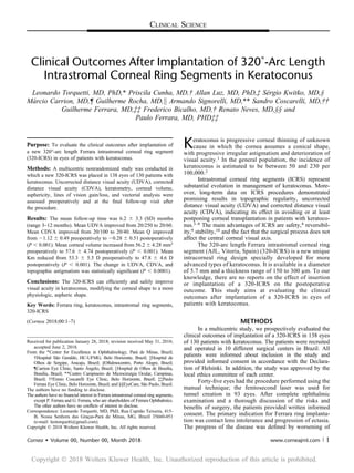
  • 1. CLINICAL SCIENCE Clinical Outcomes After Implantation of 320˚-Arc Length Intrastromal Corneal Ring Segments in Keratoconus Leonardo Torquetti, MD, PhD,* Priscila Cunha, MD,† Allan Luz, MD, PhD,‡ Sérgio Kwitko, MD,§ Márcio Carrion, MD,¶ Guilherme Rocha, MD,k Armando Signorelli, MD,** Sandro Coscarelli, MD,†† Guilherme Ferrara, MD,‡‡ Frederico Bicalho, MD,† Renato Neves, MD,§§ and Paulo Ferrara, MD, PHD‡‡ Purpose: To evaluate the clinical outcomes after implantation of a new 320°-arc length Ferrara intrastromal corneal ring segment (320-ICRS) in eyes of patients with keratoconus. Methods: A multicentric nonrandomized study was conducted in which a new 320-ICRS was placed in 138 eyes of 130 patients with keratoconus. Uncorrected distance visual acuity (UDVA), corrected distance visual acuity (CDVA), keratometry, corneal volume, asphericity, lines of vision gain/loss, and vectorial analysis were assessed preoperatively and at the final follow-up visit after the procedure. Results: The mean follow-up time was 6.2 6 3.3 (SD) months (range 3–12 months). Mean UDVA improved from 20/250 to 20/60. Mean CDVA improved from 20/100 to 20/40. Mean Q improved from 21.12 6 0.49 preoperatively to 20.28 6 0.51 postoperatively (P , 0.001). Mean corneal volume increased from 56.2 6 4.28 mm3 preoperatively to 57.6 6 4.74 postoperatively (P , 0.001). Mean Km reduced from 53.3 6 5.5 D preoperatively to 47.8 6 4.6 D postoperatively (P , 0.001). The change in UDVA, CDVA, and topographic astigmatism was statistically significant (P , 0.0001). Conclusions: The 320-ICRS can efficiently and safely improve visual acuity in keratoconus, modifying the corneal shape to a more physiologic, aspheric shape. Key Words: Ferrara ring, keratoconus, intrastromal ring segments, 320-ICRS (Cornea 2018;00:1–7) Keratoconus is progressive corneal thinning of unknown cause in which the cornea assumes a conical shape, with progressive irregular astigmatism and deterioration of visual acuity.1 In the general population, the incidence of keratoconus is estimated to be between 50 and 230 per 100,000.2 Intrastromal corneal ring segments (ICRS) represent substantial evolution in management of keratoconus. More- over, long-term data on ICRS procedures demonstrated promising results in topographic regularity, uncorrected distance visual acuity (UDVA) and corrected distance visual acuity (CDVA), indicating its effect in avoiding or at least postponing corneal transplantation in patients with keratoco- nus.3–8 The main advantages of ICRS are safety,4 reversibil- ity,9 stability,10 and the fact that the surgical process does not affect the central corneal visual axis. The 320-arc length Ferrara intrastromal corneal ring segment (AJL, Vitoria, Spain) (320-ICRS) is a new unique intracorneal ring design specially developed for more advanced types of keratoconus. It is available in a diameter of 5.7 mm and a thickness range of 150 to 300 mm. To our knowledge, there are no reports on the effect of insertion or implantation of a 320-ICRS on the postoperative outcome. This study aims at evaluating the clinical outcomes after implantation of a 320-ICRS in eyes of patients with keratoconus. METHODS In a multicentric study, we prospectively evaluated the clinical outcomes of implantation of a 320-ICRS in 138 eyes of 130 patients with keratoconus. The patients were recruited and operated in 10 different surgical centers in Brazil. All patients were informed about inclusion in the study and provided informed consent in accordance with the Declara- tion of Helsinki. In addition, the study was approved by the local ethics committee of each center. Forty-five eyes had the procedure performed using the manual technique; the femtosecond laser was used for tunnel creation in 93 eyes. After complete ophthalmic examination and a thorough discussion of the risks and benefits of surgery, the patients provided written informed consent. The primary indication for Ferrara ring implanta- tion was contact lens intolerance and progression of ectasia. The progress of the disease was defined by worsening of Received for publication January 28, 2018; revision received May 31, 2018; accepted June 2, 2018. From the *Center for Excellence in Ophthalmology, Pará de Minas, Brazil; †Hospital São Geraldo, HC-UFMG, Belo Horizonte, Brazil; ‡Hospital de Olhos de Sergipe, Aracaju, Brazil; §Oftalmocentro, Porto Alegre, Brazil; ¶Carrion Eye Clinic, Santo Ângelo, Brazil; kHospital de Olhos de Brasília, Brasília, Brazil; **Centro Campineiro de Microcirurgia Ocular, Campinas, Brazil; ††Ennio Coscarelli Eye Clinic, Belo Horizonte, Brazil; ‡‡Paulo Ferrara Eye Clinic, Belo Horizonte, Brazil; and §§EyeCare, São Paulo, Brazil. The authors have no funding to disclose. The authors have no financial interest in Ferrara intrastromal corneal ring segments, except P. Ferrara and G. Ferrara, who are shareholders of Ferrara Ophthalmics. The other authors have no conflicts of interest to disclose. Correspondence: Leonardo Torquetti, MD, PhD, Rua Capitão Teixeira, 415- B, Nossa Senhora das Graças-Pará de Minas, MG, Brazil 35660-051 (e-mail: leotorquetti@gmail.com). Copyright © 2018 Wolters Kluwer Health, Inc. All rights reserved. Cornea Volume 00, Number 00, Month 2018 www.corneajrnl.com | 1 Copyright Ó 2018 Wolters Kluwer Health, Inc. Unauthorized reproduction of this article is prohibited.
  • 2. UDVA and CDVA, increasing intolerance to contact lens wear, and progressive corneal steepening documented by topography. CLINICAL MEASUREMENTS Preoperative and Postoperative Evaluation All patients had complete ophthalmologic examination preoperatively and postoperatively. The outcome analysis comprised UDVA, CDVA, spherical equivalent (SE), central pachymetry, corneal volume, refractive error, corneal topo- graphic astigmatism, and minimum and maximum keratom- etry (K) values. All data were obtained before and at the final visit after ICRS implantation. CDVA, slip-lamp evaluation, refraction, corneal topography, fundoscopy, and tonometry were performed at each control visit. Anterior segment parameters were obtained by a corneal tomographer (Oculus Pentacam, Wetzlar, Germany). All clinical examinations were performed in a standardized manner according to the guide- lines of the multicentric study. On the first postoperative day, slit-lamp biomicro- scopic examination was performed. Healing of the wound and migration of the segments were evaluated. At final follow-up examination, manifest refraction, UDVA and CDVA, and slit-lamp and topographic examinations were performed. Surgical Technique The 320-ICRS was implanted using the manual tech- nique or femtosecond laser–assisted procedure. Manual Technique Surgery was performed under topical anesthesia after miosis was achieved with 2% pilocarpine. An eyelid specu- lum was used to expose the eye, and 2.5% povidone–iodine eye drops were instilled onto the cornea and conjunctival cul- de-sac. The visual axis was marked by pressing the Sinskey hook on the central corneal epithelium while asking the patient to fixate on the corneal light reflex of the microscope light. Using a marker tinted with gentian-violet tinted, a 5.0- mm optical zone and incision site were aligned to the desired axis in which the incision would be made. The depth of a 1.0-mm, square diamond blade was set at 80% of corneal thickness at the incision site, and this knife is used to make the incision. Using a “stromal spreader,” a pocket was formed on each side of the incision. Specific 380-degree circular dissecting spatulas were inserted through the incision and gently pushed with some quick, rotary “back and forth” tunneling movements. After channel creation, the 320-ICRS was inserted using a modified McPherson forceps. The ICRS was adequately positioned with the aid of a Sinskey hook.7 Femtosecond Laser Technique The surgical procedure was performed under sterile conditions and topical anesthesia. Purkinje reflex was chosen as the central point and was marked. A 5-mm marker was used to locate the exact ring channel. The tunnel depth was set at 75% of the thinnest corneal thickness on the tunnel location in the femtosecond laser cases.11–16 A 60-kHz femtosecond laser (LDV; Ziemer, Switzer- land) or the IntraLase was used to create the ring channel. Particular attention was given to centralizing the suction ring to mark the central point to minimize decentration. The channel’s inner diameter was set to 4.4 mm, the outer diameter was 5.6 mm, the ring energy used for channel creation was 1.30 J, and the entry cut energy was 1.30 J. Channel creation timing with the femtosecond laser was 15 seconds. The 320- ICRS was implanted immediately after channel creation before the bubbles disappeared, which reveals the exact tunnel location. After channel creation, the 320-ICRS was inserted using a modified McPherson forceps. The 320-ICRS was properly positioned with the aid of a Sinskey hook. The postoperative regimen consisted of moxifloxacin 0.5% (Vigamox; Alcon, Forth Worth, TX) and dexametha- sone 0.1% (Maxidex; Alcon, Forth Worth, TX) eye drops 4 times daily for 2 weeks. The patients were instructed to avoid rubbing the eye and to use preservative-free artificial tears frequently—polyethylene glycol 400 0.4% (Systane; Alcon, Forth Worth, TX). Statistical Analysis Statistical analysis was performed using Minitab soft- ware (2007, Minitab Inc.). The Student t test for paired data was used to compare preoperative and postoperative data. RESULTS There were 62 females and 68 males with a mean age of 27 6 9.9 years (range 10–63 years). The mean follow-up time was 6.2 6 3.3 (SD) months (range 3–12 months). Pre- operative and postoperative UDVA, CDVA, SE, and pachy- metry were obtained from all patients (Table 1). Median UDVA improved from 20/250 preoperatively to 20/60 post- operatively (P , 0.001). Mean CDVA improved from 20/100 TABLE 1. Preoperative and Postoperative Evaluated Parameters Preoperative Postoperative P UDVA* 20/250 20/60 ,0.001 CDVA 20/100 20/40 ,0.001 CDVA* 20/60 20/30 ,0.001 SE 27.02 (64.8) 23.29 (63.8) ,0.001 Pachymetry 430 (647.8) 447 (653.8) ,0.001 Q 21.25 (60.49) 20.31 (60.51) ,0.001 Corneal volume 56.2 (64.28) 57.6 (64.74) ,0.001 Topographic astigmatism 4.62 (62.87) 2.74 (62.07) ,0.001 K1 50.5 (64.9) 46.0 (64.4) ,0.001 K2 55.2 (65.6) 49.0 (64.6) ,0.001 Km 53.3 (65.5) 47.8 (64.6) ,0.001 *Median DQ = 0.94. Torquetti et al Cornea Volume 00, Number 00, Month 2018 2 | www.corneajrnl.com Copyright © 2018 Wolters Kluwer Health, Inc. All rights reserved. Copyright Ó 2018 Wolters Kluwer Health, Inc. Unauthorized reproduction of this article is prohibited.
  • 3. preoperatively to 20/40 postoperatively (P , 0.001). Median CDVA improved from 20/60 preoperatively to 20/30 post- operatively (P , 0.001). Mean preoperative SE was 27.02 6 4.80 D preoperatively and 23.29 6 3.8 D postoperatively (P , 0.001). Mean central pachymetry increased from 430 6 47.8 D preoperatively to 447 6 53.8 D postoperatively (P , 0.001). Two or more lines of CDVA were gained in 87% of eyes. Four percent of eyes lost 1 line of vision. All these eyes had preoperative CDVA better than 20/40. There was progressive improvement in CDVA and lines of CDVA gained from 3 to 6 months of follow-up (Figs. 1, 2). During the follow-up period after ICRS implantation, there were no cases of segment extrusion. There were 4 cases of migration, which were excluded from the statis- tical analysis. All these cases were performed with the manual technique (8.8% of manual cases). No intraoper- ative complications were observed with the femtosecond laser technique. There were 2 (4.4%) cases of intraoper- ative perforation with the manual method, which were excluded from the study. Preoperative and postoperative anterior segment param- eters are described in Table 1. Mean Q improved from 21.12 6 0.49 preoperatively to 20.28 6 0.51 postoperatively. Mean corneal volume increased from 56.2 6 4.28 pre- operatively to 57.6 6 4.74 mm3 postoperatively (P , 0.001). Mean K1 reduced from 50.5 6 4.9 D preoperatively to 46.0 6 4.4 D postoperatively. Mean K2 decreased from 55.2 6 5.6 D preoperatively to 49.0 6 4.6 D postoperatively. Mean Km reduced from 53.3 6 5.5 D preoperatively to 47.8 6 4.6 D postoperatively (Fig. 3). The change in Q, corneal volume, topographic astigmatism, K1, K2, and Km was statistically significant (P , 0.0001). We evaluated the keratometry change after implanta- tion of the 320-ICRS, compared to preoperative keratom- etry. Steep corneas had a more significant flattening effect compared with flat corneas (Table 2). When considering the keratoconus type (oval, nipple, or astigmatic), we did not find significant differences among them, regarding the change in keratometry after 320-ICRS implantation (Table 3). The topographic astigmatism change was more significant in astigmatic types of keratoconus (Table 4). The vectorial analysis of refraction and keratometry showed poor predictability of results after implantation of the 320-ICRS (Fig. 4). FIGURE 1. CDVA after implantation of 320-ICRS (3 and 6 months of follow-up). Left = 3 months; right = 6 months. FIGURE 2. Lines of CDVA gain/lost after implantation of 320-ICRS (3 and 6 months of follow-up). Left = 3 months; right = 6 months. Cornea Volume 00, Number 00, Month 2018 320˚-Arc Length Ferrara ICRS Copyright © 2018 Wolters Kluwer Health, Inc. All rights reserved. www.corneajrnl.com | 3 Copyright Ó 2018 Wolters Kluwer Health, Inc. Unauthorized reproduction of this article is prohibited.
  • 4. DISCUSSION The 3 main advantages of the 320-ICRS over the standard 160-degree arc length ICRS are 1) significant asphericity change,17,18 2) significant corneal flattening,17,18 and 3) implantation of a single segment. ICRSs are available from 90-arc length to 210-arc length (90 degrees, 120 degrees, 140 degrees, 160 degrees, and 210 degrees) with different thicknesses ranging from 150 to 350 mm in 50-mm steps. Several studies have shown the role and behavior of each arc length and thickness on ICRS sur- gery.14,17,18 It is accepted that the goals of ICRS implantation are 1) reduction of corneal irregularity and irregular astigmatism; 2) corneal flattening and myopia corneal prolatism reduction; and 3) better visual performance expressed through visual acuity lines gained. For achieving these goals, ICRSs in different arc lengths and thickness combinations lead to corneal changes and desired improvement. Reintroduction of a long-arc ICRS has the primary objective of increasing corneal flattening and prolatism reduction, especially for the nipple type of keratoconus and advanced cases.19 Few publications are available, and first results were recently published. Jadidi et al19,20 published their results and complications using a 355-degree arc ICRS, and recently, Sado- ughi et al21 released their results using a 340-degree arc ICRS. Kwitko and Severo22 reported that keratoconus with central ectasia had significantly better results after sym- metrical implantation. This is true, and probably, these cases may have done better if implanted with the 320- ICRS, which was not available when that study was published. Implantation of a single segment is advanta- geous because of less corneal trauma, less risk of infection and extrusion, besides less chance of haloes in the postoperative period. A positive correlation was found between ring thick- ness and DQ; that is, the thicker the ring, the more reduction of the excess of prolatism effect on the cornea. A positive correlation was also found between ring thickness and DK; that is, the thicker the ring, the more flattening effect on the cornea. We observed a progressive flattening effect of the 320- ICRS. This can be explained by its significant arc length, which produces a “new limbus” in the cornea, which changes progressively after implantation. This behavior is desirable in advanced keratoconus cases but not in mild ones, which are a relative contraindication for its use. The corneal changes after 320-ICRS occur in the whole area central to the ICRS; for this reason, we could consider this segment as a “new limbus.” Colin et al,6 in a 2-year follow-up study after implan- tation of Intacs, described a gain of 1 or more lines of CDVA in 61.0% eyes at 1 year and 68.3% eyes at 2 years. Fewer than 15% of eyes experienced a CDVA loss of 1 or more lines FIGURE 3. Preoperative (left) and postoperative (right) corneal tomography. TABLE 2. Evaluation of Variation of Mean Postoperative Keratometry According to Preoperative Keratometry Preoperative Postoperative P DKm ,50.00 D 47.77 (61.67) 44.24 (62.63) ,0.001 3.70 50.00–60.00 D 52.9 (62.37) 47.7 (63.07) ,0.001 5.19 .60.00 D 63.3 (63.2) 55.3 (64.7) ,0.001 8.23 TABLE 3. Evaluation of Variation of Mean Postoperative Keratometry According to Keratoconus Type Preoperative Postoperative P DKm Oval 50.8 (64.83) 46.1 (64.61) ,0.001 4.64 Nipple 51.7 (62.45) 46.9 (62.81) ,0.001 4.78 Astigmatic 55.1 (64.98) 51.09 (66.17) ,0.001 4.07 Torquetti et al Cornea Volume 00, Number 00, Month 2018 4 | www.corneajrnl.com Copyright © 2018 Wolters Kluwer Health, Inc. All rights reserved. Copyright Ó 2018 Wolters Kluwer Health, Inc. Unauthorized reproduction of this article is prohibited.
  • 5. during the 2 years after Intacs implantation. There was CDVA loss of 1 or more lines in 14.6% of eyes. In the present study, we observed that 3 months after ICRS implantation, 76% of eyes gained 1 or more lines of CDVA. Six months after surgery, 92.4% of eyes gained 1 or more lines of CDVA (Fig. 2). These data reinforce the progressive effect of 320-ICRS along the time. Our results are similar to the 355-degree arc report,19 with significant improvement in UDVA, CDVA, and SE. On the other hand, the 340-degree arc report21 showed different results, with a lower percentage of patients gaining at least 2 lines of UDVA and no statistical significance in CDVA improvement (P = 0.09), with only 50.0% of patients gaining at least 2 lines of CDVA. Sadoughi et al21 related this poor result to the advanced stage of the disease (patients with Km . 55.0 D and K2 . 57.0 D). In the current study, there were 4 cases of postoperative ICRS migration and 2 cases of intraoperative perforation. Jadidi et al19 enrolled 5 patients in a study of the Keraring 355-degree arc ICRS using a femtosecond laser for tunnel creation. In all cases, postoperative complications happened, and the worst one was corneal melting, near the incision site. Proximity between the ICRS tip and the incision is the leading risk factor for extrusion, infection, and corneal melting. The 320-ICRS, in theory, can provide similar corneal changes (compared with the 355-ICRS); the main advantage of this ICRS is to be 20 degree gap on each side, far from the incision, what makes it safer to be used. This study suggests that the ideal patients to be implanted with the 320-ICRS are those with central cones, TABLE 4. Evaluation of Variation of Mean Postoperative Topographic Astigmatism According to Keratoconus Type Preoperative Postoperative P D Topographic Astigmatism Oval 4.8 (62.12) 2.84 (62.13) ,0.001 1.95 Nipple 2.03 (61.09) 1.74 (61.13) 0.284 0.29 Astigmatic 8.50 (61.55) 5.01 (62.50) ,0.001 3.43 FIGURE 4. Vectorial analysis (vector analysis). Cornea Volume 00, Number 00, Month 2018 320˚-Arc Length Ferrara ICRS Copyright © 2018 Wolters Kluwer Health, Inc. All rights reserved. www.corneajrnl.com | 5 Copyright Ó 2018 Wolters Kluwer Health, Inc. Unauthorized reproduction of this article is prohibited.
  • 6. hyperprolate corneas, low-to-moderate astigmatism, and CDVA worse than 20/40. Because it causes significant changes in Q and Km, it should be reserved for moderate to advanced cases of keratoconus. The more advanced the case, the more significant the change after 320-ICRS implantation (Table 2). It should be avoided in asymmetric and mild23 keratoconus because it can overcorrect and produce unpredictable/unreasonable results (Fig. 5) because of its uniform flattening effect and persistence of asymmetry after its implantation in asymmetric ectasias. We found a significant increase in corneal thickness after ICRS implantation. This can be explained by theoretical corneal collagen remodeling induced by implantation of the ICRS. Acting as “spacers,” the ring segments could interfere in corneal collagen turnover, with consequent increase in central corneal pachymetry.24 One limitation of the study was the lack of a control group. However, several studies in the literature describe the results of other ICRS designs and arc length that could be compared with these first results of the 320-ICRS. The 320-ICRS improved all parameters studied. Despite that, it does not provide a predictable refractive result, as demonstrated by the vectorial analysis (Fig. 4). Therefore, it should be used as a means to restore the cornea to a more physiologic shape, and a second refractive pro- cedure (eg, phakic intraocular lens) may be required to achieve a better refractive outcome.25 In summary, our results suggest that the 320-ICRS might improve visual acuity in keratoconus, modifying the corneal shape to a more physiologic, aspheric shape. Further randomized controlled studies are needed to confirm our findings and also the safety of this new ring segment. REFERENCES 1. Espandar L, Meyer J. Keratoconus: overview and update on treatment. Middle East Afr J Ophthalmol. 2010;17:15–20. 2. Gordon-Shaag A, Millodot M, Shneor E. The epidemiology and etiology of keratoconus. Int J Keratoconus Ectatic Corneal Dis. 2012;1:7–15. 3. Colin J, Velou S. Implantation of Intacs and a refractive intraocular lens to correct keratoconus. J Cataract Refract Surg. 2003;29:832–834. 4. Kymionis GD, Siganos CS, Tsiklis NS, et al. Long-term follow-up of Intacs in keratoconus. Am J Ophthalmol. 2007;143:236–244. 5. Colin J, Cochener B, Savary G, et al. Correcting keratoconus with intracorneal rings. J Cataract Refract Surg. 2000;26:1117–1122. 6. Colin J, Cochener B, Savary G, et al. INTACS inserts for treating keratoconus: one-year results. Ophthalmology. 2001;108:1409–1414. 7. Siganos CS, Kymionis GD, Kartakis N, et al. Management of keratoconus with Intacs. Am J Ophthalmol. 2003;135:64–70. 8. Siganos D, Ferrara P, Chatzinikolas K, et al. Ferrara intrastromal corneal rings for the correction of keratoconus. J Cataract Refract Surg. 2002;28: 1947–1951. 9. Piñero DP, Alió JL, El Kady B, et al. Refractive and aberrometric outcomes of intracorneal ring segments for keratoconus: mechanical versus femtosecond-assisted procedures. Ophthalmology. 2009;116: 1675–1687. 10. Torquetti L, Berbel RF, Ferrara P. Long-term follow-up of intrastromal corneal ring segments in keratoconus. J Cataract Refract Surg. 2009;35: 1768–1773. 11. Kubaloglu A, Sari ES, Cinar Y, et al. Comparison of mechanical and femtosecond laser tunnel creation for intrastromal corneal ring segment implantation in keratoconus: prospective randomized clinical trial. J Cataract Refract Surg. 2010;36:1556–1561. 12. Lisa C, García-Fernandez M, Madrid-Costa D, et al. Femtosecond laser- assisted intrastromal corneal ring segment implantation for high astig- matism correction after penetrating keratoplasty. J Cataract Refract Surg. 2013;39:1660–1667. 13. Coimbra CC, Gomes MT, Campos M, et al. Femtosecond assisted intrastromal corneal ring (ISCR) implantation for the treatment of corneal ectasia. Arq Bras Oftalmol. 2012;75:126–130. 14. Coskunseven E, Kymionis GD, Bouzoukis DI, et al. Single intrastromal corneal ring segment implantation using the femtosecond laser after radial keratotomy in a keratoconic patient. J Cataract Refract Surg. 2009;35:197–199. 15. Yeung SN, Ku JYF, Lichtinger A, et al. Efficacy of single or paired intrastromal corneal ring segment implantation combined with collagen crosslinking in keratoconus. J Cataract Refract Surg. 2013;39:1146–1151. 16. Pinero DP, Alió JL, El Kady B, et al. Corneal aberrometric and refractive performance of 2 intrastromal corneal ring segment models in early and moderate ectatic disease. J Cataract Refract Surg. 2010;36:102–109. FIGURE 5. Preoperative (left) and postoperative (right) corneal tomography in asymmetrical keratoconus. Torquetti et al Cornea Volume 00, Number 00, Month 2018 6 | www.corneajrnl.com Copyright © 2018 Wolters Kluwer Health, Inc. All rights reserved. Copyright Ó 2018 Wolters Kluwer Health, Inc. Unauthorized reproduction of this article is prohibited.
  • 7. 17. Ferrara P, Torquetti L. Clinical outcomes after implantation of a new intrastromal corneal ring with a 210-degree arc length. J Cataract Refract Surg. 2009;35:1604–1608. 18. Ferrara G, Torquetti L, Ferrara P, et al. Intrastromal corneal ring segments: visual outcomes from a large case series. Clin Exp Oph- thalmol. 2012;40:433–439. 19. Jadidi K, Mosavi SA, Nejat F, et al. Intrastromal corneal ring segment implantation (keraring 355°) in patients with central keratoconus: 6- month follow-up. J Ophthalmol. 2015;2015:916385. 20. Jadidi K, Mosavi SA, Nejat F. Complications of intracorneal ring implantation (keraring 355°) using a femtosecond laser for channel creation. Int J Kerat Ectatic Cor Dis. 2014;3:53–56. 21. Sadoughi MM, Einollahi B, Veisi AR, et al. Femtosecond laser implantation of a 340-degree intrastromal corneal ring segment in keratoconus: short-term outcomes. J Cataract Refract Surg. 2017; 43:1251–1256. 22. Kwitko S, Severo NS. Ferrara intracorneal ring segments for keratoco- nus. J Cataract Refract Surg. 2004;30:812–820. 23. Rodrigues P, Ferrara G, Ferrara P, et al. Intrastromal corneal ring segments implantation in patients with mild keratoconus. Int J Kerat Ectatic Cor Dis. 2014:122–126. 24. Flynn BP, Bhole AP, Saeidi N, et al. Mechanical strain stabilizes reconstituted collagen fibrils against enzymatic degradation by mamma- lian collagenase matrix metalloproteinase 8 (MMP-8). PLoS One. 2010; 5:e12337. 25. Alfonso JF, Lisa C, Fernandez-Vega L, et al. Intrastromal corneal ring segments and posterior chamber phakic intraocular lens implantation for keratoconus correction. J Cataract Refract Surg. 2011;37:706–713. Cornea Volume 00, Number 00, Month 2018 320˚-Arc Length Ferrara ICRS Copyright © 2018 Wolters Kluwer Health, Inc. All rights reserved. www.corneajrnl.com | 7 Copyright Ó 2018 Wolters Kluwer Health, Inc. Unauthorized reproduction of this article is prohibited.