Call Girls Siliguri Just Call 9907093804 Top Class Call Girl Service Available
Scrotal ultrasound
1. Scrotal ultrasound
Indication/Technique
Scrotal ultrasound is requested when pathology is suspected.
This may include:
palpable swellings
pain
asymmetry
trauma
Infection
Pain
Haematospermia
Infertility
Preparation of the patient
No special preparation is required
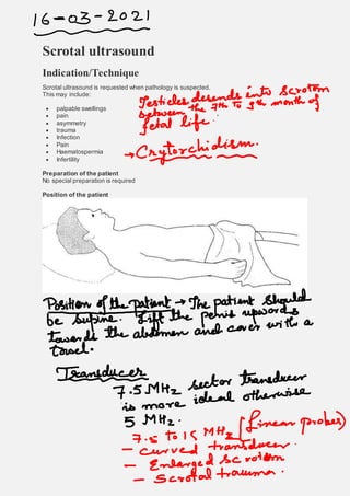
Position of the patient
2. Ultrasound is a safe and quick way to distinguish various pathologies. Pathology may vary from
Patients are examined in supine position.
The scrotum is evaluated transversally (fig. 1) and sagittally (fig. 2) using a transducer (=
ultrasound probe).
Figure 1. Technique for scrotal ultrasound in the transversal plane.
Figure 2. Technique for scrotal ultrasound in the sagittal plane.
3. By moving and rotating the transducer, each part of the scrotum is assessed systematically.
When the transducer is tipped, it remains in the same location and only the sound beam changes
direction. This allows you to scan in the craniocaudal direction (= transversal plane) and left-right
direction (= sagittal plane).
Important: location and direction of the transducer on the patient's skin determine
anterior/posterior and left/right on the image.
In general, scrotal ultrasound (as other ultrasound examinations) is performed in the transversal
plane (fig. 3):
the top of the ultrasound image is the anterior side; the bottom is the posterior side.
left on the image is actually right and vice versa. You are looking at the body from below, as
it were (as in a transversal section of a CT scan).
Figure 3. Transversal image of the right testicle.
As a general rule, in ultrasound in the sagittal plane (fig. 4):
the top of the ultrasound image is the anterior side and the bottom is the posterior side.
right on the image is the foot side (= caudal) and left is the head side (= cranial).
4. Figure 4. Sagittal image of the right testicle.
The images can be read immediately from the screen during the examination.
Orientation tip when attending a live examination: the top of the image is the location where the
sound waves enter the patient first. So irrespective of position and tipping, the top is always the
skin side.
Ultrasound uses sound waves, which are reflected, deflected or absorbed in the body. The
reflected sound waves produce the ultrasound image. The more sound waves are reflected, the
more hyperechogenic (= whiter) the tissue is imaged. Reduced reflection will cause the image to
be hypoechogenic, and anechogenic (= black) if reflection is absent (fig. 5).
Figure 5. Echogenicity with corresponding terms.
The speed of sound through tissue, and tissue density and structure all influence the image
obtained. For instance, high-density tissue generates relatively many echo reflections (e.g.
bone/calcium) and produces a hyperechogenic image.
Fluid does not reflect sound waves and is therefore anechogenic (= black). Soft tissues (e.g.
organs) are somewhere between hyperechogenic and anechogenic.
Isoechogenic: tissue with the same echogenicity as the surrounding tissue.
7. epididymis and the funiculus spermaticus (= spermatic cord). The epididymis can be subdivided
into the caput, corpus and cauda (fig. 6).
At the bottom of the testicle, the epididymal cauda changes into the ductus deferens.
Figure 6. Normal anatomy of the testicle/epididymis/spermatic cord.
Arteries, veins, lymph vessels, nerves and the ductus deferens (ejaculatory duct) run through the
spermatic cord. Venous drainage is through the pampiniformis plexus, which in turn drains into
the testicular vein (= internal spermatic vein).
The rete testis is a network of small canals that join in the hilus of the testicle (= testicular
mediastinum). From the highly coiled seminiferous tubules, sperm cells will be transported via
the rete testis to the epididymis.
At the bottom of the testicle, the epididymal cauda eventually changes into the ductus deferens.
The testicles and epididymis are surrounded by the tunica vaginalis.
8. The tunica vaginalis forms a protective layer for the testicles and consists of two layers: the
visceral and parietal layer.
The visceral plate is on the inside and keeps the testicles and the epididymis together.
During ultrasound examination, the testicles are evaluated transversally and sagittally.
Normal testicles have an oval shape and are virtually homogeneous (fig. 8).
9.
10. ♦ Figure 8. Normal right testicle and epididymis (*) in the sagittal (a) and transversal plane (b).
At the cranial side (= always LEFT on sagittal ultrasound images) is the epididymal head. It has a
triangular shape and always has the same echogenicity as the testicles or slightly more (fig. 9).
11. ♦ Figure 9. Sagittal plane of the right testicle. Normal epididymis.
♦ Figure 10. Sagittal plane of the right testicle. The intratesticular linear hyperechogenic structure
is the mediastinum.
A normal testicle is vascularized. The blood stream patterns may be evaluated using Doppler
ultrasound. One of the applications of Doppler ultrasound is color Doppler (fig. 11). This device
images frequency changes in the colors blue and red. Blue means the blood is streaming away
from the transducer and red means the blood is moving towards the transducer (Note: blue and
red does not necessarily mean low-oxygen and high-oxygen blood).
12. ♦ Figure 11. The color Doppler shows normal vascularization of the right testicle.
Back to top
Checklist
The following points may be used as a guide to assess scrotal ultrasound.
1. Are the testicles within the scrotum?
2. Is the testicle homogeneous? Focal abnormalities? Asymmetry?
3. Symmetric flow (= vascularization) present in the testicles?
4. Changes caused by the Valsava maneuver?
Back to top
Pathology
Extratesticular scrotal masses
Testicular torsion
Epididymitis
Extratesticular scrotal masses
The most common extratesticular masses are hydrocele, varicocele and spermatocele.
Hydrocele
Hydrocele is fluid accumulation in the tunica vaginalis covering the testicles.
Ultrasound characteristics:
13. anechogenic collection in the tunica vaginalis
the collection shows NO color Doppler flow
♦ Figure 12. Left testicle in the sagittal plane. Hydrocele.
Causes:
idiopathic.
congenital; communicating hydrocele in persistent vaginal process (fig. 13).
imbalance of fluid production in the tunica vaginalis, e.g. due to reduced lymphatic drainage,
infection or tumor (fig. 14).
14. Figure 13. Normal: obliteration of vaginal process. Communicating hydrocele: persistent
presence of the vaginal process causing the abdominal fluid to move downward towards the
tunica vaginalis space.
15. ♦ Figure 14. Right testicle in the transversal and sagittal plane. Hydrocele with enlarged non-
homogeneous right testicle (testicular tumor).
This disorder is generally asymptomatic and is felt as a painless swelling around the testicle.
The fluid frequently accumulates at the ventral side and presses the testicle towards posterior.
If the collection contains septa (= partitions), consider a hematoma (e.g. after trauma), a so-
called hematocele.
Hydrocele generally does not require treatment.
Varicocele
In varicocele, peritesticular veins are dilated (‘varicose veins’) due to reduced function of the
spermatic vein. Varicocele generally develops in the left side of the scrotum.
Particularly when standing and with increased pressure (coughing/straining) a 'sausage-like’
swelling can be felt. Varicocele is generally asymptomatic, but can sometimes produce pain on
exertion.
Ultrasound characteristics (fig. 15/16):
multiple tortuous dilated veins (lateral, posterior or superior of the testicle).
increased venous flow in the Valsava maneuver (straining/coughing).
16. ♦ Figure 15. Color Doppler of left testicle in the sagittal plane. Posterior of the testicle is a dilated
tortuous peritesticular vein. Picture consistent with varicocele.
♦ Figure 16. Color Doppler of left testicle in the transversal plane. There are multiple veins lateral
of the left testicle. Marked increased flow and dilatation during Valsava maneuver, picture of
varicocele.
Spermatocele
Spermatoceles are the most common scrotal masses. They are bony cystic structures filled with
(dead) sperm cells. They are located in the epididymal head. Spermatocele cannot be accurately
distinguished by ultrasound from an epididymal cyst.
Spermatoceles are rarely symptomatic.
Ultrasound characteristics (fig. 17/18):
sharply delineated hypoechogenic lesions.
may contain echogenic debris (= sperm cells).
17. ♦ Figure 17. A large spermatocele/epididymal cyst.
♦ Figure 18. A small spermatocele/epididymal cyst.
Extratesticular scrotal masses
Testicular torsion
Epididymitis
Testicular torsion
The testicles may become twisted around the funiculus spermaticus (= spermatic cord). As a
result, testicular circulation will be compromised; starting with venous obstruction followed by
18. arterial obstruction. The testicles will eventually die off. For this reason, it is important to detorse
the testicles (surgically where necessary) within +-6 hours after the start of symptoms (fig. 19).
|
Figure 19. Twisted funiculus spermaticus; testicular torsion.
Classical:
age: puberty – 25 years.
acute severe pain.
painful (bluish) swollen half of the scrotum.
The most sensitive finding of ischemia is reduced/absent color Doppler signal. The color Doppler
signal of the other (non-painful) testicle is used as reference (fig. 20).
19. ♦ Figure 20. Testicles in transversal plane with color Doppler. Swollen right testicle with virtually
absent color Doppler signal; testicular torsion.
If the torsion persists, the soft tissues around the testicle may become inflamed leading to a
hyperemic scrotal wall, which manifests in a high color Doppler signal.
In addition to abnormal vascularization, there may also be non-specific abnormalities, such as
swelling, reduced echogenicity (= blacker) and reactive hydrocele. In some cases, the twisted
spermatic cord can be seen cranial from the testicle (fig. 21/22).
♦ Figure 21. Right testicle in transversal plane with color Doppler. Marked tortuous epididymis
and absent color Doppler signal of the epididymis and testicle; testicular torsion.
20. ♦ Figure 22. Testicles in the transversal plane. The yellow dotted line shows twisted left
spermatic cord. Note also the mild swelling of the left testicle with a trace of hydrocele. Picture of
testicular torsion.
Extratesticular scrotal masses
Testicular torsion
Epididymitis
Epididymitis
In the differential diagnosis of acute pain and swelling of the scrotum, scrotal inflammation is the
most common disorder in addition to testicular torsion.
A scrotal infection usually involves the epididymis and then spreads to the testicles and scrotal
wall. The infection may affect the entire epididymis or only a focal area.
Patients are usually treated with antibiotics.
Ultrasound characteristics of epididymitis (fig. 23):
enlarged epididymis.
reduced echogenicity of the epididymis.
increased color Doppler signal (vascularization!).
Comment: vascularization may be the only ultrasound abnormality in epididymitis.
21.
22. ♦ Figure 23. Right and left testicle in sagittal plane with color Doppler. Enlarged hyperechogenic
left cauda epididymis with increased echo Doppler signal. Epididymitis left.
In epididymo-orchitis, both the epididymis and testicle are affected (fig. 24).
23. ♦ Figure 24. Left and right testicles in sagittal plane (a/b) and transversal plane (c) Enlarged right
epididymis. Increased echo Doppler signal of the right epididymis and right testicle. Picture
consistent with right-sided epididymo-orchitis.