SlideShare a Scribd company logo
1 of 82
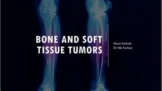
BONE AND SOFT
TISSUE TUMORS
Nurul Azimah
Dr Nik Farhan
BONE TUMORS
­ Imaging features
­ Benign
­ Malignant
­ Primary or secondary tumor (i.e Radiation, Pagets)
IMAGING FEATURES
vMorphology
vAge
vLocation
vGrowth pattern/ margin/ zone of transition
vPeriosteal reaction
vMatrix mineralization
vSize and no
vSoft tissue component
AGE OF PATIENT
Patient age is of huge importance in suggesting a differential diagnosis of a
focal bone lesion.
Primary bone tumors are rare below age 5yo and over the age of 40yo, with
exception of myeloma and chondrosarcoma.
Metastasis are the commonest lesions over the age of 40yo
AGE OF PATIENT
­ Simple bone cyst and chondroblastoma occur in
skeletally immature people
­ Ewing sarcoma typically in 10-20 years old
patients
­ Osteosarcoma has two age peaks; in teenagers
and in pagetic bone or previously irradiated
bone (>50 years old)
­ Malignant bone lesion in adult >40 years old
are metastatic carcinoma, multiple myeloma
and metastatic non-Hodgkin lymphoma rather
than primary bone sarcoma.
LOCATION: EPIPHYSIS -
METAPHYSIS - DIAPHYSIS
Epiphysis
Only a few lesions are located in the epiphysis.
In young patients - either a chondroblastoma or an
infection.
In patients over 20, a giant cell tumor has to be
included in the differential diagnosis.
In older patients a geode (degenerative subchondral
bone cyst) must be added to the differential
diagnosis.
Metaphysis
NOF, SBC, Osteosarcoma, Chondrosarcoma,
Enchondroma and infections.
Diaphysis
Ewing's sarcoma, SBC, ABC, Enchondroma, Fibrous
dysplasia and Osteoblastoma.
LOCATION WITHIN
THE BONE
Eccentric within bone
giant-cell tumor, chondroblastoma, aneurysmal
bone cyst, non-ossifying broma, and the rare
chondromyxoid broma are located
eccentrically within the bone.
Central (in the middle of a long bone)
Simple bone cyst, enchondroma, and fibrous
dysplasia are located centrally within the bone.
RATE OF GROWTH
Most important feature is nature of the
margin
Lodwick classification
Type 1: geographic (benign and low grade
malignant tumor)
­ 1A: thin, rim sclerotic margin
­ 1B: well-defined lesion, but no marginal sclerosis
­ 1C: indistinct border
Type 2: moth-eaten
type 3: permeative (most aggressive)
RATE OF GROWTH
MARGIN/ ZONE OF TRANSITION
§ In order to classify osteolytic lesions as well-defined or ill-defined, we
need to look at the zone of transition between the lesion and the
adjacent normal bone.
§The zone of transition is the most reliable indicator in determining
whether an osteolytic lesion is benign or malignant.
§The zone of transition only applies to osteolytic lesions since sclerotic
lesions usually have a narrow transition zone.
MARGIN/ ZONE OF TRANSITION
Wide transition zone
Ill-defined border + broad TZ = aggressive
growth
Narrow transition zone
Sharp, well-defined, sign of slow growth
NOF SBC ABC
PERIOSTEAL REACTION
§Non-specific reaction and will occur
whenever the periosteum is irritated by
a malignant tumor, benign tumor,
infection or trauma.
§2 types:
­ Benign – benign lesion and following
trauma
­ Aggressive – malignant tumors, benign
lesions with aggrasive behavior eg.
Infection or EG
*Malignant lesions never cause benign
periosteal reaction
PERIOSTEAL
REACTION
Benign periosteal reaction in osteoid
osteoma
PERIOSTEAL
REACTION
Osteosarcoma with Codman's triangle
proximally.
.
PERIOSTEAL
REACTION
Ewing sarcoma with lamellated and
focally interrupted periosteal reaction
PERIOSTEAL
REACTION
Osteosarcoma with sunburst periosteal
reaction
MATRIX
Matrix – type of tissue of the tumor
­ Osteoid, chondral, fibrous, adipose
­ All of these are radiolucent
Mineralization – calcification of the matrix
Certain tumours have characteristic matrix mineralization, which are radiographically visibleand
allows the histological cell type to be predicted.
PATTERNS OF MINERALIZATION
Chondral calcification – linear, curvilinear, punctate,
ring-like or nodular
* image: enchondroma with punctate and arc-like
mineralization
PATTERN OF MINERALIZATION
Osseous calcification – fluffy,
amorphous, cloud-like, poorly defined
SIZE AND NUMBER
•Size can be a clue to diagnosis
•Osteoid osteoma vs osteoblastoma = histologically similar lesion
• nidus of osteoid osteoma <1.5cm , osteoblastoma >1.5cm in diameter
•Primary bone tumors are solitary
•Multiple sclerotic lesions might represent metastasis or osteopoikilocytosis (multiple
bone islands)
•Most common causes of multiple lucencies in >40 years old are metastatic carcinoma,
multiple myeloma and metastatic non-Hodgkin lymphoma
• benign entity like multiple brown tumors may look similar.
SOFT TISSUE COMPONENT
Soft tissue component with a bone lesion
suggest malignant process.
May have destroyed the cortex as the tumor
expanded.
Eg:
Osteosarcoma, Ewing sarcoma, lymphoma.
BENIGN BONE TUMOR
BENIGN LYTIC BONE LESIONS *FEGNOMASHIC*
BENIGN BONE TUMORS
1. Fibrous Dysplasia
2. Osteochondroma
3. Enchondroma
4. Osteoma
5. Osteoid Osteoma
6. Aneurysmal Bone Cyst
7. Giant Cell Tumour / Osteoclastoma
8. Osteoblastoma
9. Non Ossifying Fibroma(NOF)
FIBROUS DYSPLASIA
§ Developmental disorder, 7% of benign bone
tumor
§ Monostotic (70-85%) or polyostotic
§<30 years old (75%)
§ Usually painless, unless fracture
§Common sites for monostotic are the ribs, proximal
femur, and craniofacial bones.
§Has predilection for the pelvis, proximal femur,
ribs, and skull
§Malignant change in fibrous dysplasia is rare
§Association
§McCune–Albright syndrome :
§Polyostotic fibrous dysplasia (typically
unilateral) + ipsilateral cafe ́ au lait spots
(coast of Maine) + endocrine disturbances
(commonly precocious puberty in girls)
§Mazabraud’s syndrome:
§FD (commonly polyostotic) + soft tissue
myxomata
FIBROUS DYSPLASIA –
RADIOGRAPHIC FEATURES
§No periosteal reaction
§ Often purely lytic with bone expansion + ground
glass matrix mineralization
§ Endosteal scalloping
§ Rind sign is characteristic – thick sclerotic margin
§ Shepherd's crook deformity – Varus deformity of
proximal femur; characteristic late finding
§ Typical location: metadiaphyseal region
§ Discriminator: No periosteal reaction
RIND SIGN
SHEPHERD'S CROOK DEFORMITY
When fibrous dysplasia affects the ribs,
the posterior ribs often demonstrate a
lytic expansile appearance.
When the anterior ribs are involved, they
are most often sclerotic in appearance.
BENIGN BONE TUMORS
1. Fibrous Dysplasia
2. Osteochondroma
3. Enchondroma
4. Osteoma
5. Osteoid Osteoma
6. Aneurysmal Bone Cyst
7. Giant Cell Tumour / Osteoclastoma
8. Osteoblastoma
9. Non Ossifying Fibroma(NOF)
OSTEOCHONDROMA
§ Benign cartilage-capped bony growth projecting outward from
bone, often pedunculated. It is the most common benign bone
lesion.
§ Present from 2 to 60 years, highest incidence is in the second
decade
§ Arises from the metaphysis and grows away from the epiphysis.
§ Key features are the continuity of cortex of host bone with the
cortex of the osteochondroma and communication of the
medullary cavities.
§Location:
§ Metaphysis of long bones (70%) – femur, tibia, humerus
§ Hands and feet
§ Pelvis
§ Present as a palpable mass, which usually stops growing at
skeletal maturity.
§ Growth after maturity suggest malignancy degeneration (1%)
OSTEOCHONDROMA –
RADIOGRAPHIC FEATURES
§ Cartilage-capped bony projection + calcification of hyaline
cartilage cap
§ Pedunculated type – pedicle directed away from joint
§ Sessile type - arise from a broad base
§ Malignant degeneration
§ thick bulky cartilaginous cap (thickness >1cm by CT, >2cm by
MRI)
§ dispersed calcifications within cartilaginous cap
§ soft tissue mass
BENIGN BONE TUMORS
1. Fibrous Dysplasia
2. Osteochondroma
3. Enchondroma
4. Osteoma
5. Osteoid Osteoma
6. Aneurysmal Bone Cyst
7. Giant Cell Tumour / Osteoclastoma
8. Osteoblastoma
9. Non Ossifying Fibroma(NOF)
ENCHONDROMA
• Enchondroma is an intramedullary neoplasm
comprising lobules of benign hyaline
cartilage
• Age range: 0–80 years, with most
presenting in 20-40s
•Location:
• Proximal phalanges (40-50%)
• Metacarpals (15-30%)
• Middle phalanges (20-30%)
• Femur, tibia, humerus (25%)
• Ollier's disease is multiple enchondromas.
• Maffucci's syndrome - multiple enchondromas
with soft tissue hemangiomas.
ENCHONDROMA –
RADIOGRAPHIC FEATURES
• Well-circumscribed, lobular or oval lytic lesions
• Expansion of cortex without cortical break
• Chondral-type mineralization may be identified within the
matrix.
• Present centrally in metaphyseal or diaphyseal
• No periosteal reaction or soft tissue mass
•Discriminator:
1. Must have calcification (except in phalanges)
2. No periosteal reaction
ENCHONDROMA
Ollier’s and Maffucci’s syndrome
ENCHONDROMA VS LOW GRADE CHONDROSARCOMA
§ Distinguishing between enchondromas and low-grade chondrosarcomas is a frequent
difficulty as the lesions are both histologically and radiographically very similar.
§ However, this differentiation may not be of clinical relevance, since both can either be closely
followed up clinically and radiologically or treated if symptomatic.
§ Size: over 5-6 cm favors chondrosarcomas
§ Cortical breach seen in 88% of long bone chondrosarcomas
§ Deep endosteal scalloping involving > 2/3 of cortical thickness seen in 90% of
chondrosarcomas
§ Permeative or moth-eaten bone appearance seen in high-grade chondrosarcomas
§ Chondrosarcomas almost always present with pain
§ Location: Hands and feet are uncommon locations for chondrosarcoma
BENIGN BONE TUMORS
1. Fibrous Dysplasia
2. Osteochondroma
3. Enchondroma
4. Osteoma
5. Osteoid Osteoma
6. Aneurysmal Bone Cyst
7. Giant Cell Tumour / Osteoclastoma
8. Osteoblastoma
9. Non Ossifying Fibroma(NOF)
OSTEOMA
• Slow-growing tumour representing dysplastic
developmental anomaly.
• Cortical > cancellous bones.
•Location:
• Cortical osteomas commonly affect PNS
• May interfere with drainage of PNS
• Frontal and ethmoidal > sphenoidal
• Multiple osteomas in Gardner’s syndrome.
•Histological patterns
•1. ivory osteoma
• dense bone lacking Haversian system
•2. mature osteoma
• resembles 'normal' bone, including
trabecular bone often with marrow
•3. mixed osteoma
• a mixture of ivory and mature histology
OSTEOMA
Ivory osteoma Mature osteoma
OSTEOMA – RADIOGRAPHIC
FEATURES
• Dense ivory-like sclerotic
mass
• Well-defined margin
• No associated osseous
destruction
• Rarely exceeds 2-3cm in
diameter
BENIGN BONE TUMORS
1. Fibrous Dysplasia
2. Osteochondroma
3. Enchondroma
4. Osteoma
5. Osteoid Osteoma
6. Aneurysmal Bone Cyst
7. Giant Cell Tumour / Osteoclastoma
8. Osteoblastoma
9. Non Ossifying Fibroma(NOF)
OSTEOID OSTEOMA
Benign osteoblastic lesion characterised by a nidus of osteoid
tissue surrounded by reactive bone sclerosis.
Clinical Features:
­ 2nd and 3rd decade, M: F = 2-3:1
­ Night pain relieved by aspirin
Location:
­ >50% in diaphysis or metaphysis of tibia and femur
OSTEOID OSTEOMA –
RADIOGRAPHIC FEATURES
• Nidus ; can be lucent, sclerotic
or mixed density
• < 10–15mm in diameter
(mostly < 5 mm)
• Surrounding reactive medullary
sclerosis and solid periosteal
reaction
OSTEOID OSTEOMA
AP plain film of the femur in a child with hip pain.
Area of sclerosis medially near the lesser trochanter
with a small lucency (arrow), which is the nidus of
an osteoid osteoma.
CT scan of the femur shows the sclerosis medially and the
lucent nidus (arrow)
Osteoid osteoma.
Lateral radiograph of the tibial diaphysis shows
solid thickening of the cortex, within which is a
small calcified nidus (arrow).
OSTEOID OSTEOMA VS OSTEMYELITIS
Because an osteoid osteoma resembles osteomyelitis, regardless of the appearance of the nidus, it can
be difficult to differentiate the two radiographically. In fact, it cannot be done with plain films, CT scan,
or MRI scans.
However, because the nidus is extremely vascular, it avidly accumulates radiopharmaceutical bone
scanning agents. An osteoid osteoma will have an area of increased uptake corresponding to the area
of reactive sclerosis, but, in addition, it will demonstrate a second area of increased uptake
corresponding to the nidus (Figures 8-13 to 8-15). This has been termed the double-density sign.
In contrast, osteomyelitis has a photopenic area corresponding to the plain film lucency, which represents
an avascular focus of purulent material.
BENIGN BONE TUMORS
1. Fibrous Dysplasia
2. Osteochondroma
3. Enchondroma
4. Osteoma
5. Osteoid Osteoma
6. Aneurysmal Bone Cyst
7. Giant Cell Tumour / Osteoclastoma
8. Osteoblastoma
9. Non Ossifying Fibroma(NOF)
ANEURYSMAL BONE CYST (ABC)
• Expansile multicystic lesion seen in children and adolescents (<30 years old).
• Consists of blood filled sinusoids and solid fibrous elements.
• May arise secondarily within a pre-existing tumor. (eg. in non-ossifying fibroma,
chondroblastoma, giant cell tumour, fibrous dysplasia, osteoblastoma and
osteosarcoma)
• Location:
• Long bones (> 50%)
• Spine (20%)
• Pelvis (5–10%)
•Diff dx: osteoblastoma, telangiectatic osteosarcoma
ABC – RADIOLOGICAL
FEATURES
§ The classical lesion (accounting for 75–80%) is a purely
lytic, expansile intramedullary lesion
§ Metaphysis (most cases), diaphysis (20%)
§ Extending to growth plate.
§ Extends to the growth plate (rarely extension to the
articular surface)
§ May be centrally or eccentrically (more common)
§ Sclerotic margin
§ Trabeculations within the lesion
§Discriminators:
§ Lesion must be expansile
§ Patient must be younger than 30
ABC
ABC
CT showing a proximal tibial aneurysmal
bone cyst with evidence of faint septal
ossification (arrowheads)
ABC
Xray tibia/fibula – eccentric lucent bone lesion at metaphysis tibia.
Axial T2 fatsat MRI images show multiple fluid-fluid levels within the
expansile bone lesion in the proximal tibia.
ABC
ABC involving posterior elements of thoracic
vertebral body with fluid-fluid levels.
DDx Expansile Lytic Lesion of
Posterior Element of Spine
• ABC
• Osteoblastoma
• TB
BENIGN BONE TUMORS
1. Fibrous Dysplasia
2. Osteochondroma
3. Enchondroma
4. Osteoma
5. Osteoid Osteoma
6. Aneurysmal Bone Cyst
7. Giant Cell Tumour / Osteoclastoma
8. Osteoblastoma
9. Non Ossifying Fibroma(NOF)
GIANT CELL TUMOR / OSTEOCLASTOMA
• Aggressive benign neoplasm arising from osteoclasts.
• Almost exclusively in adults
• 5% of all primary bone tumors, 20% of benign tumors
• 5-10% of GTC are malignant
• Common sites:
• Distal femur and proximal tibia (55%)
• Distal radius (10%)
• Proximal humerus (6%)
• Sacrum - the most frequent site in spine
• Multifocal, metachronous GCT has also been reported, which is associated with
hyperparathyroidism.
GCT – RADIOGRAPHIC
FEATURES
4 classic features in long bone
★ occurs only with a closed epiphyses
★ must be epiphyseal and abuts articular surface: 98-
99%
★ eccentric: if large this may be difficult to assess
★ well-defined with non-sclerotic margin (does not apply
in flat bones eg pelvis and calcaneus)
• Pathological fracture may be present
• No matrix calcification/mineralisation
GCT
A large, well-defined lytic lesion in the iliac wing with a
sclerotic margin and does not appear to abut any
articular surface.
The usual rules for giant cell tumors such as the presence
of a nonsclerotic margin do not apply in flat bones.
BENIGN BONE TUMORS
1. Fibrous Dysplasia
2. Osteochondroma
3. Enchondroma
4. Osteoma
5. Osteoid Osteoma
6. Aneurysmal Bone Cyst
7. Giant Cell Tumour / Osteoclastoma
8. Osteoblastoma
9. Non Ossifying Fibroma(NOF)
OSTEOBLASTOMA
• Possesses histological similarities to osteoid osteoma and is differentiated primarily by its size
(>1.5 cm)
• Rare 1-3% of all primary bone tumors
• Occurs mainly in children and adolescents (Over 80% <30 years)
• Presentation : painful scoliosis, dull pain (worse at night, rarely relieved by aspirin), swelling, tender, reduced
ROM.
• Locations:
• Spine (40%) – posterior elements >60%
• Long bones 30% (most common humerus)
• Hands and feet 15%
• Usually monostotic (single bone involvement)
• Metaphyseal or diaphyseal of long bones
• Lesion arises in the medullary cavity, although a periosteal location has also been described.
OSTEOBLASTOMA –
RADIOGRAPHIC FEATURES
q Predominantly lytic, measuring over
2cm in diameter
q Larger lesions showing a greater
degree of matrix mineralization (50%)
and bone expansion with or without a
surrounding shell of reactive bone.
q Can cause extracortical mass
Have 2 appearances:
1. Look like large osteoid osteoma
2. Simulate aneurysmal bone
cysts(ABCs). Expansile, often soap
bubble appearance. If ABC is being
considered, so should an osteoblastoma.
BENIGN BONE TUMORS
1. Fibrous Dysplasia
2. Osteochondroma
3. Enchondroma
4. Osteoma
5. Osteoid Osteoma
6. Aneurysmal Bone Cyst
7. Giant Cell Tumour / Osteoclastoma
8. Osteoblastoma
9. Non Ossifying Fibroma(NOF)
NON OSSIFYING FIBROMA (NOF)
§ Benign well-defined, solitary lesion due to proliferation of fibrous tissue.
It is the most common bone lesion.
§ Patients usually present in the second decade of life
§ The majority (~90%) involve the lower limbs, particularly the tibia and distal end of the
femur.
§ Multiple lesions is associated with neurofibromatosis (5%). The Jaffe–Campanacci syndrome
consists of multiple (usually unilateral) NOFs with café-au-lait spots but no other stigmata of
neurofibromatosis.
§ NOF can usually be diagnosed radiologically, in which case biopsy is unnecessary.
Discriminators:
Must be under age 30.
No periostitis or pain.
NON-OSSIFYING FIBROMA
– RADIOGRAPHIC
FEATURES
- Multiloculated lucent lesion with a sclerotic
rim.
- Located eccentrically in the metaphysis,
adjacent to the physis.
- As the patient ages, they seem to migrate
away from the physis.
- No associated periosteal reaction, cortical
breach or associated soft tissue mass.
- Frequently a coincidental finding with or
without a fracture.
NOF
FURTHER READING WELL DEFINED OSTEOLYTIC
TUMOR & TUMOR LIKE LESION
Eosinophilic granuloma
Solitary bone cyst
Chondroblastoma
Chrondromyxoid fibroma
Infection
Hyperparathyroidism
TAKE HOME MESSAGE
FEGNOMASHIC
REFERENCES
1. Clyde A Helms, Fundamental Skeletal Radiology
2. Diagnostic Imaging Orthopedics
3. Grainger and Allisos Diagnostic Radiology
4. Radiologyassistant
5. Radiopedia

More Related Content

What's hot

Osteolytic lesions of Bone
Osteolytic lesions of BoneOsteolytic lesions of Bone
Osteolytic lesions of BoneVineel Bezawada
 
Benign bone tumors an approach
Benign  bone tumors  an approachBenign  bone tumors  an approach
Benign bone tumors an approachRafeevudh Dharaja
 
Bone tumour , enchondroma , osteochondroma
Bone tumour , enchondroma , osteochondromaBone tumour , enchondroma , osteochondroma
Bone tumour , enchondroma , osteochondromaSagar Savsani
 
radiographic analysis of bone tumors
radiographic analysis of bone tumorsradiographic analysis of bone tumors
radiographic analysis of bone tumorsNilesh Kucha
 
Xray bone tumor UG lecture
Xray  bone tumor UG lectureXray  bone tumor UG lecture
Xray bone tumor UG lectureDhananjaya Sabat
 
Systemic approach to bone tumor radiology
Systemic approach to bone tumor radiologySystemic approach to bone tumor radiology
Systemic approach to bone tumor radiologyRamin Sadeghi
 
Tumor like lesions of bone
Tumor like lesions of boneTumor like lesions of bone
Tumor like lesions of boneSwati Wadhai
 
Benign bone tumours
Benign bone tumoursBenign bone tumours
Benign bone tumoursArif S
 
Benign bone tumors
Benign bone tumorsBenign bone tumors
Benign bone tumorsmacshrestha
 
Avascular necrosis Radiology
Avascular necrosis RadiologyAvascular necrosis Radiology
Avascular necrosis Radiologyrajss007
 
Cystic lesion of bones
Cystic lesion of bonesCystic lesion of bones
Cystic lesion of bonesSidharth Yadav
 
Radiological Approach To Bone Tumours
Radiological Approach To Bone TumoursRadiological Approach To Bone Tumours
Radiological Approach To Bone TumoursDr. Soe Moe Htoo
 
Presentation1.pptx, radiological imaging of benign bone tumour.
Presentation1.pptx, radiological imaging of benign bone tumour.Presentation1.pptx, radiological imaging of benign bone tumour.
Presentation1.pptx, radiological imaging of benign bone tumour.Abdellah Nazeer
 
Bone tumors part one
Bone tumors part oneBone tumors part one
Bone tumors part oneRamin Sadeghi
 
Benign Bone Tumors and Tumor Like Conditions
Benign Bone Tumors and Tumor Like Conditions Benign Bone Tumors and Tumor Like Conditions
Benign Bone Tumors and Tumor Like Conditions priyanka rana
 

What's hot (20)

Radiology of Bone Tumours
Radiology of Bone TumoursRadiology of Bone Tumours
Radiology of Bone Tumours
 
Osteolytic lesions of Bone
Osteolytic lesions of BoneOsteolytic lesions of Bone
Osteolytic lesions of Bone
 
Benign bone tumors an approach
Benign  bone tumors  an approachBenign  bone tumors  an approach
Benign bone tumors an approach
 
Bone tumour , enchondroma , osteochondroma
Bone tumour , enchondroma , osteochondromaBone tumour , enchondroma , osteochondroma
Bone tumour , enchondroma , osteochondroma
 
radiographic analysis of bone tumors
radiographic analysis of bone tumorsradiographic analysis of bone tumors
radiographic analysis of bone tumors
 
Xray bone tumor UG lecture
Xray  bone tumor UG lectureXray  bone tumor UG lecture
Xray bone tumor UG lecture
 
Systemic approach to bone tumor radiology
Systemic approach to bone tumor radiologySystemic approach to bone tumor radiology
Systemic approach to bone tumor radiology
 
Chondrosarcoma
ChondrosarcomaChondrosarcoma
Chondrosarcoma
 
Tumor like lesions of bone
Tumor like lesions of boneTumor like lesions of bone
Tumor like lesions of bone
 
Benign bone tumours
Benign bone tumoursBenign bone tumours
Benign bone tumours
 
Benign bone tumors
Benign bone tumorsBenign bone tumors
Benign bone tumors
 
Avascular necrosis Radiology
Avascular necrosis RadiologyAvascular necrosis Radiology
Avascular necrosis Radiology
 
Cystic lesion of bones
Cystic lesion of bonesCystic lesion of bones
Cystic lesion of bones
 
Radiological Approach To Bone Tumours
Radiological Approach To Bone TumoursRadiological Approach To Bone Tumours
Radiological Approach To Bone Tumours
 
Presentation1.pptx, radiological imaging of benign bone tumour.
Presentation1.pptx, radiological imaging of benign bone tumour.Presentation1.pptx, radiological imaging of benign bone tumour.
Presentation1.pptx, radiological imaging of benign bone tumour.
 
Malignant bone tumours
Malignant bone tumoursMalignant bone tumours
Malignant bone tumours
 
Bone tumors part one
Bone tumors part oneBone tumors part one
Bone tumors part one
 
Bone tumors
Bone tumorsBone tumors
Bone tumors
 
Benign Bone Tumors and Tumor Like Conditions
Benign Bone Tumors and Tumor Like Conditions Benign Bone Tumors and Tumor Like Conditions
Benign Bone Tumors and Tumor Like Conditions
 
Benign bone tumors
Benign bone tumorsBenign bone tumors
Benign bone tumors
 

Similar to BONE AND SOFT TISSUE TUMORS: KEY IMAGING FEATURES

Radiological and pathological correlation of bone tumours Dr.Argha Baruah
Radiological and pathological correlation of bone tumours  Dr.Argha BaruahRadiological and pathological correlation of bone tumours  Dr.Argha Baruah
Radiological and pathological correlation of bone tumours Dr.Argha BaruahArgha Baruah
 
Benign bone disease spotters
Benign bone disease spottersBenign bone disease spotters
Benign bone disease spottersAGRAWAL14
 
Benign bone tumors imaging
Benign bone tumors imagingBenign bone tumors imaging
Benign bone tumors imagingRiyaz Ahmed
 
Benign bone tumors - Dr. Sachin M
Benign bone tumors - Dr. Sachin MBenign bone tumors - Dr. Sachin M
Benign bone tumors - Dr. Sachin MSachinMalayaiah1
 
Benign bone disease
Benign bone diseaseBenign bone disease
Benign bone diseaseAGRAWAL14
 
benign bone tumors contd...
benign bone tumors contd...benign bone tumors contd...
benign bone tumors contd...Anand Dev
 
anandbenignbonetumors-150803083037-lva1-app6892.pptx
anandbenignbonetumors-150803083037-lva1-app6892.pptxanandbenignbonetumors-150803083037-lva1-app6892.pptx
anandbenignbonetumors-150803083037-lva1-app6892.pptxasdgja
 
Classification of bone tumors
Classification of bone tumorsClassification of bone tumors
Classification of bone tumorsDpt Memon
 
Bone tumors and tumor-like lesions.ppt
Bone tumors and tumor-like lesions.pptBone tumors and tumor-like lesions.ppt
Bone tumors and tumor-like lesions.pptdrqazi7777
 
Benign bone lesion 1
Benign bone lesion 1Benign bone lesion 1
Benign bone lesion 1sabir khadka
 
Lytic lesions of bone
Lytic lesions of boneLytic lesions of bone
Lytic lesions of bonedevrajkandel1
 
Tumor and tumor like conditions 2.
Tumor and tumor like conditions 2.Tumor and tumor like conditions 2.
Tumor and tumor like conditions 2.Ali Jiwani
 

Similar to BONE AND SOFT TISSUE TUMORS: KEY IMAGING FEATURES (20)

Radiological and pathological correlation of bone tumours Dr.Argha Baruah
Radiological and pathological correlation of bone tumours  Dr.Argha BaruahRadiological and pathological correlation of bone tumours  Dr.Argha Baruah
Radiological and pathological correlation of bone tumours Dr.Argha Baruah
 
Radiology Malignant bone tumors
Radiology Malignant bone tumorsRadiology Malignant bone tumors
Radiology Malignant bone tumors
 
Malignant bone tumor
Malignant bone tumorMalignant bone tumor
Malignant bone tumor
 
Bone tumor
Bone tumorBone tumor
Bone tumor
 
Osteosarcoma ppt
Osteosarcoma pptOsteosarcoma ppt
Osteosarcoma ppt
 
Bone tumors
Bone tumorsBone tumors
Bone tumors
 
Benign bone disease spotters
Benign bone disease spottersBenign bone disease spotters
Benign bone disease spotters
 
Benign bone tumors imaging
Benign bone tumors imagingBenign bone tumors imaging
Benign bone tumors imaging
 
Benign bone tumors - Dr. Sachin M
Benign bone tumors - Dr. Sachin MBenign bone tumors - Dr. Sachin M
Benign bone tumors - Dr. Sachin M
 
Benign bone disease
Benign bone diseaseBenign bone disease
Benign bone disease
 
Bone tumors
Bone tumorsBone tumors
Bone tumors
 
benign bone tumors contd...
benign bone tumors contd...benign bone tumors contd...
benign bone tumors contd...
 
anandbenignbonetumors-150803083037-lva1-app6892.pptx
anandbenignbonetumors-150803083037-lva1-app6892.pptxanandbenignbonetumors-150803083037-lva1-app6892.pptx
anandbenignbonetumors-150803083037-lva1-app6892.pptx
 
FOLs
FOLs FOLs
FOLs
 
Classification of bone tumors
Classification of bone tumorsClassification of bone tumors
Classification of bone tumors
 
Bone tumors and tumor-like lesions.ppt
Bone tumors and tumor-like lesions.pptBone tumors and tumor-like lesions.ppt
Bone tumors and tumor-like lesions.ppt
 
Benign bone lesion 1
Benign bone lesion 1Benign bone lesion 1
Benign bone lesion 1
 
Lytic lesions of bone
Lytic lesions of boneLytic lesions of bone
Lytic lesions of bone
 
Tumor and tumor like conditions 2.
Tumor and tumor like conditions 2.Tumor and tumor like conditions 2.
Tumor and tumor like conditions 2.
 
Bone tumour
Bone tumourBone tumour
Bone tumour
 

More from FaizahMohdZakiPPUKM

More from FaizahMohdZakiPPUKM (8)

Peadiatric msk 031220
Peadiatric msk 031220Peadiatric msk 031220
Peadiatric msk 031220
 
Paediatric chest 031220
Paediatric chest 031220Paediatric chest 031220
Paediatric chest 031220
 
Neuroradiology 1a
Neuroradiology 1a Neuroradiology 1a
Neuroradiology 1a
 
Pancreas and spleen 04022021
Pancreas and spleen 04022021Pancreas and spleen 04022021
Pancreas and spleen 04022021
 
Abdominal disease
Abdominal diseaseAbdominal disease
Abdominal disease
 
Abdominal radiology pattern recognition
Abdominal radiology pattern recognitionAbdominal radiology pattern recognition
Abdominal radiology pattern recognition
 
Cardiac disease
Cardiac diseaseCardiac disease
Cardiac disease
 
Chest diseases
Chest diseasesChest diseases
Chest diseases
 

Recently uploaded

(Rocky) Jaipur Call Girl - 9521753030 Escorts Service 50% Off with Cash ON De...
(Rocky) Jaipur Call Girl - 9521753030 Escorts Service 50% Off with Cash ON De...(Rocky) Jaipur Call Girl - 9521753030 Escorts Service 50% Off with Cash ON De...
(Rocky) Jaipur Call Girl - 9521753030 Escorts Service 50% Off with Cash ON De...indiancallgirl4rent
 
Bangalore Call Girls Nelamangala Number 7001035870 Meetin With Bangalore Esc...
Bangalore Call Girls Nelamangala Number 7001035870  Meetin With Bangalore Esc...Bangalore Call Girls Nelamangala Number 7001035870  Meetin With Bangalore Esc...
Bangalore Call Girls Nelamangala Number 7001035870 Meetin With Bangalore Esc...narwatsonia7
 
Aspirin presentation slides by Dr. Rewas Ali
Aspirin presentation slides by Dr. Rewas AliAspirin presentation slides by Dr. Rewas Ali
Aspirin presentation slides by Dr. Rewas AliRewAs ALI
 
Call Girl Number in Vashi Mumbai📲 9833363713 💞 Full Night Enjoy
Call Girl Number in Vashi Mumbai📲 9833363713 💞 Full Night EnjoyCall Girl Number in Vashi Mumbai📲 9833363713 💞 Full Night Enjoy
Call Girl Number in Vashi Mumbai📲 9833363713 💞 Full Night Enjoybabeytanya
 
Bangalore Call Girls Majestic 📞 9907093804 High Profile Service 100% Safe
Bangalore Call Girls Majestic 📞 9907093804 High Profile Service 100% SafeBangalore Call Girls Majestic 📞 9907093804 High Profile Service 100% Safe
Bangalore Call Girls Majestic 📞 9907093804 High Profile Service 100% Safenarwatsonia7
 
Russian Call Girls in Pune Tanvi 9907093804 Short 1500 Night 6000 Best call g...
Russian Call Girls in Pune Tanvi 9907093804 Short 1500 Night 6000 Best call g...Russian Call Girls in Pune Tanvi 9907093804 Short 1500 Night 6000 Best call g...
Russian Call Girls in Pune Tanvi 9907093804 Short 1500 Night 6000 Best call g...Miss joya
 
Call Girls Service In Shyam Nagar Whatsapp 8445551418 Independent Escort Service
Call Girls Service In Shyam Nagar Whatsapp 8445551418 Independent Escort ServiceCall Girls Service In Shyam Nagar Whatsapp 8445551418 Independent Escort Service
Call Girls Service In Shyam Nagar Whatsapp 8445551418 Independent Escort Serviceparulsinha
 
VIP Call Girls Tirunelveli Aaradhya 8250192130 Independent Escort Service Tir...
VIP Call Girls Tirunelveli Aaradhya 8250192130 Independent Escort Service Tir...VIP Call Girls Tirunelveli Aaradhya 8250192130 Independent Escort Service Tir...
VIP Call Girls Tirunelveli Aaradhya 8250192130 Independent Escort Service Tir...narwatsonia7
 
VIP Call Girls Indore Kirti 💚😋 9256729539 🚀 Indore Escorts
VIP Call Girls Indore Kirti 💚😋  9256729539 🚀 Indore EscortsVIP Call Girls Indore Kirti 💚😋  9256729539 🚀 Indore Escorts
VIP Call Girls Indore Kirti 💚😋 9256729539 🚀 Indore Escortsaditipandeya
 
Vip Call Girls Anna Salai Chennai 👉 8250192130 ❣️💯 Top Class Girls Available
Vip Call Girls Anna Salai Chennai 👉 8250192130 ❣️💯 Top Class Girls AvailableVip Call Girls Anna Salai Chennai 👉 8250192130 ❣️💯 Top Class Girls Available
Vip Call Girls Anna Salai Chennai 👉 8250192130 ❣️💯 Top Class Girls AvailableNehru place Escorts
 
Kesar Bagh Call Girl Price 9548273370 , Lucknow Call Girls Service
Kesar Bagh Call Girl Price 9548273370 , Lucknow Call Girls ServiceKesar Bagh Call Girl Price 9548273370 , Lucknow Call Girls Service
Kesar Bagh Call Girl Price 9548273370 , Lucknow Call Girls Servicemakika9823
 
(👑VVIP ISHAAN ) Russian Call Girls Service Navi Mumbai🖕9920874524🖕Independent...
(👑VVIP ISHAAN ) Russian Call Girls Service Navi Mumbai🖕9920874524🖕Independent...(👑VVIP ISHAAN ) Russian Call Girls Service Navi Mumbai🖕9920874524🖕Independent...
(👑VVIP ISHAAN ) Russian Call Girls Service Navi Mumbai🖕9920874524🖕Independent...Taniya Sharma
 
VIP Mumbai Call Girls Hiranandani Gardens Just Call 9920874524 with A/C Room ...
VIP Mumbai Call Girls Hiranandani Gardens Just Call 9920874524 with A/C Room ...VIP Mumbai Call Girls Hiranandani Gardens Just Call 9920874524 with A/C Room ...
VIP Mumbai Call Girls Hiranandani Gardens Just Call 9920874524 with A/C Room ...Garima Khatri
 
💎VVIP Kolkata Call Girls Parganas🩱7001035870🩱Independent Girl ( Ac Rooms Avai...
💎VVIP Kolkata Call Girls Parganas🩱7001035870🩱Independent Girl ( Ac Rooms Avai...💎VVIP Kolkata Call Girls Parganas🩱7001035870🩱Independent Girl ( Ac Rooms Avai...
💎VVIP Kolkata Call Girls Parganas🩱7001035870🩱Independent Girl ( Ac Rooms Avai...Taniya Sharma
 
VIP Call Girls Pune Sanjana 9907093804 Short 1500 Night 6000 Best call girls ...
VIP Call Girls Pune Sanjana 9907093804 Short 1500 Night 6000 Best call girls ...VIP Call Girls Pune Sanjana 9907093804 Short 1500 Night 6000 Best call girls ...
VIP Call Girls Pune Sanjana 9907093804 Short 1500 Night 6000 Best call girls ...Miss joya
 
College Call Girls Pune Mira 9907093804 Short 1500 Night 6000 Best call girls...
College Call Girls Pune Mira 9907093804 Short 1500 Night 6000 Best call girls...College Call Girls Pune Mira 9907093804 Short 1500 Night 6000 Best call girls...
College Call Girls Pune Mira 9907093804 Short 1500 Night 6000 Best call girls...Miss joya
 
Call Girls Service Surat Samaira ❤️🍑 8250192130 👄 Independent Escort Service ...
Call Girls Service Surat Samaira ❤️🍑 8250192130 👄 Independent Escort Service ...Call Girls Service Surat Samaira ❤️🍑 8250192130 👄 Independent Escort Service ...
Call Girls Service Surat Samaira ❤️🍑 8250192130 👄 Independent Escort Service ...CALL GIRLS
 
VIP Call Girls Pune Vani 9907093804 Short 1500 Night 6000 Best call girls Ser...
VIP Call Girls Pune Vani 9907093804 Short 1500 Night 6000 Best call girls Ser...VIP Call Girls Pune Vani 9907093804 Short 1500 Night 6000 Best call girls Ser...
VIP Call Girls Pune Vani 9907093804 Short 1500 Night 6000 Best call girls Ser...Miss joya
 
Call Girls Colaba Mumbai ❤️ 9920874524 👈 Cash on Delivery
Call Girls Colaba Mumbai ❤️ 9920874524 👈 Cash on DeliveryCall Girls Colaba Mumbai ❤️ 9920874524 👈 Cash on Delivery
Call Girls Colaba Mumbai ❤️ 9920874524 👈 Cash on Deliverynehamumbai
 

Recently uploaded (20)

(Rocky) Jaipur Call Girl - 9521753030 Escorts Service 50% Off with Cash ON De...
(Rocky) Jaipur Call Girl - 9521753030 Escorts Service 50% Off with Cash ON De...(Rocky) Jaipur Call Girl - 9521753030 Escorts Service 50% Off with Cash ON De...
(Rocky) Jaipur Call Girl - 9521753030 Escorts Service 50% Off with Cash ON De...
 
Bangalore Call Girls Nelamangala Number 7001035870 Meetin With Bangalore Esc...
Bangalore Call Girls Nelamangala Number 7001035870  Meetin With Bangalore Esc...Bangalore Call Girls Nelamangala Number 7001035870  Meetin With Bangalore Esc...
Bangalore Call Girls Nelamangala Number 7001035870 Meetin With Bangalore Esc...
 
Aspirin presentation slides by Dr. Rewas Ali
Aspirin presentation slides by Dr. Rewas AliAspirin presentation slides by Dr. Rewas Ali
Aspirin presentation slides by Dr. Rewas Ali
 
Call Girl Number in Vashi Mumbai📲 9833363713 💞 Full Night Enjoy
Call Girl Number in Vashi Mumbai📲 9833363713 💞 Full Night EnjoyCall Girl Number in Vashi Mumbai📲 9833363713 💞 Full Night Enjoy
Call Girl Number in Vashi Mumbai📲 9833363713 💞 Full Night Enjoy
 
Bangalore Call Girls Majestic 📞 9907093804 High Profile Service 100% Safe
Bangalore Call Girls Majestic 📞 9907093804 High Profile Service 100% SafeBangalore Call Girls Majestic 📞 9907093804 High Profile Service 100% Safe
Bangalore Call Girls Majestic 📞 9907093804 High Profile Service 100% Safe
 
Russian Call Girls in Pune Tanvi 9907093804 Short 1500 Night 6000 Best call g...
Russian Call Girls in Pune Tanvi 9907093804 Short 1500 Night 6000 Best call g...Russian Call Girls in Pune Tanvi 9907093804 Short 1500 Night 6000 Best call g...
Russian Call Girls in Pune Tanvi 9907093804 Short 1500 Night 6000 Best call g...
 
Call Girls Service In Shyam Nagar Whatsapp 8445551418 Independent Escort Service
Call Girls Service In Shyam Nagar Whatsapp 8445551418 Independent Escort ServiceCall Girls Service In Shyam Nagar Whatsapp 8445551418 Independent Escort Service
Call Girls Service In Shyam Nagar Whatsapp 8445551418 Independent Escort Service
 
VIP Call Girls Tirunelveli Aaradhya 8250192130 Independent Escort Service Tir...
VIP Call Girls Tirunelveli Aaradhya 8250192130 Independent Escort Service Tir...VIP Call Girls Tirunelveli Aaradhya 8250192130 Independent Escort Service Tir...
VIP Call Girls Tirunelveli Aaradhya 8250192130 Independent Escort Service Tir...
 
VIP Call Girls Indore Kirti 💚😋 9256729539 🚀 Indore Escorts
VIP Call Girls Indore Kirti 💚😋  9256729539 🚀 Indore EscortsVIP Call Girls Indore Kirti 💚😋  9256729539 🚀 Indore Escorts
VIP Call Girls Indore Kirti 💚😋 9256729539 🚀 Indore Escorts
 
Vip Call Girls Anna Salai Chennai 👉 8250192130 ❣️💯 Top Class Girls Available
Vip Call Girls Anna Salai Chennai 👉 8250192130 ❣️💯 Top Class Girls AvailableVip Call Girls Anna Salai Chennai 👉 8250192130 ❣️💯 Top Class Girls Available
Vip Call Girls Anna Salai Chennai 👉 8250192130 ❣️💯 Top Class Girls Available
 
Kesar Bagh Call Girl Price 9548273370 , Lucknow Call Girls Service
Kesar Bagh Call Girl Price 9548273370 , Lucknow Call Girls ServiceKesar Bagh Call Girl Price 9548273370 , Lucknow Call Girls Service
Kesar Bagh Call Girl Price 9548273370 , Lucknow Call Girls Service
 
(👑VVIP ISHAAN ) Russian Call Girls Service Navi Mumbai🖕9920874524🖕Independent...
(👑VVIP ISHAAN ) Russian Call Girls Service Navi Mumbai🖕9920874524🖕Independent...(👑VVIP ISHAAN ) Russian Call Girls Service Navi Mumbai🖕9920874524🖕Independent...
(👑VVIP ISHAAN ) Russian Call Girls Service Navi Mumbai🖕9920874524🖕Independent...
 
VIP Mumbai Call Girls Hiranandani Gardens Just Call 9920874524 with A/C Room ...
VIP Mumbai Call Girls Hiranandani Gardens Just Call 9920874524 with A/C Room ...VIP Mumbai Call Girls Hiranandani Gardens Just Call 9920874524 with A/C Room ...
VIP Mumbai Call Girls Hiranandani Gardens Just Call 9920874524 with A/C Room ...
 
💎VVIP Kolkata Call Girls Parganas🩱7001035870🩱Independent Girl ( Ac Rooms Avai...
💎VVIP Kolkata Call Girls Parganas🩱7001035870🩱Independent Girl ( Ac Rooms Avai...💎VVIP Kolkata Call Girls Parganas🩱7001035870🩱Independent Girl ( Ac Rooms Avai...
💎VVIP Kolkata Call Girls Parganas🩱7001035870🩱Independent Girl ( Ac Rooms Avai...
 
VIP Call Girls Pune Sanjana 9907093804 Short 1500 Night 6000 Best call girls ...
VIP Call Girls Pune Sanjana 9907093804 Short 1500 Night 6000 Best call girls ...VIP Call Girls Pune Sanjana 9907093804 Short 1500 Night 6000 Best call girls ...
VIP Call Girls Pune Sanjana 9907093804 Short 1500 Night 6000 Best call girls ...
 
sauth delhi call girls in Bhajanpura 🔝 9953056974 🔝 escort Service
sauth delhi call girls in Bhajanpura 🔝 9953056974 🔝 escort Servicesauth delhi call girls in Bhajanpura 🔝 9953056974 🔝 escort Service
sauth delhi call girls in Bhajanpura 🔝 9953056974 🔝 escort Service
 
College Call Girls Pune Mira 9907093804 Short 1500 Night 6000 Best call girls...
College Call Girls Pune Mira 9907093804 Short 1500 Night 6000 Best call girls...College Call Girls Pune Mira 9907093804 Short 1500 Night 6000 Best call girls...
College Call Girls Pune Mira 9907093804 Short 1500 Night 6000 Best call girls...
 
Call Girls Service Surat Samaira ❤️🍑 8250192130 👄 Independent Escort Service ...
Call Girls Service Surat Samaira ❤️🍑 8250192130 👄 Independent Escort Service ...Call Girls Service Surat Samaira ❤️🍑 8250192130 👄 Independent Escort Service ...
Call Girls Service Surat Samaira ❤️🍑 8250192130 👄 Independent Escort Service ...
 
VIP Call Girls Pune Vani 9907093804 Short 1500 Night 6000 Best call girls Ser...
VIP Call Girls Pune Vani 9907093804 Short 1500 Night 6000 Best call girls Ser...VIP Call Girls Pune Vani 9907093804 Short 1500 Night 6000 Best call girls Ser...
VIP Call Girls Pune Vani 9907093804 Short 1500 Night 6000 Best call girls Ser...
 
Call Girls Colaba Mumbai ❤️ 9920874524 👈 Cash on Delivery
Call Girls Colaba Mumbai ❤️ 9920874524 👈 Cash on DeliveryCall Girls Colaba Mumbai ❤️ 9920874524 👈 Cash on Delivery
Call Girls Colaba Mumbai ❤️ 9920874524 👈 Cash on Delivery
 

BONE AND SOFT TISSUE TUMORS: KEY IMAGING FEATURES

  • 1. BONE AND SOFT TISSUE TUMORS Nurul Azimah Dr Nik Farhan
  • 2. BONE TUMORS ­ Imaging features ­ Benign ­ Malignant ­ Primary or secondary tumor (i.e Radiation, Pagets)
  • 3. IMAGING FEATURES vMorphology vAge vLocation vGrowth pattern/ margin/ zone of transition vPeriosteal reaction vMatrix mineralization vSize and no vSoft tissue component
  • 4.
  • 5. AGE OF PATIENT Patient age is of huge importance in suggesting a differential diagnosis of a focal bone lesion. Primary bone tumors are rare below age 5yo and over the age of 40yo, with exception of myeloma and chondrosarcoma. Metastasis are the commonest lesions over the age of 40yo
  • 6. AGE OF PATIENT ­ Simple bone cyst and chondroblastoma occur in skeletally immature people ­ Ewing sarcoma typically in 10-20 years old patients ­ Osteosarcoma has two age peaks; in teenagers and in pagetic bone or previously irradiated bone (>50 years old) ­ Malignant bone lesion in adult >40 years old are metastatic carcinoma, multiple myeloma and metastatic non-Hodgkin lymphoma rather than primary bone sarcoma.
  • 7. LOCATION: EPIPHYSIS - METAPHYSIS - DIAPHYSIS Epiphysis Only a few lesions are located in the epiphysis. In young patients - either a chondroblastoma or an infection. In patients over 20, a giant cell tumor has to be included in the differential diagnosis. In older patients a geode (degenerative subchondral bone cyst) must be added to the differential diagnosis. Metaphysis NOF, SBC, Osteosarcoma, Chondrosarcoma, Enchondroma and infections. Diaphysis Ewing's sarcoma, SBC, ABC, Enchondroma, Fibrous dysplasia and Osteoblastoma.
  • 8. LOCATION WITHIN THE BONE Eccentric within bone giant-cell tumor, chondroblastoma, aneurysmal bone cyst, non-ossifying broma, and the rare chondromyxoid broma are located eccentrically within the bone. Central (in the middle of a long bone) Simple bone cyst, enchondroma, and fibrous dysplasia are located centrally within the bone.
  • 9. RATE OF GROWTH Most important feature is nature of the margin Lodwick classification Type 1: geographic (benign and low grade malignant tumor) ­ 1A: thin, rim sclerotic margin ­ 1B: well-defined lesion, but no marginal sclerosis ­ 1C: indistinct border Type 2: moth-eaten type 3: permeative (most aggressive)
  • 10.
  • 11.
  • 13. MARGIN/ ZONE OF TRANSITION § In order to classify osteolytic lesions as well-defined or ill-defined, we need to look at the zone of transition between the lesion and the adjacent normal bone. §The zone of transition is the most reliable indicator in determining whether an osteolytic lesion is benign or malignant. §The zone of transition only applies to osteolytic lesions since sclerotic lesions usually have a narrow transition zone.
  • 14. MARGIN/ ZONE OF TRANSITION Wide transition zone Ill-defined border + broad TZ = aggressive growth Narrow transition zone Sharp, well-defined, sign of slow growth NOF SBC ABC
  • 15.
  • 16. PERIOSTEAL REACTION §Non-specific reaction and will occur whenever the periosteum is irritated by a malignant tumor, benign tumor, infection or trauma. §2 types: ­ Benign – benign lesion and following trauma ­ Aggressive – malignant tumors, benign lesions with aggrasive behavior eg. Infection or EG *Malignant lesions never cause benign periosteal reaction
  • 19. PERIOSTEAL REACTION Ewing sarcoma with lamellated and focally interrupted periosteal reaction
  • 21.
  • 22. MATRIX Matrix – type of tissue of the tumor ­ Osteoid, chondral, fibrous, adipose ­ All of these are radiolucent Mineralization – calcification of the matrix Certain tumours have characteristic matrix mineralization, which are radiographically visibleand allows the histological cell type to be predicted.
  • 23. PATTERNS OF MINERALIZATION Chondral calcification – linear, curvilinear, punctate, ring-like or nodular * image: enchondroma with punctate and arc-like mineralization
  • 24. PATTERN OF MINERALIZATION Osseous calcification – fluffy, amorphous, cloud-like, poorly defined
  • 25.
  • 26. SIZE AND NUMBER •Size can be a clue to diagnosis •Osteoid osteoma vs osteoblastoma = histologically similar lesion • nidus of osteoid osteoma <1.5cm , osteoblastoma >1.5cm in diameter •Primary bone tumors are solitary •Multiple sclerotic lesions might represent metastasis or osteopoikilocytosis (multiple bone islands) •Most common causes of multiple lucencies in >40 years old are metastatic carcinoma, multiple myeloma and metastatic non-Hodgkin lymphoma • benign entity like multiple brown tumors may look similar.
  • 27. SOFT TISSUE COMPONENT Soft tissue component with a bone lesion suggest malignant process. May have destroyed the cortex as the tumor expanded. Eg: Osteosarcoma, Ewing sarcoma, lymphoma.
  • 28.
  • 30. BENIGN LYTIC BONE LESIONS *FEGNOMASHIC*
  • 31.
  • 32. BENIGN BONE TUMORS 1. Fibrous Dysplasia 2. Osteochondroma 3. Enchondroma 4. Osteoma 5. Osteoid Osteoma 6. Aneurysmal Bone Cyst 7. Giant Cell Tumour / Osteoclastoma 8. Osteoblastoma 9. Non Ossifying Fibroma(NOF)
  • 33. FIBROUS DYSPLASIA § Developmental disorder, 7% of benign bone tumor § Monostotic (70-85%) or polyostotic §<30 years old (75%) § Usually painless, unless fracture §Common sites for monostotic are the ribs, proximal femur, and craniofacial bones. §Has predilection for the pelvis, proximal femur, ribs, and skull §Malignant change in fibrous dysplasia is rare §Association §McCune–Albright syndrome : §Polyostotic fibrous dysplasia (typically unilateral) + ipsilateral cafe ́ au lait spots (coast of Maine) + endocrine disturbances (commonly precocious puberty in girls) §Mazabraud’s syndrome: §FD (commonly polyostotic) + soft tissue myxomata
  • 34. FIBROUS DYSPLASIA – RADIOGRAPHIC FEATURES §No periosteal reaction § Often purely lytic with bone expansion + ground glass matrix mineralization § Endosteal scalloping § Rind sign is characteristic – thick sclerotic margin § Shepherd's crook deformity – Varus deformity of proximal femur; characteristic late finding § Typical location: metadiaphyseal region § Discriminator: No periosteal reaction
  • 37. When fibrous dysplasia affects the ribs, the posterior ribs often demonstrate a lytic expansile appearance. When the anterior ribs are involved, they are most often sclerotic in appearance.
  • 38. BENIGN BONE TUMORS 1. Fibrous Dysplasia 2. Osteochondroma 3. Enchondroma 4. Osteoma 5. Osteoid Osteoma 6. Aneurysmal Bone Cyst 7. Giant Cell Tumour / Osteoclastoma 8. Osteoblastoma 9. Non Ossifying Fibroma(NOF)
  • 39. OSTEOCHONDROMA § Benign cartilage-capped bony growth projecting outward from bone, often pedunculated. It is the most common benign bone lesion. § Present from 2 to 60 years, highest incidence is in the second decade § Arises from the metaphysis and grows away from the epiphysis. § Key features are the continuity of cortex of host bone with the cortex of the osteochondroma and communication of the medullary cavities. §Location: § Metaphysis of long bones (70%) – femur, tibia, humerus § Hands and feet § Pelvis § Present as a palpable mass, which usually stops growing at skeletal maturity. § Growth after maturity suggest malignancy degeneration (1%)
  • 40. OSTEOCHONDROMA – RADIOGRAPHIC FEATURES § Cartilage-capped bony projection + calcification of hyaline cartilage cap § Pedunculated type – pedicle directed away from joint § Sessile type - arise from a broad base § Malignant degeneration § thick bulky cartilaginous cap (thickness >1cm by CT, >2cm by MRI) § dispersed calcifications within cartilaginous cap § soft tissue mass
  • 41.
  • 42. BENIGN BONE TUMORS 1. Fibrous Dysplasia 2. Osteochondroma 3. Enchondroma 4. Osteoma 5. Osteoid Osteoma 6. Aneurysmal Bone Cyst 7. Giant Cell Tumour / Osteoclastoma 8. Osteoblastoma 9. Non Ossifying Fibroma(NOF)
  • 43. ENCHONDROMA • Enchondroma is an intramedullary neoplasm comprising lobules of benign hyaline cartilage • Age range: 0–80 years, with most presenting in 20-40s •Location: • Proximal phalanges (40-50%) • Metacarpals (15-30%) • Middle phalanges (20-30%) • Femur, tibia, humerus (25%) • Ollier's disease is multiple enchondromas. • Maffucci's syndrome - multiple enchondromas with soft tissue hemangiomas.
  • 44. ENCHONDROMA – RADIOGRAPHIC FEATURES • Well-circumscribed, lobular or oval lytic lesions • Expansion of cortex without cortical break • Chondral-type mineralization may be identified within the matrix. • Present centrally in metaphyseal or diaphyseal • No periosteal reaction or soft tissue mass •Discriminator: 1. Must have calcification (except in phalanges) 2. No periosteal reaction
  • 46.
  • 47. ENCHONDROMA VS LOW GRADE CHONDROSARCOMA § Distinguishing between enchondromas and low-grade chondrosarcomas is a frequent difficulty as the lesions are both histologically and radiographically very similar. § However, this differentiation may not be of clinical relevance, since both can either be closely followed up clinically and radiologically or treated if symptomatic. § Size: over 5-6 cm favors chondrosarcomas § Cortical breach seen in 88% of long bone chondrosarcomas § Deep endosteal scalloping involving > 2/3 of cortical thickness seen in 90% of chondrosarcomas § Permeative or moth-eaten bone appearance seen in high-grade chondrosarcomas § Chondrosarcomas almost always present with pain § Location: Hands and feet are uncommon locations for chondrosarcoma
  • 48. BENIGN BONE TUMORS 1. Fibrous Dysplasia 2. Osteochondroma 3. Enchondroma 4. Osteoma 5. Osteoid Osteoma 6. Aneurysmal Bone Cyst 7. Giant Cell Tumour / Osteoclastoma 8. Osteoblastoma 9. Non Ossifying Fibroma(NOF)
  • 49. OSTEOMA • Slow-growing tumour representing dysplastic developmental anomaly. • Cortical > cancellous bones. •Location: • Cortical osteomas commonly affect PNS • May interfere with drainage of PNS • Frontal and ethmoidal > sphenoidal • Multiple osteomas in Gardner’s syndrome. •Histological patterns •1. ivory osteoma • dense bone lacking Haversian system •2. mature osteoma • resembles 'normal' bone, including trabecular bone often with marrow •3. mixed osteoma • a mixture of ivory and mature histology
  • 51. OSTEOMA – RADIOGRAPHIC FEATURES • Dense ivory-like sclerotic mass • Well-defined margin • No associated osseous destruction • Rarely exceeds 2-3cm in diameter
  • 52. BENIGN BONE TUMORS 1. Fibrous Dysplasia 2. Osteochondroma 3. Enchondroma 4. Osteoma 5. Osteoid Osteoma 6. Aneurysmal Bone Cyst 7. Giant Cell Tumour / Osteoclastoma 8. Osteoblastoma 9. Non Ossifying Fibroma(NOF)
  • 53. OSTEOID OSTEOMA Benign osteoblastic lesion characterised by a nidus of osteoid tissue surrounded by reactive bone sclerosis. Clinical Features: ­ 2nd and 3rd decade, M: F = 2-3:1 ­ Night pain relieved by aspirin Location: ­ >50% in diaphysis or metaphysis of tibia and femur
  • 54. OSTEOID OSTEOMA – RADIOGRAPHIC FEATURES • Nidus ; can be lucent, sclerotic or mixed density • < 10–15mm in diameter (mostly < 5 mm) • Surrounding reactive medullary sclerosis and solid periosteal reaction
  • 55. OSTEOID OSTEOMA AP plain film of the femur in a child with hip pain. Area of sclerosis medially near the lesser trochanter with a small lucency (arrow), which is the nidus of an osteoid osteoma. CT scan of the femur shows the sclerosis medially and the lucent nidus (arrow)
  • 56. Osteoid osteoma. Lateral radiograph of the tibial diaphysis shows solid thickening of the cortex, within which is a small calcified nidus (arrow).
  • 57. OSTEOID OSTEOMA VS OSTEMYELITIS Because an osteoid osteoma resembles osteomyelitis, regardless of the appearance of the nidus, it can be difficult to differentiate the two radiographically. In fact, it cannot be done with plain films, CT scan, or MRI scans. However, because the nidus is extremely vascular, it avidly accumulates radiopharmaceutical bone scanning agents. An osteoid osteoma will have an area of increased uptake corresponding to the area of reactive sclerosis, but, in addition, it will demonstrate a second area of increased uptake corresponding to the nidus (Figures 8-13 to 8-15). This has been termed the double-density sign. In contrast, osteomyelitis has a photopenic area corresponding to the plain film lucency, which represents an avascular focus of purulent material.
  • 58.
  • 59. BENIGN BONE TUMORS 1. Fibrous Dysplasia 2. Osteochondroma 3. Enchondroma 4. Osteoma 5. Osteoid Osteoma 6. Aneurysmal Bone Cyst 7. Giant Cell Tumour / Osteoclastoma 8. Osteoblastoma 9. Non Ossifying Fibroma(NOF)
  • 60. ANEURYSMAL BONE CYST (ABC) • Expansile multicystic lesion seen in children and adolescents (<30 years old). • Consists of blood filled sinusoids and solid fibrous elements. • May arise secondarily within a pre-existing tumor. (eg. in non-ossifying fibroma, chondroblastoma, giant cell tumour, fibrous dysplasia, osteoblastoma and osteosarcoma) • Location: • Long bones (> 50%) • Spine (20%) • Pelvis (5–10%) •Diff dx: osteoblastoma, telangiectatic osteosarcoma
  • 61. ABC – RADIOLOGICAL FEATURES § The classical lesion (accounting for 75–80%) is a purely lytic, expansile intramedullary lesion § Metaphysis (most cases), diaphysis (20%) § Extending to growth plate. § Extends to the growth plate (rarely extension to the articular surface) § May be centrally or eccentrically (more common) § Sclerotic margin § Trabeculations within the lesion §Discriminators: § Lesion must be expansile § Patient must be younger than 30
  • 62. ABC
  • 63. ABC CT showing a proximal tibial aneurysmal bone cyst with evidence of faint septal ossification (arrowheads)
  • 64. ABC Xray tibia/fibula – eccentric lucent bone lesion at metaphysis tibia. Axial T2 fatsat MRI images show multiple fluid-fluid levels within the expansile bone lesion in the proximal tibia.
  • 65. ABC ABC involving posterior elements of thoracic vertebral body with fluid-fluid levels. DDx Expansile Lytic Lesion of Posterior Element of Spine • ABC • Osteoblastoma • TB
  • 66. BENIGN BONE TUMORS 1. Fibrous Dysplasia 2. Osteochondroma 3. Enchondroma 4. Osteoma 5. Osteoid Osteoma 6. Aneurysmal Bone Cyst 7. Giant Cell Tumour / Osteoclastoma 8. Osteoblastoma 9. Non Ossifying Fibroma(NOF)
  • 67. GIANT CELL TUMOR / OSTEOCLASTOMA • Aggressive benign neoplasm arising from osteoclasts. • Almost exclusively in adults • 5% of all primary bone tumors, 20% of benign tumors • 5-10% of GTC are malignant • Common sites: • Distal femur and proximal tibia (55%) • Distal radius (10%) • Proximal humerus (6%) • Sacrum - the most frequent site in spine • Multifocal, metachronous GCT has also been reported, which is associated with hyperparathyroidism.
  • 68. GCT – RADIOGRAPHIC FEATURES 4 classic features in long bone ★ occurs only with a closed epiphyses ★ must be epiphyseal and abuts articular surface: 98- 99% ★ eccentric: if large this may be difficult to assess ★ well-defined with non-sclerotic margin (does not apply in flat bones eg pelvis and calcaneus) • Pathological fracture may be present • No matrix calcification/mineralisation
  • 69. GCT A large, well-defined lytic lesion in the iliac wing with a sclerotic margin and does not appear to abut any articular surface. The usual rules for giant cell tumors such as the presence of a nonsclerotic margin do not apply in flat bones.
  • 70. BENIGN BONE TUMORS 1. Fibrous Dysplasia 2. Osteochondroma 3. Enchondroma 4. Osteoma 5. Osteoid Osteoma 6. Aneurysmal Bone Cyst 7. Giant Cell Tumour / Osteoclastoma 8. Osteoblastoma 9. Non Ossifying Fibroma(NOF)
  • 71. OSTEOBLASTOMA • Possesses histological similarities to osteoid osteoma and is differentiated primarily by its size (>1.5 cm) • Rare 1-3% of all primary bone tumors • Occurs mainly in children and adolescents (Over 80% <30 years) • Presentation : painful scoliosis, dull pain (worse at night, rarely relieved by aspirin), swelling, tender, reduced ROM. • Locations: • Spine (40%) – posterior elements >60% • Long bones 30% (most common humerus) • Hands and feet 15% • Usually monostotic (single bone involvement) • Metaphyseal or diaphyseal of long bones • Lesion arises in the medullary cavity, although a periosteal location has also been described.
  • 72. OSTEOBLASTOMA – RADIOGRAPHIC FEATURES q Predominantly lytic, measuring over 2cm in diameter q Larger lesions showing a greater degree of matrix mineralization (50%) and bone expansion with or without a surrounding shell of reactive bone. q Can cause extracortical mass Have 2 appearances: 1. Look like large osteoid osteoma 2. Simulate aneurysmal bone cysts(ABCs). Expansile, often soap bubble appearance. If ABC is being considered, so should an osteoblastoma.
  • 73.
  • 74. BENIGN BONE TUMORS 1. Fibrous Dysplasia 2. Osteochondroma 3. Enchondroma 4. Osteoma 5. Osteoid Osteoma 6. Aneurysmal Bone Cyst 7. Giant Cell Tumour / Osteoclastoma 8. Osteoblastoma 9. Non Ossifying Fibroma(NOF)
  • 75. NON OSSIFYING FIBROMA (NOF) § Benign well-defined, solitary lesion due to proliferation of fibrous tissue. It is the most common bone lesion. § Patients usually present in the second decade of life § The majority (~90%) involve the lower limbs, particularly the tibia and distal end of the femur. § Multiple lesions is associated with neurofibromatosis (5%). The Jaffe–Campanacci syndrome consists of multiple (usually unilateral) NOFs with café-au-lait spots but no other stigmata of neurofibromatosis. § NOF can usually be diagnosed radiologically, in which case biopsy is unnecessary. Discriminators: Must be under age 30. No periostitis or pain.
  • 76. NON-OSSIFYING FIBROMA – RADIOGRAPHIC FEATURES - Multiloculated lucent lesion with a sclerotic rim. - Located eccentrically in the metaphysis, adjacent to the physis. - As the patient ages, they seem to migrate away from the physis. - No associated periosteal reaction, cortical breach or associated soft tissue mass. - Frequently a coincidental finding with or without a fracture.
  • 77. NOF
  • 78.
  • 79.
  • 80. FURTHER READING WELL DEFINED OSTEOLYTIC TUMOR & TUMOR LIKE LESION Eosinophilic granuloma Solitary bone cyst Chondroblastoma Chrondromyxoid fibroma Infection Hyperparathyroidism
  • 82. REFERENCES 1. Clyde A Helms, Fundamental Skeletal Radiology 2. Diagnostic Imaging Orthopedics 3. Grainger and Allisos Diagnostic Radiology 4. Radiologyassistant 5. Radiopedia