Instrument-Assisted Soft-Tissue Mobilization
•
1 like•453 views

Instrument-Assisted Soft-Tissue Mobilization

Recommended
More Related Content
What's hot
What's hot (20)
Similar to Instrument-Assisted Soft-Tissue Mobilization
Similar to Instrument-Assisted Soft-Tissue Mobilization (20)
Recently uploaded
Recently uploaded (20)
Instrument-Assisted Soft-Tissue Mobilization
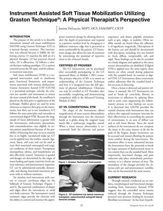
- 1. 32 Orthopaedic Practice Vol. 18;3:06 Instrument Assisted Soft Tissue Mobilization Utilizing Graston Technique® : A Physical Therapist’s Perspective Justine DeLuccio, MSPT, OCS, FAAOMPT, CKTP INTRODUCTION The purpose of this article is to describe instrument assisted soft tissue mobilization (IASTM) using Graston Technique (GT) in a manual therapy construct. This interven- tion was selected because it fulfills the fol- lowing criteria: (1) matches the skill set of a physical therapist, (2) has practical clinical value, (3) is efficacious, (4) follows a clini- cal decision making paradigm, (5) is patient centered across the life span, and (6) enhanc- es treatment outcomes. Soft tissue mobilization (STM) is a rec- ognized intervention used to ameliorate pain, functional limitations, and impair- ments associated with somatic dysfunction. Graston Instrument Assisted STM (GISTM) is a specialized technique whereby the clini- cian uses stainless steel instruments to contact the tissue instead of the hands. An emollient is placed on the skin prior to application of the technique. Rubber gloves are used by some clinicians to improve their grip on the instru- ments, but their use is not mandatory. GT follows the same principles and rationales as conventional digital STM. Because the mag- nitude of tissue deformation is greater with the Instruments, indications, precautions, and contraindications vary slightly in cer- tain patient populations because of the pos- sibility of bruising that may occur in muscles due to its highly vascularized make up. GT can be applied over the tendon, muscle, liga- ment, fascia, and scars and can be used to treat their associated nonsurgical and surgi- cal conditions of these tissues. Entrapment neuropathies, edema, and lymphedema are also effectively treated. Treatment is guided and dosages are determined by the stages of tissue healing and repair, reactivity levels, pa- tient tolerance, and posttreatment responses. GT can be administered statically, dynami- cally, and during functional movement pat- terns with or without resistance. Six stainless steel instruments with con- vex or concave curvilinear edges are the hallmark of Graston Technique (Figures 1 and 2). The patented combination of shapes and edges allow the instruments to mold over body contours. The Instrument’s varied treatment edges provide the clinician with the ability to control and monitor the appro- priate treatment dosage by allowing them to alter the depth of penetration and respond to a patients comfort level by changing to a different treatment edge that is perceived as more comfortable by the patient. GT Instru- ment design also allows for ease of treatment by minimizing the potential of repetitive stress to the clinician’s hands. CERTIFIED GT PROVIDER The GT Instruments can be purchased after successfully participating in the GT sponsored Basic, or Module 1 (M1) course. The primary objective of M1 is to assure an understanding of the Graston Technique and how it is integrated into the full spec- trum of physical rehabilitation. Clinicians can only be certified as GT Providers after successfully completing and demonstrating compulsory skills in the Advanced Training, Module II (M2). GT VS. CONVENTIONAL STM The shape of the Instruments allows magnification of tissue texture abnormalities through the Instrument into the clinician’s hands as it glides along the targeted tissue much like a stethoscope magnifies sound. When a tissue texture abnormality is en- countered, both the clinician and patient experience and detect palpable sensations such as grit, ridges, or nodules. Often au- dible sounds are heard when the adhesion is of significant magnitude. Descriptions of the lesions can and should be documented with descriptors including, but not limited to: focal, diffuse, compressible, soft, and rigid. These findings can be also be recorded on a body diagram and updated as the tissue texture abnormalities are abolished. If digital STM is applied over the same region, these lesions are often missed during palpation with the unaided hand. In contrast to digi- tal STM, GT Instruments detect restrictions and/or adhesions that the unaided hand is less accurate in detecting. Once a lesion is detected and patient tol- erance is assessed, the GT Instruments are used to ‘break up’ cross-links, fibrosis, or restrictions or adhesions by splaying fibers and in some cases augmenting the inflam- matory process so that healing can occur. It is theorized that GISTM provides the trained clinician with Instruments that can achieve this expected treatment outcome by their effectiveness in controlling the amount of microtrauma in an area of diffuse scar and or soft tissue fibrosis. Since the metal surfaces of the instruments do not compress the tissue in the same manner as do the fat pads of the fingers, deeper restrictions can be accessed and treated affording the patient more comfort during the intervention. The treatment effect is more substantial because the Instruments have the potential to break up larger amounts of dysfunctional tissue in one session than can the unaided hand. Most importantly, functional changes and pain reduction take place immediately postinter- vention or in a shorter amount of time. The immediacy of the changes provides the cli- nician with pre and posttreatment variables that can be documented the same session. CURRENT RESEARCH Through research conducted on rat ten- dons, morphological and functional changes resulting from Instrument Assisted STM suggests that the controlled micro trauma induced through the Graston Technique protocol may promote healing by increased fibroblast recruitment.1 As has been hypoth- Figure 1. Graston Technique® Instruments Figure 2. GT treatment s/p lateral meniscus transplant, osteochondral autograft lateral femoral condyle.
- 2. 33Orthopaedic Practice Vol. 18;3:06 esized with transverse friction massage, it is theorized that the controlled micro trauma induced through GT also initiates the in- flammatory cascade to start the healing pro- cess. Results from a recent unpublished study on animal ligaments reveals that ligaments treated with Instrument-Assisted Cross Fi- ber Massage (IACFM) were found to be 31% stronger (p < 0.01) and 34% stiffer (p < 0.001) than untreated ligaments indicat- ing that IACFM is a beneficial intervention for providing mechanical stimulation to re- pairing ligaments to accelerate and re-gain ligament strength.2 Following treatment with GT, adaptive stress is paramount dur- ing the reparative process to promote proper tissue healing and alignment. Stretching and ROM activities are of equal importance and used to increase and maintain movement gained during the GT intervention. Recent research explaining inflammation at the molecular level and the histopathology underlying tendon disorders reveals degen- erative changes vs. the previously assumed presence of inflammation or inflammatory cells which are not present upon examina- tion.3-6 These findings explain why somatic pain associated with injury, repetitive stress or the like often prove recalcitrant to phar- macological and manual therapy treatment. This new information provides the ground- work for changing contemporary models of care and how it affects clinical management of various conditions. GT is a reasonable choice based on recent histological findings. It is being determined through research that GT enhances the adaptive potential of CT structure. More importantly, it is a least in- vasive alternative and more practical than pharmacological management in certain cases based on current clinical management guidelines. GT’S EFFECT ON MOVEMENT IMPAIRMENTS AND PAIN BEHAVIORS Because most changes in movement and pain are immediate my ability to identify and treat movement impairments and as- sociated pain behaviors has improved since incorporating GT into my daily patient care regimen. I have always critically analyzed the elusive nature of the CT system, especially muscles and fascia due to their multidimen- sional nature. Take for example the helix configuration of the levator scapulae which changes shape and form based on the posi- tion of the head and scapula. Using GT, I am better able to conceptualize this system and where lesions might be found because I can see changes that might include improved ROM or the amelioration of pain simultane- ously during treatment. Because treatment effects are usually immediately observable, GT has afforded me the opportunity to iden- tify trends in pain behaviors that are caused by CT dysfunction in regions I would not have addressed during digital STM because I was not aware that region, remotely distant to the region of perceived CT pain, was the target tissue. I am hypothesizing that CT restrictions or adhesions in the muscle and fascial system produce tension points capa- ble of causing stress and subsequent over use symptoms including pain and movement loss above or below the area of restriction. This hypothesis might be elucidated further by the work of Thomas Myers who describes the interconnectedness of the linkages of the muscular and fascial systems and offers clini- cal insights as to how any alterations in the balance of this system may make contribu- tions to pains and dysfunctions consistent with somatic dysfunction.7 RECIDIVISM AND SOMATIC PAIN As clinicians we can generalize that a ma- jority of our patients achieve favorable out- comes. We can also generalize that we have discharged patient’s who only achieved par- tial restoration of function, still had pain, or participated in a longer episode of care than projected. Recidivism is another variable of somatic pain and dysfunction that affects long-term favorable treatment outcomes. Like most clinicians, I am always asking questions and critically appraising my suc- cess and failures. Time and clinic tenure en- hance effectiveness but sometimes we are still left with limited treatment successes. I have always questioned why in certain cases my interventions did not resolve or completely ameliorate somatic dysfunction related to connective tissue. It was not until I imple- mented GT that I realized my hands were the confounding variable limiting my treat- ment effectiveness when addressing certain CT dysfunctions. In some scenarios I was not identifying the correct tissues or did not use my hands effectively enough to produce a meaningful treatment outcome. There are also times in a clinician’s tenure when a patient’s pain cannot be reproduced by any test or movement rendering them unable to identify the connective tissue lesion. GT can be used diagnostically to identify lesions be- cause of the Instruments’ inherent ability to tease out lesions when a muscle is ‘scanned’ or examined better than the unaided hand. Once the lesion is localized a patient’s usu- al response is “That’s it! That IS my pain.” While routinely implementing GT, I have found and continue to find the pieces of the treatment puzzle that I have been searching for. Due to the immediate changes that occur in movements and pain while implementing GT, documentation that reflects efficacy of care can be used more easily. I have become more effective in changing and implement- ing interventions due to these immediate changes. Prior to starting any treatment, my patients and I have a dialoged about their response to the last treatment, their current functional status, and symptom behavior. When they don’t have an objective measur- able variable to offer I ask them “What do we still need to get better?” When movements or functional activities are provocative they are used as pretreatment and posttreatment measures during GT. Although subjective, the patient can quantify the percentage of change in pain. Range of motion can be doc- umented pre- and posttreatment and quali- tative variables about the change in the func- tional task can be documented as evidence of change. This immediate change enhances patient satisfaction and increases compliance with their self management efforts. As they see the changes, they become more active in their care reporting and quantifying change without prompting. This team effort by the patient and clinician enhances the ability to document changes in function and impair- ments. THE CURRENT EVOLUTION OF GT GT was developed and evaluated in clini- cal trials at Ball Memorial Hospital and Ball State University in Muncie, Indiana. The GT is part of the curriculum at 4 colleges/uni- versities. Research is ongoing and includes current projects at Texas Back Institute, New York Chiropractic College and St. Vincent’s Hospital in Indianapolis. GT has been vis- ible in the literature.8-34 GT is now present in the work force and used currently by 4 major companies for the care of their injured employees. More than 40 major professional amateur sports organizations currently uti- lize GT. GT is evolving as an effective inter- vention in many settings. CONCLUSION GT is a technique that meets my clinical expectations. It has plausible explanations for its effects and has predictable outcomes. GT makes practical clinical sense to me. Effects can be documented and it actively involves the patient. The pt can be an active partici-
- 3. 34 Orthopaedic Practice Vol. 18;3:06 pant in the treatment based on how they localize the lesion, by position or activity etc…they become more active in the treat- ment. The GT has the potential to enhance the effectiveness of other interventions such as muscle energy techniques (MET), high velocity low amplitude thrust techniques (HVLAT), and mobilization due to its effect on CT. I currently use GT on at least 95% of my patients; however, I have not abandoned any prior treatment interventions and I still apply digital STM. GT is not the answer to all clinical shortcomings such as recidivism, and partial recovery rather the technique represents part of the solution for treating tissue dysfunction. The effectiveness of GT is enhanced by a clinician’s skills. This treat- ment approach has the potential to decrease recidivism, improve patient compliance and produce more favorable outcomes in a short- er episode of care. I recommend and urge clinicians to learn more about this technique because it contributes to our clinical knowl- edge and supports clinical practice. REFERENCES 1. Craig JD, Ganion LR, Gehlsen GM, et al. Rat tendon morphologic and functional changes resulting from soft tissue mobilization. Med Sci Sports Exer. 1997;313-319. 2. Loghmani MT, Avin K, Burr D, Stuart W. Instrument-assisted cross fiber massage accelerates knee ligament healing. Submitted abstract for platform presentation APTA-CSM 2006. 3. Khan KM, Cook JL, Bonar F, et al. Histopathology of common tendinopathies: update and implications for clinical management. Sports Med. 1999;27:393-408. 4. Khan KM, Cook JL, Maffulli N, Kannus P. Where is the pain coming from in tendinopathy? It may be biochemical, not only structural, in origin. Br J Sports Med. 2000:34: 81-83. 5. Scott A, Khan KM, Roberts CR, et al. What do we mean by the term “inflam- mation”? A contemporary basic science update for sports medicine. Br J Sports Med. 2004;38;372-380. 6. Scott A, Khan KM, Cook JL, Duronio V. What is “inflammation”? Are we ready to move beyond celsus? Br J Sports Med. 2004;38:248-249. 7. Myers TW. Anatomy Trains Myofascial Meridians for Manual and Movement Therapists. Churchill Livingstone; 2001. 8. Davidson CJ, Ganion L, Gehlsen G, Roepke J, Verhoestra B, Sevier TL: Morphologic and functional changes in rat achilles tendon following collage- nase injury and GASTM. J Am College Sports Med. 1995;27. 9. Sevier TL, Gehlsen GM, Wilson JK, Stover SA, Helfst RH. Traditional physical therapy vs. Graston Technique Augmented Soft Tissue Mobilization in treatment of lateral epicondylitis. J Am College Sports Med. 1995;27. 10. 10 More Great Techniques. Feature Cover Article: The American Chiropractor. Aug., 2003. 11. Duffy M, Greenapple S. Graston Technique: The (non-) Cutting Edge of Treatment. Carolina SportsLink Maga- zine. 2004:May 18th . 12. Falvey M. Repetitive stress injuries: fighting back with the Graston Technique. Claims Magazine. 2000;4:38-46. 13. Falvey M. The promise of tissue remodeling. Dynamic Chiropractic. 2001;March 26. 14. Focht D. Augmented soft tissue mobilization: A new frontier in treating soft tissue conditions. OT Practice. 1998;3:49-50. 15. Gehlsen GM, Ganion LR, Helfst RH. Fibroblast responses to variation in soft tissue mobilization pressure. Med Sci Sports Exer. 1999;31:531-535. 16. Hammer WI. Applying the Graston Technique: An update. Dynamic Chiropractic. 2003;21. 17. Hammer WI. Graston Technique: a necessary piece of the puzzle. Dynamic Chiropractic. 2001;19. 18. Hammer WI. Instrument-Assisted soft tissue mobilization: a scientific and clinical perspective. Dynamic Chiroprac- tic. 2004;22. 19. Hammer WI, Pfefer MT. Treatment of a case of subacute lumbar compartment syndrome using the Graston Technique. J Manip Physiological Therapeutics. 2005;28:199-204. 20. Henry P, Panawitz B, Wilson JK. Rehabilitation of a post-surgical patella fracture: A case study. Physiotherapy. 2000;86:139-142. 21. Henry P, Panwitz B, Wilson JK. Treatment of a bilateral total knee replacement using Augmented Soft Tissue Mobilization. Phys Ther Case Reports. 1999;2:27-30. 22. Hyde TE. Graston Technique for Ath- letic Injuries. D.C. Tracts. 2003;15:2-4. 23. Hyde TE. The Graston Technique: a new manual therapy for back pain. Spine-health website. Posted August 5, 2003. 24. Martinez R. Graston Instrument As- sisted Soft Tissue Mobilization. Integra- tive Med. 2003;2:18-23. 25. Melham TJ, Sevier TL, Malnofski MJ, Wilson JK, Helfst RH. Chronic ankle pain and fibrosis successfully treated with a new non-invasive Augmented Soft Tissue Mobilization (ASTM): A case report. Med Sci Sports Exerc. 1998;30:801-804. 26. O’Connor SM. New soft tissue mobili- zation process claims success in decreas- ing scar tissue and repetitive motion symptoms. Work Injury Management News and Digest. 1988;7:3. 27. Perle SM. The Leading Edge. Training Conditioning XIII. 2003:27-31. 28. Perle SM, Lawson G. Stimulating healing by initiating inflamma- tory response. Canadian Chiropractor. 2004;9:10-13. 29. Roush MB, Sevier TL, Wilson JK, Helfst RH, Gehlsen GM, Bassey AL. Anterior Knee Pain: A Clinical Com- parison of Rehabilitation. Clin J Sports Med. 2000;10:22-28. 30. Sevier TL, Helfst RH, Stover SA, Wilson JK. Clinical trends on tendinitis. WORK: J Preven Assessment Rehabil. 2000;14:123-126. 31. Smith S. Faster, better, easier soft-tissue mobilization. Exploring Hand Ther. 2005;5:1,3,4,9,13,18. 32. Smith S. A growing trend in hand therapy: Instrument-assisted soft tissue mobilization helps therapist work smarter, not harder. Phys Ther Products. April/May 2005. 33. Thomann A, Sevier TL, Wilson JK. Treating soft tissue fibrosis: A new re- habilitation technique for the treatment of various soft tissue injuries. Phys Ther Products. 1999;10:56-58. 34. Wilczewski K. The Graston Technique: Changing the treatment of soft tissue injuries. The American Chiropractor. 2002;5. Justine Deluccio is with Physical Therapy Services of Morristown in Morristown, NJ.