Fireside chat: Newton Howard, Director of the MIT Synthetic Intelligence Lab ...
Clinical Sensoring and Monitoring Faculty Infrared Analysis Brain Tumor Tissue
1. Faculty of Medicine Carl Gustav Carus, Clinical Sensoring and Monitoring, Prof. Edmund Koch
1,5
2
2,5
3
3,5
4
4,5
5
50 250 450 650 850
Acknowledgements:
This project was supported by SAB (Saechsische Aufbaubank),
the BMBF (Bundesministerium für Bildung und Forschung:
NBL3) and the MeDDrive program of the Faculty of Medicine
of the TU Dresden.
Rapid intra-operative analysis of human brain tumor tissue with infrared
spectroscopy
Jelena Tavkina, Allison L. Stellinga, Ortrud Uckermannb, Elke Leipnitzb, Holger Cramma, Kathrin D. Geigerc, Matthias Kirschb, Gerald Steinera
aFaculty of Medicine, Clinical Sensing and Monitoring, Dresden University of Technology
bFaculty of Medicine and University Hospital, Neurosurgery, Dresden University of Technology
cFaculty of Medicine, Department of Neuropathology, Institute for Pathology, Dresden University of Technology
Contact:
Dr. Allison L. Stelling
Dr. rer. nat. habil. Gerald Steiner
Clinical Sensoring and Monitoring
Faculty of Medicine, TU Dresden
Fetscherstraße 74, 01307 Dresden, Germany
julia.walther@mailbox.tu-dresden.de
www.tu-dresden.de/medksm
Is this spectral region a
biomarker for increased
RNA in the tumor tissue?
Infrared analysis of glioblastoma cell lines
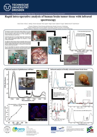
Human brain tumor tissue
Histopathology of the ATR-
measured tissue
Intra-operative assessment of brain tumor margins continues to be an active
area of clinical research, as tumor borders are altered during neurosurgery
from the pre-operative images. This work investigated the suitability of
infrared spectroscopy for inter-operative use as a fast, chemically based
method for delineating brain tumor borders.
Excised brain tumor tissue was investigated during surgery. Infrared spectra
could be obtained from wet tissue within minutes after removal from the
patient. Parallel histopathological diagnoses were obtained by a board
certified neuropathologist.
The differences observed between tumor and non-tumor tissues may arise
from higher levels of RNA present in freshly excised tissue. RNA in particular
is known to degrade within minutes after excision. To interpret these spectral
differences, additional infrared studies were performed in an animal model,
on tumor cell lines, and on yeast RNA.
This work both highlights the importance of acquiring the infrared spectra
within ten minutes after removal from the brain, and demonstrates the high
clinical potential of infrared spectroscopy for rapid intra-operative analysis.
Infrared analysis of freshly extracted mouse brain tissue
RNA extract from yeast (PBS buffer, pH 7.5)
No. GBM cells
IntegratedATRabsorbance(X1000)
Mouse brain
Glioblastoma multiform
(GBM) spheroid cells
Cell concentration vs. ATR absorbance in
cell culture media
1. Intra-operational spectroscopy
2. Matched histopathology
ATRabsorbance
Wavenumbers, cm-1
ATRabsorbance
Wavenumbers, cm-1
Cell
pellet
Time-resolved ATR
subtraction spectra of brain
tissue death after extraction
(A and B)
ATRabsorbance
Wavenumbers, cm-1
Wavenumbers, cm-1
ATRabsorbance
-1220
-1070