SlideShare a Scribd company logo
1 of 10
Download to read offline
INTRODUCTION
Over the past three decades, bonding of resin-based
composite restorations has been revolutionized by
continuing advances in dental adhesive technology. The
ultimate goal in the design and development of dental
adhesives is to render a stronger and more durable
adhesion to hard dental tissues —despite the severe
conditions in the oral environment. Improvements
have been made in the areas of aesthetic appeal, ease
of use and reduction of technique sensitivity1)
. However,
the process was, and still is, not without its faults,
and chief among those faults is the reduced durability
of resin-dentine bonds compared with resin-enamel
bonds, owing to the fact that dentine bonding relies on
organic components2)
. The infiltration of hydrophilic
resin monomers into demineralized collagen matrix,
to produce a hybrid layer (HL) that couples adhesives/
resin composites to the underlying mineralized dentine,
provides ample opportunity for nano-leakage to occur
beneath the restoration: oral fluids penetrate any poorly
infiltrated area and serve as functional medium for the
functioning of host-derived, collagen degrading matrix
metalloproteinases (MMPs)3)
. While each experimental
strategy that attempted to overcome these problems has
its own benefits and reciprocal limitations4)
, progressive
water replacement by hydroxyapatite generated during
dentine remineralization may be a suitable strategy for
extending the service life of resin-based dentine bonding
procedures and its actualization has been a source of
conjecture until now5)
. In this case, nano-leakage might
only be a temporary phenomenon that could be solved
by new hard-tissue formation6)
. Water may provide the
aqueous environment for the infiltration of calcium
phosphate (Ca/P) precursors into the gap zones of
collagen fibrils to initiate nucleation and growth of
newly formed mineral crystals. As remineralization
proceeds, free and loosely bound water becomes
less readily available to MMPs to initiate autolytic
phenomena. Moreover, catalytic and allosteric domains
of these enzymes may be fossilized and inactivated by
the deposition of hydroxyapatite crystallites between
polymer matrices and exposed collagen fibrils5)
.
It has been suggested that molecular immobilization
of the functional activity of collagenolytic enzymes
could be accomplished in dental restorations with
a guided tissue approach7)
. Previous research has
concentrated on nanotechnology principles and the use
of biomimetic analogs of matrix phosphoproteins to
mimic what occurs in biomineralization, particularly in
areas devoid of seed crystallites8)
.
An excellent review paper was presented by
Cochrane et al.9)
to highlight recent nanotechnological
developments in the non-invasive management of
incipient caries lesions, with a specific focus on bottom-
up remineralization strategies for controlled enamel
synthesis.
This narrative article provides thorough
information about the development of new approaches
to enhanced remineralization of tooth tissues from a
different perspective, namely the aim was to answer
questions about whether the presence of reactive
silicate compounds at the resin bonded-dentine
interface could have a significant impact on the
processes occurring in their vicinity, and the formation
of hydroxyapatite deposits on hypomineralized adjacent
Dentine bonding agents comprising calcium-silicates to support proactive
dental care: origins, development and future
Andrea Corrado PROFETA
Department of Restorative Dentistry, Biomaterials Science, Biomimetics and Biophotonics (B3) Research Group, King’s College London Dental
Institute, Floor 17, Tower Wing, Guy’s Dental Hospital,Great Maze Pond SE1 9RT, London, England
Corresponding author, Andrea Corrado PROFETA; E-mail: andrea.profeta@kcl.ac.uk
The origin of ion-releasing dentine bonding agents lies in a change in attitude regarding the qualities demanded of a restorative
dental material. The objectives of this paper are to review recent studies on novel hybrid adhesives comprising bioactive fillers based
on information from original research papers, reviews, and patent literatures. Literature searches of free text and MeSH terms were
performed by using MedLine (PubMed), Web of Science, Scopus, Scielo and the Cochrane Library (6th November, 2013). Reference
lists of primary research reports and eligible systematic reviews were cross-checked in an attempt to identify additional studies.
Experimental methacrylate-based adhesives, either when incorporating calcium/sodium phosphate-phyllosilicates or calcium silicate
cements, demonstrated to promote therapeutic/protective effects on the micro-mechanical and ultramorphological properties of resin
bonded-dentine interfaces associated with mineral deposition over time. Further randomized control trials are needed in order to
confirm these initial results in vivo.
Keywords: Etch-and-rinse adhesives, Bioactive micro-fillers, Adhesion durability, Nano-leakage
Color figures can be viewed in the online issue, which is avail-
able at J-STAGE.
Received Sep 26, 2013: Accepted Dec 24, 2013
doi:10.4012/dmj.2013-267 JOI JST.JSTAGE/dmj/2013-267
Review
Dental Materials Journal 2014; 33(2): 1–10
Fig. 1	 Crystal structure of biogenic HCAp.
dentine surfaces might take place in presence of
phosphate-containing solutions that closely mimic
physiological fluids present in the oral environment.
With information sources collated from original
scientific papers, reviews, and patent literatures,
this paper tracked and arranged the early to recent
developments in this field into three sections as follows:
I) Pathways in dentinal mineralization; II) Top-down
remineralization of hybrid layer collagen matrix; III)
Development of dentine bonding systems comprising
bioactive particles.
PATHWAYS IN DENTINAL MINERALIZATION
Biologically mineralized tissues have remarkable
hierarchical structures that have evolved over time to
achieve great functions in a large variety of organisms.
Enormous progress has been made over the last few
decades in understanding the mineralization processes in
mammalian tissues such as bone, enamel and dentine.
Dentine is mainly composed of type-I collagen with
associated noncollagenous proteins, to form a highly
cross-linked three-dimensional matrix reinforced by
mineral crystallites10)
.
The quality of dentine is dependent upon the total
sum of characteristics of the tissue that influence
its competence: microstructure, mineral density and
composition, along with the particular location of the
mineral phase with respect to the organic structures.
This mineral-reinforced composite was described by
Weiner and Wagner as containing oriented plate-like
crystals with their c-axis aligned with the long axis of
each collagen fibril11)
. Their sizes, ranging in length
from 20 Å to 1100 Å12)
, is not arbitrary; rather, it seems
to give them remarkable physical characteristics13,14)
,
exposing a very large surface area to the extracellular
fluids, which is critically important for rapid ion
exchange. Minerals start to nucleate within or
immediately adjacent to the holes and pores present in
the collagen fibrils15)
. This intrafibrillar heterogeneous
nucleation is catalyzed by the presence of phosphated
esters groups16)
and carboxylate groups17)
present
in the collagen fibrils. Subsequently, the growth, or
mineralization, takes place along the collagen fibrils,
eventually occupying all of the interstitial extrafibrillar
spaces (20 nm). The location of these crystals was
demonstrated in a study by Traub et al.18)
in which
mineralized collagen fibrils had the same banded pattern
as negatively stained collagen fibrils, with domains of
charged amino acids at the border of the gap and overlap
zones acting as nucleation sites. Modelling of collagen
fibrils showed that these nanosized, positively charged
regions are used for mineral infiltration as well as
charge-charge attraction. It was proposed that mineral
platelets were arranged in parallel within the gap zones
and interstices of the fibril like a stack of cards19)
.
The principal mineral component is carbonated
nanocrystalline hydroxyapatite (HCAp) whose structure
is far different from stoichiometric hydroxyapatite
(HAp), represented by the formula:
Me10(XO4)6Y2
Where Me is a divalent metal (e.g. calcium, Ca2+
), XO4
is a trivalent anion (e.g. phosphate, PO4
−3
), and Y is a
monovalent anion (e.g. hydroxyl, OH−
).
Given the unique mechanisms involved in its
formation, biogenic HCAp varies in several ways from
the corresponding geologically produced mineral. First,
HCAp has a hexagonal lattice structure, having a strong
ability to form solid solutions, and to accept numerous
substitutions (Fig. 1). These substitutions affect the
apatitic lattice parameters: the crystal size is decreased,
and thereby the surface area is increased compared
to stoichiometric HAp, thus permitting additional
adsorption of ions and molecules20)
. HCAp contains in
fact various trace elements from intrinsic or extrinsic
origins, namely significant carbonate substitutions, OH−
deficiencies, and imperfections in the crystal lattice21)
.
This phenomenon provides certain physico-chemical
and functional features important in the formation and
dissolution of crystals in dental tissues. For example,
F−
ions are readily incorporated into HCAp, forming
fluoroapatite, a less soluble phase of Ca/P as compared to
HAp, confering to enamel its low dissolution properties to
resist acidic attacks. With respect to dentinal structure,
the peculiarity is represented by numerous substitutions
(i) by hydrogenophosphate (HPO4
2−
) of XO4 groups and
(ii) by carbonate (CO3
2−
) of Y2 and XO4 groups.
Lastly, biological minerals tend to attain high
crystallinity and a more organized structure on the time
scale of days or months rather than years22)
. To this end,
Lowenstam and Weiner23)
used transmission electron
microscopy to evaluate the ultrastructure of HCAp
crystallites and confirmed that the average length and
width of crystallites was 20 nm, with an approximate
thickness of 2–3 nm, resembling plate-like structures.
TOP-DOWN REMINERALIZATION OF HYBRID
LAYER COLLAGEN MATRIX
Despite their complex hierarchical structures, the basic
building blocks of bones and teeth during both initial
2 Dent Mater J 2014; 33(2): 1–10
and later formation stages are nanosized mineral
particles. Although extensive investigations of Ca/P
crystallization have been performed, many have studied
the final structures and morphologies and have not
emphasised the need to consider the early contacts
between mineral nuclei and organic matrix, or the
thermodynamic and kinetic controls imposed by the
latter and soluble proteins. Current results and concepts
of crystal nucleation and growth at the molecular
level, and the role of site-specific interactions, provide
possible mechanisms of Ca/P crystallization that are
related to the remineralization of dentine13,14)
. However,
the detailed physical and chemical processes by which
nucleation control is established and the thermodynamic
and kinetic parameters that define those processes
remain largely unknown.
The site and the amount of mineral deposition are
probably determined by the chemical process (deposition)
and by the physical condition (mineral distribution
profile and transport mechanism).
Organic phases play an initial role in templating
the structure of mineralized tissues; therefore, their
matrices should be sound as a scaffold for the mineral
crystals to grow24,25)
. Structurally intact collagen
fibrils that retain their banded characteristics
and intermolecular crosslinks are considered to be
physiologically remineralizable26,27)
. Also, non-denatured
intact collagen is remineralizable as long as seed
crystallites are still present28)
. Partial demineralization
of the collagen matrix by acids derived from bacteria
or dentine bonding procedures leaves remnant seed
crystallites and noncollagenous phosphoproteins
that can act as heterogeneous nucleation sites29,30)
. In
nanotechnology terminology, this represents a top-down
approach via epitaxial growth31)
.
Mineral/matrix contact is a key factor. This leads
to the deposition of a dense network of pre-nucleation
clusters bound by polyanions within any nanosized
region of the organic template and their subsequent
growth involves various possible precursor phases to the
final mineral phase32)
.
Classic crystallization theory assumes that crystals
nucleate and grow from elementary building blocks
(ions, molecules) in a supersaturated solution, although
phase transformations may also occur at later stages.
The association of solution species to form “metastable
intermediate precursors”33)
, or “growth units”, that
subsequently dissolve as the precipitation reactions
proceed, is a crucial step34)
.
The nature of the primary mineral phase remains
controversial. Dicalcium-phosphate salts have been
implicated as possible precursors to the formation of
biogenic HCAp with trace of carbonate and fluoride35)
.
Moreover, in vivo calcifications have also suggested the
involvement of an initial amorphous Ca/P phase (ACP)
followed by transformation to the final HCAp product36)
.
According to Posner, HCAp derives from Ca/P clusters
[Ca9
(PO4
)6
] packing randomly with interfacial water
to form ACP precursors37)
. This theory is supported by
the presence of several Ca/P growth inhibitors such as
magnesium that stabilize the amorphous state38,39)
.
These precursor nanoparticle phases grow rapidly
in size via ion-by-ion attachment40)
and aggregation,
with local loss of solvent, undergoing amorphous-
crystalline transformations or phase transformations
en route to a thermodynamically stable macro-crystal.
Unlike biomimetic bottom-up remineralization
strategies that take months of protracted chemical
reactions to complete, top-down mineral fabrication
of HCAp crystals proceeds fast. Thus, while the
mineral constituents is being restored, the region
of the exposed collagen fibrils is less susceptible
to hydrolytic degradation by MMPs. Furthermore,
traditional remineralization by epitaxial growth is a
thermodynamically favorable process that overcomes
the energy barrier of homogeneous nucleation41)
.
According to Ostwald’s rule42)
, normally the first
occurring phase in polymorphism is the least stable
and closest in free energy to the mother-phase, followed
by other phases in order of increasing stability. The
formation, dissolution, and transformation of Ca/P
bodies depend on their nature (particle size,
crystallographic features, density) and the nature of
the solution (composition, pH, temperature). Most Ca/
Ps are sparingly soluble in water, and some are very
insoluble, but all dissolve in acids. Their solubility,
defined as the amount of dissolved solute contained
in a saturated solution when particles of solute are
continually passing into solution (dissolving) while
other particles are returning to the solid solute phase
(growth) at exactly the same rate43)
, decreases with
increments in temperature and in pH44)
. Each Ca/P
phase possesses its own thermodynamical solubility.
For example, at pH=7 and 37ºC, HCAp is the most
stable phase32)
. However, these thermodynamical
considerations are under equilibrium conditions, and
therefore they do not take into account kinetics that
dictate the formation of one or the other phase under
dynamic conditions. In vivo, the interactions between
Ca/Ps and their “biological surroundings” are highly
complex due to the non-equilibrium conditions and due
to the undefined amount of compounds playing a role in
these interactions. The second important factor in the
stability of Ca/Ps is the characteristics of the solution
in which these salts are formed or placed, namely the
solution supersaturation in free Ca2+
and PO4
−3
ions45)
.
At a given pH and temperature, a free Ca2+
and PO4
−3
ion containing solution can be categorized in three
different states: (i) the stable (undersaturated)
condition, when crystallization is impossible; (ii) the
metastable condition, when spontaneous crystallization
of Ca/P salt is improbable, although the concentrations
are higher than the ones corresponding to the salt
solubility. If a crystal seed were placed in such a
metastable solution, growth would occur on the seed;
(iii) the unstable condition, when spontaneous
crystallization of Ca/P is probable, but not inevitable32)
.
Extracellular fluids that are supersaturated for Ca2+
and PO4
−3
under normal conditions or, alternatively,
by means of external sources with tailored treatments
3Dent Mater J 2014; 33(2): 1–10
Fig. 2	 Schematic diagram of bioactive micro-fillers conversion within the mineral-depleted resin-
dentine interface.
	 It is believed that throughout the course of nano-leakage events HCAp can form from dissolved
precursor species on top of a silica nucleation layer.
	 In presence of salivary or dentinal fuid, providing a natural source of Ca2+
and PO4
−3
ions,
Bioglass®
45S5 and the silicate phases of PCs undergo a series of physicochemical reactions
with subsequent precipitation of an amorphous silica-rich layer “Si-gel” or resulting in the
formation of a nanoporous matrix/gel of calcium silicate hydrates “CSH phase”. Either way,
silica nucleation layers provide the negatively charged sites for the migration and incorporation
of mineral ions (e.g. Ca2+
), which in turn lead to an over-saturated solution that exceeds the
solubility product constants for a number of mineral forms, inducing crystal growth.
that deliver the same ions (Fig. 2), may induce the
heterogeneous nucleation and growth of new Ca/P
crystals32)
. The newly formed minerals, in their turn,
may act as sites for further nucleation promoting a
continuous remineralization over time in presence of
environmental mineral ions.
DEVELOPMENT OF DENTINE BONDING SYSTEMS
COMPRISING BIOACTIVE PARTICLES
Many modifications and improvements have been
made to dentine bonding agents in the years since
their inception. Several “generations” of products
have been developed in the last few decades, but the
advancements have mainly increased ease of use and
reduced technique sensitivity. To date, however, little
progress has been made with regard to reducing the
propensity of the adhesive towards nano-leakage (Fig.
3), though the achievement of improved bond strengths
has significantly reduced gross gaps from occurring46)
.
One possible modification that may significantly
reduce the occurrence and extent of nano-leakage is
represented by the incorporation of biologically active
agents into the resin-bonding procedure to repair any
nanometre-sized void that remains within the HL after
polymerization of the adhesive.
The development of self-sealing dentine bonding
systems with therapeutic ability to create a chemical
bond in addition to the micromechanical one, enabling
dental restorations to have longer lifetimes, is
currently one of the main targets of dental biomaterials
research47)
.
Key objectives in the design of ion-releasing interfacial
materials for dental restoration
In view of the clinical demand on engineered dental
tissue, new biologically active adhesive/primers
formulations have to exhibit:
Ⅰ.	 A hydrophilic nature to interact with oral fluids,
tolerate moisture and absorb water for the
necessary ion movement.
Ⅱ.	 Light-curable chemistry characteristics with
controlled water sorption and solubility
behaviour of the cross-linked network.
Ⅲ.	 Ability to release mineral ions in a sustained
manner (bioavailability) and crystallographic
inclusion of foreign ions from aqueous
environments that contain Ca2+
and PO4
−3
ions,
such as saliva and dentinal fluid.
Ⅳ.	 Appropriate filler size, or fineness, to influence
hydration rate: the smaller the size, the greater
the surface-area-to-volume ratio, and thus,
the more area available for water-particle
interaction per unit volume48)
.
Ⅴ.	 Potential to completely replace water from the
resin-sparse regions of the HL with redeposition
of thermodynamically stable, apatitic tooth
mineral (bioactivity).
Ⅵ.	 Antibacterial properties at the dentine-
restoration interfacial region to reduce the risk
of reinfection and secondary caries, in particular
when using dental composites lacking any
antimicrobial activity.
These said features thus form the requisites for
the improvement of dentine bonding technology with
regard to the hydrolytic enzyme-related loss of HL’s
4 Dent Mater J 2014; 33(2): 1–10
Fig. 3	 Confocal laser scanning microscopy (CLSM) characterization (single-projection images,
fluorescence mode) and nano-leakage (fluorescein dye) of resin-dentine interfaces
created with a typical 3-step etch-and-rinse adhesive.
	 The adhesive is labeled with rhodamin B to reveal the extent of resin diffusion within
the demineralized (acid-etched) dentine at the conclusion of the bonding procedure
(left). Note the presence of intense fluorescein dye uptake within the HL and at bottom
of the adhesive layer after the ageing period (right). The presence of gaps is due to HL
degradation [Adapted from Profeta et al., Dent Mater 2013; 29: 729-741].
organic components and bond strength.
An initial appraisal of the effects of calcium/sodium
phosphosilicates on the resin bonded- dentine interface
Recently, calcium/sodium phosphosilicates have been
successfully used to reduce tooth sensitivity due to their
ability to act as biomimetic mineralizers, promoting
Ca/P precipitation and subsequent crystallization into
carbonated HCAp on the glass/tissue interface without
toxicological consequences49)
.
The chemical composition is significant. The
components are basically oxides of calcium, sodium,
phosphorus, and silicon at certain weight ratios. Silica
maintains the glass structure, while sodium aids in
maintenance of physiological ionic balance and pH50)
.
More than three decades of study have revealed
a distinct, though as yet not completely understood,
series of chemical reactions that takes place after
few minutes of exposure to calcium-phosphate-
rich environments, beginning with exchange of
cations followed by loss of soluble silica. Both steps
are characterized by formation of silanols (SiOH).
Polycondensation of SiOH to form a hydrated silica gel
precedes the formation of an ACP layer. The reaction
is concluded with crystallization into HCAp51)
. More in
detail, the first phase is characterized by rapid release
of alkali or alkaline earth elements (Na+
or K+
), usually
through cation exchange with H+
or H3O+
ions, and de-
alkalinization of the glass surface. This ion exchange
process leads to an increase in interfacial pH, to values
>7.4. Network dissolution occurs concurrently by
breaking of -S-O-Si-O-Si- bonds of the glass structure
through the action of OH−
ions (base catalyzed
hydrolysis of -S-O-Si-O-Si- bonds). Breakdown of
the network is localized and releases silica into the
solution in the form of silicic acid (SiOH4). Last stages
involve the development of silica rich and ACP layers
respectively. Hydrated silica formed on the glass
surface by these reactions undergoes rearrangement
by polycondenzation of neighboring SiOH, culminating
in a silica-rich gel layer. During the precipitation
reaction, Ca2+
and PO4
−3
ions liberated from the glass
along with those of the solution combine to create a
calcia-phosphate-rich (Ca/P) layer on the silica gel. The
Ca/P phase that stratifies in the gel surface is initially
amorphous. Within 3–6 h, the ACP will crystallize
into HCAp by incorporating carbonate anions from the
surroundings51)
.
It is expected that the forming mineral would bond
to specific amino acids within exposed dentine collagen
as occurs in bone52)
. Evidence has emerged suggesting
that bonds to bone are mechanically strong and believed
to be chemical in nature; force is required to separate
the tissue from the glass53,54)
.
Confirming the relative success of surface reactive
glass-ceramic materials incorporation in dentine
bonding procedures required assessment of the
potential to reduce nano-leakage, as well as their
effect upon the strength of the bond over time. In
a study by Profeta et al.35)
when 0.05 g of bioactive
glass of the well-characterized 45S5 formulation (45
wt% SiO2; 24.5 wt% Na2O and CaO; 6 wt% P2O5) (Sylc;
OSspray, London, UK) and of an average particle size
of <10 μm were applied directly onto H3PO4-etched wet
mid-coronal dentine before bonding, a commercially
available adhesive was able to form an apparently
5Dent Mater J 2014; 33(2): 1–10
Fig. 4	 Interfacial characterization (CLSM single-projection images, fluorescence/reflection
mode) and nano-leakage (fluorescein dye) of resin-dentine interfaces created with a
3-step etch-and-rinse adhesive comprising Bioglass®
45S5.
	 The adhesive layer shows a strong fluorescent signal due to added dye (rhodamin
B) before application (left). It is also possible to observe intense fluorescein signal
from the HL (pointer) located underneath a thick adhesive layer characterized by
the presence of mineral micro-fillers. After 6 months of artificial ageing (right), it is
possible to note reduced fluorescein diffusion within a HL characterized by a strong
reflective signal (pointer), indicating a change in dentine’s mineral content within this
zone [Adapted from Profeta et al., Eur J Oral Sci 2012; 120: 353-362].
normal HL. Notably, after 6 months of storage in PO4
−3
buffered saline, the occurrence and extent of nanometre-
sized voids within the HLs was reduced due to the
chemical nature of the mineral precipitation (dicalcium-
phosphate salts) within the resin-dentine interface.
Conversely, severe water uptake was observed within
the resin-bonded dentine interfaces created with the
control resin both after 24 h and 6 months of storage.
However, a concomitant decrease in microtensile bond
strength over time was also observed. On this account,
the crystallized bonded-dentine interfaces might have
attained mechanical characteristics comparable to
those created by conventional glass-ionomer cements
applied onto polyacrylic acid-etched dentine when
submitted to tensile tests55)
, not reflecting their true
adhesive strength. Following this promising start, it
was decided to include 30 wt% of Bioglass®
45S5 within
the composition of a resin adhesive as tailored micro-
filler (filler/resin ratio of 30/70 wt%). This approach
produced bioactive/protective effects not only in terms
of nano-leakage reduction (Fig. 4) but also preserving
high adhesive strength and joint integrity. Analysis
of the failure modes displayed a pronounced tendency
for these samples to fail cohesively; strong adhesion to
tooth substrates would shift the fracture plane from the
HL to the dentine substrate or resin composite, while
the negative controls tended towards failure at the
resin-dentine interface. Another important trend was
confirmed in this study: the ultramorphology analysis
of the fractured specimens demonstrated the formation
of mineral crystals embedded within a well-preserved
collagen network over the course of the ageing period.
The mechanical characteristics of resin-dentine
bonds are a fundamental aspect of restorative
procedures. It has been advocated that atomic
force microscope (AFM)/nano-indentation analysis
performed in hydrated dentine, with force and indenter
displacement being recorded simultaneously throughout
the indentation and where the modulus of elasticity
(Ei) and hardness (Hi) are determined from the load
displacement curve, may be a suitable method to
evaluate the visco-elasticity of the demineralized
dentine and the effective remineralization of the
collagen matrix56,57)
. In 2012, Sauro and coworkers58)
reported that experimental bonding agents formulated
on bioactive micro-fillers (Bioglass®
45S5 and
polycarboxylated zinc-modified bioactive glass) were
able to induce a significant increase of the mechanical
nano-properties (Ei and Hi) of definite mineral-depleted
areas within the bonded-dentine interface (i.e. HL and
bottom of the HL) after prolonged storage in a simulated
body fluid solution (SBFS). Presence of well shaped
indentations was observed in the crystallized SBFS-
aged bonded-dentine interfaces. Conversely, scarcely
pronounced marks were detected in the specimens
stored in SBFS for 24 h due to the visco-elastic
properties of the demineralized collagen which
prevented the plastic deformation of the HL56,59)
.
Although the reincorporation of mineral into the
demineralized dentine matrix does not represent a
6 Dent Mater J 2014; 33(2): 1–10
Fig. 5	 Interfacial characterization (CLSM single-projection images, fluorescence mode) and
nano-leakage (fluorescein dye) of resin-dentine interfaces created with a 3-step etch-
and-rinse adhesive comprising a calcium-silicate Portland-derived micro-filler.
	 At the end of the bonding procedure, it is possible to observe a clear HL with long resin
tags (rhodamin B-labeled adhesive) penetrating the dentinal tubules (left). Intense
fluorescein uptake was also observed within the entire resin-dentine interface.
Following 6 months of artificial ageing (right), the CLSM analysis reveals absence of
gaps and diminished fluorescein penetration inside the HL [Adapted from Profeta et
al., Dent Mater 2013; 29: 729-741].
full recovery of its functionality, it still plays a very
important role, since the remineralized remnant
crystallites may be much more resistant to subsequent
acid attacks60)
.
Early developments in etch-and-rinse adhesives containing
calcium-silicate Portland-derived micro-fillers
Since it was introduced in 1993, mineral trioxide
aggregate (MTA) (ProRoot; Dentsply Tulsa Dental,
Tulsa, OK) has proven to be one of the most versatile
dental materials of this epoch. Abundant research shows
excellent biocompatibility and bioactive properties
in a multitude of challenging endodontic clinical
situations61)
. As stated by the patent, MTA is a
derivative of a type I ordinary Portland cement (PC)
with 4:1 proportions of bismuth oxide just added for
radiopacity62)
. PCs are hydraulic materials mainly
composed of di- and tri-calcium silicate (2CaO•SiO2
belite and 3CaO•SiO2 alite), tricalcium aluminate
(3CaO•Al2O3) and gypsum (CaSO4•2H2O) hydrophilic
powders63)
. The setting reactions of PC remain in
many respects obscure, with much controversy and
uncertainty, despite its economic importance. Perhaps
not surprisingly, the dental literature contains
no satisfactory account, although there are a few
superficial remarks and suggestions64)
. However,
taking note of the respective chemical and condition
variations, it is possible to deduce a reaction scheme
which is probably sufficiently correct to account for
the main features of the process. When hydrated, a
solid-liquid interface forms on the silicate phases and
ion dissolution occurs almost immediately. A high-
pH solution containing Ca2+
, OH−
and silicate ions is
established65)
.
A series of physicochemical reactions (hydrolysis of
SiO4
4−
groups) result in the formation of a nanoporous
matrix/gel of calcium silicate hydrates (CSH phase),
characterized by Si-OH silanol groups whose
deprotonation leads to negatively charged SiO−
groups66)
,
and of a soluble fraction of calcium hydroxide Ca(OH)2 or
portlandite61)
. While SiO−
negative groups sets over time
to form a solid network (heterogeneous nucleation of
HCAp) by bonding Ca2+
on the CSH surface67)
, the release
of Ca(OH)2 increases the alkalinity of the surrounding
medium68)
. The soaring pH and concentration of Ca2+
ions enhance supersaturation of the solution and growth
of HCAp crystals63)
.
The similar composition and mechanism of action
suggest that additives used in PC industry might also
be used to change the physical properties of MTA. In
the same manner, this allows initial research studies
to be carried out by using PC, which is advantageous
considering the significantly lower cost of PC compared
with that of MTA. If favorable results are obtained
by using PC, then additional studies can then be
performed to determine whether the same results can
be obtained by using the more expensive MTA material.
Calcium silicates appeared to bond chemically
to dentinal walls, possibly via a diffusion-controlled
reaction63)
, but their inclusion in dentine bonding
7Dent Mater J 2014; 33(2): 1–10
Fig. 6	 Dye-assisted (xylenol orange) CLSM imaging (single-projection images, fluorescence
mode) of resin-dentine interfaces created with a 3-step etch-and-rinse adhesive
comprising a calcium-silicate Portland-derived micro-filler.
	 Fluorochrome xylenol orange is taken up at the sites of active calcification, due to its
ability to form complexes with divalent Ca2+
ions, thus labeling new mineral tissue
formation. It is possible to observe strong presence of Ca2+
-chelator dye inside adhesive,
resin tags and dentinal tubules. Note the intense Ca2+
deposition at the bottom and
within the HL following 24 h of storage in simulated body fluid solution (left). The
data from the biophotonic imaging showed a clearly outlined fluorescence signal within
the bonding interface and along the walls of dentinal tubules even after 6 months of
artificial ageing (right) [Adapted from Profeta et al., Dent Mater 2013; 29: 729-741].
systems is yet to be attempted. In a recent study
small amounts of three derivatives and proprietary
formulations based on the composition of an ordinary
PC (GB patent application no. 1118138.5) were added
to the neat resin of a typical 3-step etch-and-rinse
adhesive (30 μm average particle size; 40/60 wt% filler/
resin ratio), and teeth were bonded for the purpose of
carrying out bond strength tests, ultramorphology and
nano-leakage studies69)
. The most favorable results were
obtained with the addition of set type-I PC modified
using either montmorillonite or hydrotalcite poly-ions
silicates. The use of resin bonding agents containing
these two tailored micro-fillers promoted a therapeutic
mineral deposition mechanism within the HL: nano-
leakage did indeed appear to be reduced after soaking
in simulated body fluid for 6 months (Fig. 5), and
consistent bond strength values were maintained when
compared to negative controls. The reduced fluorescent-
dye uptake observations, along with a strong Ca2+
chelating fluorophore (xylenol orange) signal from the
HL and the dentinal tubules, clearly indicated the
remineralization of those areas which were previously
detected as mineral-deficient/porous zones (Fig. 6). A
further supporting result was found in the scanning
electron microscopy: the ultramorphological analysis
performed on fractured match-sticks demonstrated the
presence of mineral crystals within the resin-dentine
matrix subsequent to the storage period. Despite no
specific biomimetic analogue agents being used in
this study, there may have been a sort of biomimetic
activity evoked by the main hydration products of the
two silicate-based fillers. It was also hypothesized
that the alkaline condition induced during the process
may have created unfavorable conditions for MMPs
expression within the demineralized collagen matrix
and may have played a supplementary role in the
remineralization kinetics70)
. Addition of a PC-based
micro-filler containing titanium oxide to the same
adhesive was less successful. Again, it was shown that
an apparently normal HL was able to form, and the
strength of the bonds formed was comparably high
following 24 h of storage in simulated body fluid.
However, long-term evaluation of the micro-mechanical
strength was less definitive than in the other two
etch-and-rinse adhesives doped with calcium silicates.
Although nano-leakage was substantially reduced,
specimens of this experimental adhesive group showed
diminution of bond strength durability due to the high
hydrophilicity of titanium oxide.
CONCLUSIONS
Noteworthy conclusions can be drawn from this review
paper. Different studies provided preliminary evidence
that the durability of the resin-dentine interfaces may
be enhanced by modified bonding agents in a clinically
relevant manner. The inclusion of highly reactive
silicate compounds, such as commercially available
8 Dent Mater J 2014; 33(2): 1–10
bioactive glass and calcium-silicate Portland-derived
cements, within the composition of representative etch-
and-rinse bonding system conferred attractive basic
properties, such as:
Ⅰ.	 Light polymerization ability and tendency to
interact with oral fluids and wet tooth structures
(hydrophylic nature).
Ⅱ.	 High bioavailability and propensity to deliver
various remineralizing species; when hydrated,
each micro-filler releases the predominant ions
of its composition enhancing mineral delivery
within and beneath the HL (mineral enrichment
effect).
Ⅲ.	 Potential to displace water from the resin-sparse
regions of the HL with redeposition of apatitic
tooth minerals (bioactivity).
Ⅳ.	 Alkalinizing activity to buffer the environmental
acids as well as to interfere with the action of
MMPs, and antibacterial properties.
Ⅴ.	 Predisposition to reduce nano-leakage and the
rate of enzymatic degradation, two of the major
causes of restoration failure, while producing no
negative effects on bond strength over time.
Ⅵ.	 Capacity to preserve the mechanical properties
and stability of resin-dentine bonded interfaces.
The initial evaluation of bioactive silicate compounds as
a possible addition to the resin-dentine bonding process
has shown their value; a new generation of biologically
active dental materials able to induce physicochemical
reactions yielding HCAp formation in demineralized
dentine has been obtained as promising adhesives to be
tested in future studies and clinical trials.
ACKNOWLEDGMENTS
The author thanks all the research co-workers at
King’s College London Dental Institute, Department
of Biomaterials, Biomimetics and Biophotonics (B3)
Research Group, for their invaluable assistance and
discussions in the preparation of this review paper.
REFERENCES
1)	 Breschi L, Mazzoni A, Ruggeri A, Cadenaro M, Di Lenarda R,
Dorigo E. Dental adhesion review: aging and stability of the
bonded interface. Dent Mater 2008; 24: 90-101.
2)	 Marshall GW, Marshall SJ, Kinney JH, Balooch M. The
dentin substrate: structure and properties related to bonding.
J Dent Res 1997; 25: 441-458.
3)	 Sadek FT, Braga RR, Muench A, Liu Y, Pashley DH, Tay F.
Ethanol wet-bonding challenges current anti-degradation
strategy. J Dent Res 2010; 89: 1499-1504.
4)	 Liu Y, Tjäderhane L, Breschi L, Mazzoni A, Li N, Mao J,
Pashley DH, Tay FR. Limitations in bonding to dentin and
experimental strategies to prevent bond degradation. J Dent
Res 2011; 90: 953-968.
5)	 Liu Y, Li N, Qi Y, Niu LN, Elshafiy S, Mao J, Breschi L,
Pashley DH, Tay FR. The use of sodium trimetaphosphate
as a biomimetic analog of matrix phosphoproteins for
remineralization of artificial caries-like dentin. Dent Mater
2011; 27: 465-477.
6)	 Tay FR, Pashley DH. Biomimetic remineralization of resin-
bonded acid-etched dentin. J Dent Res 2009; 88: 719-724.
7)	 Tay FR, Pashley DH. Guided tissue remineralisation of
partially demineralised human dentine. Biomaterials 2008;
29: 1127-1137.
8)	 Zhang X, Neoh KG, Lin CC, Kishen A. Remineralization
of partially demineralized dentine substrate based on a
biomimetic strategy. J Mater Sci Mater Med 2012; 23: 733-
742.
9)	 Cochrane NJ, Cai F, Huq NL, Burrow MF, Reynolds EC. New
approaches to enhanced remineralization of tooth enamel. J
Dent Res 2010; 89: 1187-1197.
10)	 Kinney JH, Habelitz S, Marshall SJ, Marshall GW. The
importance of intrafibrillar mineralization of collagen on the
mechanical properties of dentin. J Dent Res 2003; 82: 957-
961.
11)	 Weiner S, Wagner HD. The material bone: structure-
mechanical function relations. Annu Rev Mater Sci 1998; 28:
271-298.
12)	 Kim HM, Rey C, Glimcher MJ. Isolation of calcium-phosphate
crystals of bone by nonaqueous methods at low-temperature.
J Bone Miner Res 1995; 10: 1589-1601.
13)	 Wang LJ, Nancollas GH. Calcium orthophosphates:
crystallization and dissolution. Chem Rev 2008; 108: 4628-
4669.
14)	 Wang LJ, Nancollas GH. Biomineralization. From Nature to
Application. Sigel A, Sigel H, Sigel RKO, editors. Metal ions
in life sciences series 4. Chichester: John Wiley & Sons; 2008.
p. 413-456.
15)	 Glimcher MJ. The nature of the mineral component of bone
and the mechanism of calcification. Instr Course Lect 1987;
36: 49-69.
16)	 GlimcherMJ,KossivaD,Brickley-ParsonsD.Phosphoproteins
of chicken bone matrix. Proof of synthesis in bone tissue. J
Biol Chem 1984; 259: 290-293.
17)	 Rhee SH, Lee JD, Tanaka J. Nucleation of hydroxyapatite
crystal through chemical interaction with collagen. J Am
Ceram Soc 2000; 83: 2890-2892.
18)	 Traub W, Arad T, Weiner S. Three-dimensional ordered
distribution of crystals in turkey tendon collagen fibers. Proc
Natl Acad Sci USA 1989; 86: 9822-9826.
19)	 Palmer LC, Newcomb CJ, Kaltz SR, Spoerke ED, Stupp
SI. Biomimetic systems for hydroxyapatite mineralization
inspired by bone and enamel. Chem Rev 2008; 108: 4754-
4783.
20)	 LeGeros RZ. Calcium phosphates in oral biology and medicine.
Myers HM, editor. Monographs in oral science. Basel: Karger;
1991.
21)	 Boskey AL. Mineralization of bones and teeth. Elements
Magazine 2007; 3: 385-391.
22)	 Verdelis K, Lukashova L, Wright JT, Mendelsohn R, Peterson
MGE, Doty S, Boskey AL. Maturational changes in dentin
mineral properties. Bone 2007; 40: 1399-1407.
23)	 Lowenstam HA, Weiner S. On biomineralization. New York:
Oxford University Press; 1980.
24)	 Kusboki Y, Ohgushi K, Fusayama T. Collagen biochemistry
of the two layers of carious dentin. J Dent Res 1977; 56: 1233-
1237.
25)	 Gonzalez-CabezasC.Thechemistryofcaries:remineralization
and demineralization events with direct clinical relevance.
Dent Clin North Am 2010; 54: 469-478.
26)	 Landis WJ, Song MJ, Leith A, McEwen L, Mc-Ewen BF.
Mineral and organic matrix interaction in normally calcifying
tendon visualized in three dimensions by high-voltage electron
microscopic tomography and graphic image reconstruction. J
Struct Biol 1993; 110: 39-54.
27)	 Landis WJ. Mineral characterization in calcifying tissues:
atomic, molecular and macromolecular perspectives. Connect
Tissue Res 1996; 34: 239-246.
28)	 Koutsoukos PG, Nancollas GH. Crystal growth of calcium
phosphates —epitaxial considerations. J Cryst Growth 1981;
9Dent Mater J 2014; 33(2): 1–10
53: 10-19.
29)	 Clarkson BH, Feagin FF, McCurdy SP, Sheetz JH, Speirs R.
Effects of phosphoprotein moieties on the remineralization of
human root caries. Caries Res 1991; 25: 166-173.
30)	 Bertassoni LE, Habelitz S, Marshall SJ, Marshall GW.
Mechanical recovery of dentin following remineralization in
vitro —an indentation study. J Biomech 2011; 44: 176-181.
31)	 Wong TS, Brough B, Ho CM. Creation of functional micro/
nano systems through top-down and bottom-up approaches.
Mol Cell Biomech 2009; 1: 1-55.
32)	 Barrère F, van Blitterswijk CA, de Groot K. Bone regeneration:
molecular and cellular interactions with calcium phosphate
ceramics. Int J Nanomedicine 2006; 1: 317-332.
33)	 Onuma K, Ito A. Cluster growth model for hydroxyapatite.
Chem Mater 1998; 10: 3346-3351.
34)	 Eanes ED, Gillessen IH, Posner AS. Intermediate states in
the precipitation of hydroxyapatite. Nature 1965; 208: 365-
367.
35)	 Profeta AC, Mannocci F, Foxton RM, Thompson I, Watson TF,
Sauro S. Bioactive effects of a calcium/sodium phosphosilicate
on the resin-dentine interface: a microtensile bond strength,
scanning electron microscopy, and confocal microscopy study.
Eur J Oral Sci 2012; 120: 353-362.
36)	 Mahamid J, Sharir A, Addadi L, Weiner S. Amorphous
calcium phosphate is a major component of the forming fin
bones of zebrafish: Indications for an amorphous precursor
phase. Proc Natl Acad Sci USA 2008; 105: 12748-12753.
37)	 Posner AS. The mineral of bone. Clin Orthop Relat Res 1985;
200: 87-99.
38)	 Barrére F, Layrolle P, van Blitterswijk CA, de Groot K.
Biomimetic calcium phosphate coatings on Ti6AI4V: a crystal
growth study of octacalcium phosphate and inhibition by
Mg2+ and HCO3−
. Bone 1999; 25: 107S-111S.
39)	 Root MJ. Inhibition of the amorphous calcium phosphate
phase transformation reaction by polyphosphates and metal
ions. Calcif Tissue Int 1990; 47: 112-116.
40)	 Niederberger M, Cölfen H. Oriented attachment and
mesocrystals: non-classical crystallization mechanisms based
on nanoparticle assembly. Phys Chem Chem Phys 2006; 8:
3271-3287.
41)	 Jiang H, Liu XY. Principles of mimicking and engineering the
self-organized structure of hard tissues. J Biol Chem 2004;
297: 41286-41293.
42)	 Ostwald WF. Studies on formation and transformation of
solid materials. Z Phys Chem 1897; 22: 289-302.
43)	 Wu WJ, Nancollas GH. The dissolution and growth of
sparingly soluble inorganic salts: A kinetics and surface
energy approach. Pure Appl Chem 1998; 70: 1867-1872.
44)	 de Groot K. de Groot K, editor. Ceramics of calcium
phosphates: preparation and properties. Bioceramics of
calcium phosphate. CRC Press Inc; 1983. p.100-111.
45)	 Tang RK, Nancollas GH, Orme CA. Mechanism of dissolution
of sparingly soluble electrolytes. J Am Chem Soc 2001; 123:
5437-5443.
46)	 Pashley DH, Tay FR, Breschi L, Tjäderhane L, Carvalho RM,
Carrilho M, Tezvergil-Mutluay A. State of the art etch-and-
rinse adhesives. Dent Mater 2011; 27: 1-16.
47)	 Liu Y, Mai S, Li N, Yiu CK, Mao J, Pashley DH, Tay FR.
Differences between top-down and bottom-up approaches in
mineralizing thick, partially demineralized collagen scaffolds.
Acta Biomater 2011; 7: 1742-1751.
48)	 Hashimoto M, Iijima M, Nagano F, Ohno H, Endo K. Effect of
monomer composition on crystal growth by resin containing
bioglass. J Biomed Mater Res B Appl Biomater 2010; 94: 127-
133.
49)	 Bauer TW, Smith ST. Bioactive materials in orthopaedic
surgery: overview and regulatory considerations. Clin Orthop
Relat Res 2002; 395: 11-22.
50)	 Hench LL, Splinter RJ, Allen WC, Greenlee TK. Bonding
mechanisms at the interface of ceramic prosthetic materials.
J Biomed Mater Res 1971; 5: 117-141.
51)	 Hench LL, Andersson O. Hench LL, Wilson J, eds. Bioactive
glasses. Advanced series in ceramics. Vol. 1. An introduction
to bioceramics. Singapore: World Scientific; 1993. p. 45-47.
52)	 Efflandt SE, Magne P, Douglas WH, Francis LF. Interaction
between bioactive glasses and human dentin. J Mater Sci
Mater Med 2002; 13: 557-565.
53)	 Hench LL, Paschall HA. Direct chemical bond of bioactive
glass-ceramic materials to bone and muscle. J Biomed Mater
Res 1973; 7: 25-42.
54)	 Wilson J, Pigott GH, Schoen FJ, Hench LL. Toxicology and
biocompatibility of bioglasses. J Biomed Mater Res 1981; 15:
805-817.
55)	 Yip HK, Tay FR, Ngo HC, Smales RG, Pashley DH. Bonding
of contemporary glass ionomer cements to dentin. Dent Mater
2001; 17: 456-470.
56)	 Balooch M, Habelitz S, Kinney JH, Marshall SJ, Marshall
GW. Mechanical properties of mineralized collagen fibrils as
influenced by demineralization. J Struct Biol 2008; 162: 404-
410.
57)	 Habelitz S, Marshall SJ, Marshall GW Jr, Balooch M.
Mechanical properties of human dental enamel on the
nanometre scale. Arch Oral Biol 2001; 46: 173-183.
58)	 Sauro S, Osorio R, Watson TF, Toledano M. Therapeutic
effects of novel resin bonding systems containing bioactive
glasses on mineral-depleted areas within the bonded-dentine
interface. J Mater Sci Mater Med 2012; 23: 1521-1532.
59)	 Schulze KA, Oliveira SA, Wilson RS, Gansky SA, Marshall
GW, Marshall SJ. Effect of hydration variability on hybrid
layer properties of a self-etching versus an acid-etching
system. Biomaterials 2005; 26: 1011-1018.
60)	 Featherstone JD. Modeling the caries-inhibitory effects of
dental materials. Dent Mater 1996; 12: 194-197.
61)	 Parirokh M, Torabinejad M. Mineral trioxide aggregate:
a comprehensive literature review —part III clinical
applications, drawbacks and mechanism of action. J Endod
2010; 36: 400-413.
62)	 Torabinejad M, Hong CU, McDonald F, Pitt Ford T. Physical
and chemical properties of a new root-end filling material. J
Endod 1995; 21: 349-353.
63)	 Sarkar NK, Caicedo R, Ritwik P, Moiseyeva R, I. K.
Physicochemical basis of the biologic properties of mineral
trioxide aggregate. J Endod 2005; 31: 97-100.
64)	 Darvell BW, Wu RC. “MTA” —an Hydraulic Silicate Cement:
review update and setting reaction. Dent Mater 2011; 27:
407-422.
65)	 Taylor HFW. Cement chemistry. 2nd ed. London: Thomas
Telford Ltd,; 1997.
66)	 Sanchez F, Zhang L. Molecular dynamics modeling of the
interface between surface functionalized graphitic structures
and calcium-silicate-hydrate: interaction energies, structure,
and dynamics. J Colloid Interf Sci 2008; 323: 349-358.
67)	 Plassard C, Lesniewska E, Pochard I, Nonat A. Nanoscale
experimental investigation of particle interactions at the
origin of the cohesion of cement. Langmuir 2005; 21: 7263-
7270.
68)	 Pellenq RJM, Kushima A, Shahsavari R. A realistic molecular
model of cement hydrates. Proc Natl Acad Sci USA 2009; 106:
16102-16107.
69)	 Profeta AC, Mannocci F, Foxton R, Watson TF, Feitosa VP,
De Carlo B, Mongiorgi R, Valdré G, Sauro S. Experimental
etch-and-rinse adhesives doped with bioactive calcium
silicate-based micro-fillers to generate therapeutic resin-
dentin interfaces. Dent Mater 2013; 29: 729-741.
70)	 Somasundaran P, Ofori Amankonah J, Ananthapadmabhan
KP. Mineral—solution equilibria in sparingly soluble mineral
systems. Colloids Surf 1985; 15: 309-333.
10 Dent Mater J 2014; 33(2): 1–10

More Related Content

What's hot

ORGANICALLY MODIFIED LAYERED CLAYS LATEX STAGE MIXING MELT MIXING
ORGANICALLY MODIFIED LAYERED CLAYS    LATEX STAGE MIXING    MELT MIXINGORGANICALLY MODIFIED LAYERED CLAYS    LATEX STAGE MIXING    MELT MIXING
ORGANICALLY MODIFIED LAYERED CLAYS LATEX STAGE MIXING MELT MIXINGArjun K Gopi
 
Biodegradable polymer Matrix Nanocomposites for Tissue Engineering
Biodegradable polymer Matrix Nanocomposites for Tissue EngineeringBiodegradable polymer Matrix Nanocomposites for Tissue Engineering
Biodegradable polymer Matrix Nanocomposites for Tissue EngineeringPiyush Verma
 
Organic modification of Clay
Organic modification of ClayOrganic modification of Clay
Organic modification of Clayjeff jose
 
Review of piezodialysis
Review of piezodialysisReview of piezodialysis
Review of piezodialysisYu Mdza
 
Thin Film Materials: Alternative Methods for Creating Surface Wrinkles
Thin Film Materials: Alternative Methods for Creating Surface WrinklesThin Film Materials: Alternative Methods for Creating Surface Wrinkles
Thin Film Materials: Alternative Methods for Creating Surface WrinklesRoderik S
 
Mesochemistry lecture 1
Mesochemistry lecture 1Mesochemistry lecture 1
Mesochemistry lecture 1Aneesh Mathew
 
A recent progress in thin film composite membrane
A recent progress in thin film composite membraneA recent progress in thin film composite membrane
A recent progress in thin film composite membraneYu Mdza
 
Resistance to protein adsorption and bacterial adhesion mpc doped pes uf memb...
Resistance to protein adsorption and bacterial adhesion mpc doped pes uf memb...Resistance to protein adsorption and bacterial adhesion mpc doped pes uf memb...
Resistance to protein adsorption and bacterial adhesion mpc doped pes uf memb...Yasin Orooji博导
 
DISPERSION NUCLEATING--EFFECTS OF POLYMER NANOCOPMPOSITES
DISPERSION  NUCLEATING--EFFECTS OF POLYMER NANOCOPMPOSITESDISPERSION  NUCLEATING--EFFECTS OF POLYMER NANOCOPMPOSITES
DISPERSION NUCLEATING--EFFECTS OF POLYMER NANOCOPMPOSITESArjun K Gopi
 
1st seminar karthik
1st seminar  karthik1st seminar  karthik
1st seminar karthikKarthik SK
 
Formation of lithified micritic laminae in modern marine stromatolites
Formation of lithified micritic laminae in modern marine stromatolites Formation of lithified micritic laminae in modern marine stromatolites
Formation of lithified micritic laminae in modern marine stromatolites Omar Radwan
 
Application of bio-based material for concrete repair: case study leakage on ...
Application of bio-based material for concrete repair: case study leakage on ...Application of bio-based material for concrete repair: case study leakage on ...
Application of bio-based material for concrete repair: case study leakage on ...yane100498
 
PREPARATION OF NANOCOMPOSITES
PREPARATION OF NANOCOMPOSITESPREPARATION OF NANOCOMPOSITES
PREPARATION OF NANOCOMPOSITESArjun K Gopi
 
PolyHIPE - Sept 12
PolyHIPE - Sept 12PolyHIPE - Sept 12
PolyHIPE - Sept 12marblar
 
In-situ polymerization
In-situ polymerizationIn-situ polymerization
In-situ polymerizationArjun K Gopi
 
SIMONA CAVALU_Microstructure and bioactivity of acrylic bone cements
SIMONA CAVALU_Microstructure and bioactivity of acrylic bone cementsSIMONA CAVALU_Microstructure and bioactivity of acrylic bone cements
SIMONA CAVALU_Microstructure and bioactivity of acrylic bone cementsSimona Cavalu
 

What's hot (19)

ORGANICALLY MODIFIED LAYERED CLAYS LATEX STAGE MIXING MELT MIXING
ORGANICALLY MODIFIED LAYERED CLAYS    LATEX STAGE MIXING    MELT MIXINGORGANICALLY MODIFIED LAYERED CLAYS    LATEX STAGE MIXING    MELT MIXING
ORGANICALLY MODIFIED LAYERED CLAYS LATEX STAGE MIXING MELT MIXING
 
Biodegradable polymer Matrix Nanocomposites for Tissue Engineering
Biodegradable polymer Matrix Nanocomposites for Tissue EngineeringBiodegradable polymer Matrix Nanocomposites for Tissue Engineering
Biodegradable polymer Matrix Nanocomposites for Tissue Engineering
 
Organic modification of Clay
Organic modification of ClayOrganic modification of Clay
Organic modification of Clay
 
Review of piezodialysis
Review of piezodialysisReview of piezodialysis
Review of piezodialysis
 
Mesoporous materials
Mesoporous materialsMesoporous materials
Mesoporous materials
 
Thin Film Materials: Alternative Methods for Creating Surface Wrinkles
Thin Film Materials: Alternative Methods for Creating Surface WrinklesThin Film Materials: Alternative Methods for Creating Surface Wrinkles
Thin Film Materials: Alternative Methods for Creating Surface Wrinkles
 
Mesochemistry lecture 1
Mesochemistry lecture 1Mesochemistry lecture 1
Mesochemistry lecture 1
 
A recent progress in thin film composite membrane
A recent progress in thin film composite membraneA recent progress in thin film composite membrane
A recent progress in thin film composite membrane
 
Resistance to protein adsorption and bacterial adhesion mpc doped pes uf memb...
Resistance to protein adsorption and bacterial adhesion mpc doped pes uf memb...Resistance to protein adsorption and bacterial adhesion mpc doped pes uf memb...
Resistance to protein adsorption and bacterial adhesion mpc doped pes uf memb...
 
Resistance to protein adsorption and bacterial adhesion mpc doped pes uf memb...
Resistance to protein adsorption and bacterial adhesion mpc doped pes uf memb...Resistance to protein adsorption and bacterial adhesion mpc doped pes uf memb...
Resistance to protein adsorption and bacterial adhesion mpc doped pes uf memb...
 
DISPERSION NUCLEATING--EFFECTS OF POLYMER NANOCOPMPOSITES
DISPERSION  NUCLEATING--EFFECTS OF POLYMER NANOCOPMPOSITESDISPERSION  NUCLEATING--EFFECTS OF POLYMER NANOCOPMPOSITES
DISPERSION NUCLEATING--EFFECTS OF POLYMER NANOCOPMPOSITES
 
Prem jeet 24
Prem jeet 24Prem jeet 24
Prem jeet 24
 
1st seminar karthik
1st seminar  karthik1st seminar  karthik
1st seminar karthik
 
Formation of lithified micritic laminae in modern marine stromatolites
Formation of lithified micritic laminae in modern marine stromatolites Formation of lithified micritic laminae in modern marine stromatolites
Formation of lithified micritic laminae in modern marine stromatolites
 
Application of bio-based material for concrete repair: case study leakage on ...
Application of bio-based material for concrete repair: case study leakage on ...Application of bio-based material for concrete repair: case study leakage on ...
Application of bio-based material for concrete repair: case study leakage on ...
 
PREPARATION OF NANOCOMPOSITES
PREPARATION OF NANOCOMPOSITESPREPARATION OF NANOCOMPOSITES
PREPARATION OF NANOCOMPOSITES
 
PolyHIPE - Sept 12
PolyHIPE - Sept 12PolyHIPE - Sept 12
PolyHIPE - Sept 12
 
In-situ polymerization
In-situ polymerizationIn-situ polymerization
In-situ polymerization
 
SIMONA CAVALU_Microstructure and bioactivity of acrylic bone cements
SIMONA CAVALU_Microstructure and bioactivity of acrylic bone cementsSIMONA CAVALU_Microstructure and bioactivity of acrylic bone cements
SIMONA CAVALU_Microstructure and bioactivity of acrylic bone cements
 

Similar to Dentine bonding agents comprising calcium-silicates to support proactive dental care: origins, development and future (Andrea Corrado Profeta)

Bioactive effects of a calcium/sodium phosphosilicate on the resin–dentine in...
Bioactive effects of a calcium/sodium phosphosilicate on the resin–dentine in...Bioactive effects of a calcium/sodium phosphosilicate on the resin–dentine in...
Bioactive effects of a calcium/sodium phosphosilicate on the resin–dentine in...DrMarkHogan
 
Current Status of Nanotechnology Methods Applied For Dental Implants
Current Status of Nanotechnology Methods Applied For Dental ImplantsCurrent Status of Nanotechnology Methods Applied For Dental Implants
Current Status of Nanotechnology Methods Applied For Dental Implantsinventionjournals
 
Current Status of Nanotechnology Methods Applied For Dental Implants
Current Status of Nanotechnology Methods Applied For Dental ImplantsCurrent Status of Nanotechnology Methods Applied For Dental Implants
Current Status of Nanotechnology Methods Applied For Dental Implantsinventionjournals
 
Reaction of the pulp to various capping materials 2003
Reaction of the pulp to various capping materials 2003Reaction of the pulp to various capping materials 2003
Reaction of the pulp to various capping materials 2003Asmaa Ali
 
2014 acta odontol scand (andrea corrado profeta) ii
2014 acta odontol scand (andrea corrado profeta) ii2014 acta odontol scand (andrea corrado profeta) ii
2014 acta odontol scand (andrea corrado profeta) iiDrMarkHogan
 
Cellulose Based Materials: in-Depth Property Survey And Assessment
Cellulose Based Materials: in-Depth Property Survey And AssessmentCellulose Based Materials: in-Depth Property Survey And Assessment
Cellulose Based Materials: in-Depth Property Survey And AssessmentIRJESJOURNAL
 
His eva of caoh&bond agnt in direct pulp capping/ rotary endodontic courses b...
His eva of caoh&bond agnt in direct pulp capping/ rotary endodontic courses b...His eva of caoh&bond agnt in direct pulp capping/ rotary endodontic courses b...
His eva of caoh&bond agnt in direct pulp capping/ rotary endodontic courses b...Indian dental academy
 
Dentin bonding agents/ rotary endodontic courses by indian dental academy
Dentin bonding agents/ rotary endodontic courses by indian dental academyDentin bonding agents/ rotary endodontic courses by indian dental academy
Dentin bonding agents/ rotary endodontic courses by indian dental academyIndian dental academy
 
vital pulp therapy.pptx
vital pulp therapy.pptxvital pulp therapy.pptx
vital pulp therapy.pptxengahmedfathy5
 
Mineral Trioxide Aggregate (MTA) in apexification
Mineral Trioxide Aggregate (MTA) in apexificationMineral Trioxide Aggregate (MTA) in apexification
Mineral Trioxide Aggregate (MTA) in apexificationAbu-Hussein Muhamad
 
DENTIN BONDING AGENTS
DENTIN BONDING AGENTSDENTIN BONDING AGENTS
DENTIN BONDING AGENTSTaduri Vivek
 
Mineral trioxide (3)
Mineral   trioxide (3)Mineral   trioxide (3)
Mineral trioxide (3)Vidya Sagar
 
Biomaterials bioactive materials
Biomaterials   bioactive materialsBiomaterials   bioactive materials
Biomaterials bioactive materialsFaisal Sabit
 
Bioglass-A Miracle Material
Bioglass-A Miracle Material Bioglass-A Miracle Material
Bioglass-A Miracle Material iosrjce
 
Biocompatibility of Poly (L-Lactic Acid) Synthesized In Polymerization Unit B...
Biocompatibility of Poly (L-Lactic Acid) Synthesized In Polymerization Unit B...Biocompatibility of Poly (L-Lactic Acid) Synthesized In Polymerization Unit B...
Biocompatibility of Poly (L-Lactic Acid) Synthesized In Polymerization Unit B...IJERA Editor
 
bioceramicmaterialsinendodontics-190304205019 (1).pdf
bioceramicmaterialsinendodontics-190304205019 (1).pdfbioceramicmaterialsinendodontics-190304205019 (1).pdf
bioceramicmaterialsinendodontics-190304205019 (1).pdfVikranthRavipati1
 

Similar to Dentine bonding agents comprising calcium-silicates to support proactive dental care: origins, development and future (Andrea Corrado Profeta) (20)

Bioactive effects of a calcium/sodium phosphosilicate on the resin–dentine in...
Bioactive effects of a calcium/sodium phosphosilicate on the resin–dentine in...Bioactive effects of a calcium/sodium phosphosilicate on the resin–dentine in...
Bioactive effects of a calcium/sodium phosphosilicate on the resin–dentine in...
 
Current Status of Nanotechnology Methods Applied For Dental Implants
Current Status of Nanotechnology Methods Applied For Dental ImplantsCurrent Status of Nanotechnology Methods Applied For Dental Implants
Current Status of Nanotechnology Methods Applied For Dental Implants
 
Current Status of Nanotechnology Methods Applied For Dental Implants
Current Status of Nanotechnology Methods Applied For Dental ImplantsCurrent Status of Nanotechnology Methods Applied For Dental Implants
Current Status of Nanotechnology Methods Applied For Dental Implants
 
Reaction of the pulp to various capping materials 2003
Reaction of the pulp to various capping materials 2003Reaction of the pulp to various capping materials 2003
Reaction of the pulp to various capping materials 2003
 
2014 acta odontol scand (andrea corrado profeta) ii
2014 acta odontol scand (andrea corrado profeta) ii2014 acta odontol scand (andrea corrado profeta) ii
2014 acta odontol scand (andrea corrado profeta) ii
 
Cellulose Based Materials: in-Depth Property Survey And Assessment
Cellulose Based Materials: in-Depth Property Survey And AssessmentCellulose Based Materials: in-Depth Property Survey And Assessment
Cellulose Based Materials: in-Depth Property Survey And Assessment
 
His eva of caoh&bond agnt in direct pulp capping/ rotary endodontic courses b...
His eva of caoh&bond agnt in direct pulp capping/ rotary endodontic courses b...His eva of caoh&bond agnt in direct pulp capping/ rotary endodontic courses b...
His eva of caoh&bond agnt in direct pulp capping/ rotary endodontic courses b...
 
Advances in dental materials
Advances in dental materialsAdvances in dental materials
Advances in dental materials
 
Stem cell2
Stem cell2Stem cell2
Stem cell2
 
Dentin bonding agents/ rotary endodontic courses by indian dental academy
Dentin bonding agents/ rotary endodontic courses by indian dental academyDentin bonding agents/ rotary endodontic courses by indian dental academy
Dentin bonding agents/ rotary endodontic courses by indian dental academy
 
vital pulp therapy.pptx
vital pulp therapy.pptxvital pulp therapy.pptx
vital pulp therapy.pptx
 
Biphasic calcium phosphate ceramics for bone regeneration
Biphasic calcium phosphate ceramics for bone regenerationBiphasic calcium phosphate ceramics for bone regeneration
Biphasic calcium phosphate ceramics for bone regeneration
 
Mineral Trioxide Aggregate (MTA) in apexification
Mineral Trioxide Aggregate (MTA) in apexificationMineral Trioxide Aggregate (MTA) in apexification
Mineral Trioxide Aggregate (MTA) in apexification
 
DENTIN BONDING AGENTS
DENTIN BONDING AGENTSDENTIN BONDING AGENTS
DENTIN BONDING AGENTS
 
Adhesion in dentistry
Adhesion in dentistryAdhesion in dentistry
Adhesion in dentistry
 
Mineral trioxide (3)
Mineral   trioxide (3)Mineral   trioxide (3)
Mineral trioxide (3)
 
Biomaterials bioactive materials
Biomaterials   bioactive materialsBiomaterials   bioactive materials
Biomaterials bioactive materials
 
Bioglass-A Miracle Material
Bioglass-A Miracle Material Bioglass-A Miracle Material
Bioglass-A Miracle Material
 
Biocompatibility of Poly (L-Lactic Acid) Synthesized In Polymerization Unit B...
Biocompatibility of Poly (L-Lactic Acid) Synthesized In Polymerization Unit B...Biocompatibility of Poly (L-Lactic Acid) Synthesized In Polymerization Unit B...
Biocompatibility of Poly (L-Lactic Acid) Synthesized In Polymerization Unit B...
 
bioceramicmaterialsinendodontics-190304205019 (1).pdf
bioceramicmaterialsinendodontics-190304205019 (1).pdfbioceramicmaterialsinendodontics-190304205019 (1).pdf
bioceramicmaterialsinendodontics-190304205019 (1).pdf
 

Recently uploaded

VIP Call Girls Indore Kirti 💚😋 9256729539 🚀 Indore Escorts
VIP Call Girls Indore Kirti 💚😋  9256729539 🚀 Indore EscortsVIP Call Girls Indore Kirti 💚😋  9256729539 🚀 Indore Escorts
VIP Call Girls Indore Kirti 💚😋 9256729539 🚀 Indore Escortsaditipandeya
 
Call Girls Service Chennai Jiya 7001305949 Independent Escort Service Chennai
Call Girls Service Chennai Jiya 7001305949 Independent Escort Service ChennaiCall Girls Service Chennai Jiya 7001305949 Independent Escort Service Chennai
Call Girls Service Chennai Jiya 7001305949 Independent Escort Service ChennaiNehru place Escorts
 
Russian Call Girls in Bangalore Manisha 7001305949 Independent Escort Service...
Russian Call Girls in Bangalore Manisha 7001305949 Independent Escort Service...Russian Call Girls in Bangalore Manisha 7001305949 Independent Escort Service...
Russian Call Girls in Bangalore Manisha 7001305949 Independent Escort Service...narwatsonia7
 
Hi,Fi Call Girl In Mysore Road - 7001305949 | 24x7 Service Available Near Me
Hi,Fi Call Girl In Mysore Road - 7001305949 | 24x7 Service Available Near MeHi,Fi Call Girl In Mysore Road - 7001305949 | 24x7 Service Available Near Me
Hi,Fi Call Girl In Mysore Road - 7001305949 | 24x7 Service Available Near Menarwatsonia7
 
Russian Call Girls in Chennai Pallavi 9907093804 Independent Call Girls Servi...
Russian Call Girls in Chennai Pallavi 9907093804 Independent Call Girls Servi...Russian Call Girls in Chennai Pallavi 9907093804 Independent Call Girls Servi...
Russian Call Girls in Chennai Pallavi 9907093804 Independent Call Girls Servi...Nehru place Escorts
 
Call Girls Yelahanka Just Call 7001305949 Top Class Call Girl Service Available
Call Girls Yelahanka Just Call 7001305949 Top Class Call Girl Service AvailableCall Girls Yelahanka Just Call 7001305949 Top Class Call Girl Service Available
Call Girls Yelahanka Just Call 7001305949 Top Class Call Girl Service Availablenarwatsonia7
 
Call Girls Chennai Megha 9907093804 Independent Call Girls Service Chennai
Call Girls Chennai Megha 9907093804 Independent Call Girls Service ChennaiCall Girls Chennai Megha 9907093804 Independent Call Girls Service Chennai
Call Girls Chennai Megha 9907093804 Independent Call Girls Service ChennaiNehru place Escorts
 
Call Girls Service Noida Maya 9711199012 Independent Escort Service Noida
Call Girls Service Noida Maya 9711199012 Independent Escort Service NoidaCall Girls Service Noida Maya 9711199012 Independent Escort Service Noida
Call Girls Service Noida Maya 9711199012 Independent Escort Service NoidaPooja Gupta
 
Housewife Call Girls Hoskote | 7001305949 At Low Cost Cash Payment Booking
Housewife Call Girls Hoskote | 7001305949 At Low Cost Cash Payment BookingHousewife Call Girls Hoskote | 7001305949 At Low Cost Cash Payment Booking
Housewife Call Girls Hoskote | 7001305949 At Low Cost Cash Payment Bookingnarwatsonia7
 
Artifacts in Nuclear Medicine with Identifying and resolving artifacts.
Artifacts in Nuclear Medicine with Identifying and resolving artifacts.Artifacts in Nuclear Medicine with Identifying and resolving artifacts.
Artifacts in Nuclear Medicine with Identifying and resolving artifacts.MiadAlsulami
 
Russian Call Girls Chennai Madhuri 9907093804 Independent Call Girls Service ...
Russian Call Girls Chennai Madhuri 9907093804 Independent Call Girls Service ...Russian Call Girls Chennai Madhuri 9907093804 Independent Call Girls Service ...
Russian Call Girls Chennai Madhuri 9907093804 Independent Call Girls Service ...Nehru place Escorts
 
CALL ON ➥9907093804 🔝 Call Girls Hadapsar ( Pune) Girls Service
CALL ON ➥9907093804 🔝 Call Girls Hadapsar ( Pune)  Girls ServiceCALL ON ➥9907093804 🔝 Call Girls Hadapsar ( Pune)  Girls Service
CALL ON ➥9907093804 🔝 Call Girls Hadapsar ( Pune) Girls ServiceMiss joya
 
Low Rate Call Girls Ambattur Anika 8250192130 Independent Escort Service Amba...
Low Rate Call Girls Ambattur Anika 8250192130 Independent Escort Service Amba...Low Rate Call Girls Ambattur Anika 8250192130 Independent Escort Service Amba...
Low Rate Call Girls Ambattur Anika 8250192130 Independent Escort Service Amba...narwatsonia7
 
Vip Call Girls Anna Salai Chennai 👉 8250192130 ❣️💯 Top Class Girls Available
Vip Call Girls Anna Salai Chennai 👉 8250192130 ❣️💯 Top Class Girls AvailableVip Call Girls Anna Salai Chennai 👉 8250192130 ❣️💯 Top Class Girls Available
Vip Call Girls Anna Salai Chennai 👉 8250192130 ❣️💯 Top Class Girls AvailableNehru place Escorts
 
Call Girl Chennai Indira 9907093804 Independent Call Girls Service Chennai
Call Girl Chennai Indira 9907093804 Independent Call Girls Service ChennaiCall Girl Chennai Indira 9907093804 Independent Call Girls Service Chennai
Call Girl Chennai Indira 9907093804 Independent Call Girls Service ChennaiNehru place Escorts
 
Low Rate Call Girls Pune Esha 9907093804 Short 1500 Night 6000 Best call girl...
Low Rate Call Girls Pune Esha 9907093804 Short 1500 Night 6000 Best call girl...Low Rate Call Girls Pune Esha 9907093804 Short 1500 Night 6000 Best call girl...
Low Rate Call Girls Pune Esha 9907093804 Short 1500 Night 6000 Best call girl...Miss joya
 
Call Girls Service Pune Vaishnavi 9907093804 Short 1500 Night 6000 Best call ...
Call Girls Service Pune Vaishnavi 9907093804 Short 1500 Night 6000 Best call ...Call Girls Service Pune Vaishnavi 9907093804 Short 1500 Night 6000 Best call ...
Call Girls Service Pune Vaishnavi 9907093804 Short 1500 Night 6000 Best call ...Miss joya
 
CALL ON ➥9907093804 🔝 Call Girls Baramati ( Pune) Girls Service
CALL ON ➥9907093804 🔝 Call Girls Baramati ( Pune)  Girls ServiceCALL ON ➥9907093804 🔝 Call Girls Baramati ( Pune)  Girls Service
CALL ON ➥9907093804 🔝 Call Girls Baramati ( Pune) Girls ServiceMiss joya
 
Call Girls Service In Shyam Nagar Whatsapp 8445551418 Independent Escort Service
Call Girls Service In Shyam Nagar Whatsapp 8445551418 Independent Escort ServiceCall Girls Service In Shyam Nagar Whatsapp 8445551418 Independent Escort Service
Call Girls Service In Shyam Nagar Whatsapp 8445551418 Independent Escort Serviceparulsinha
 

Recently uploaded (20)

VIP Call Girls Indore Kirti 💚😋 9256729539 🚀 Indore Escorts
VIP Call Girls Indore Kirti 💚😋  9256729539 🚀 Indore EscortsVIP Call Girls Indore Kirti 💚😋  9256729539 🚀 Indore Escorts
VIP Call Girls Indore Kirti 💚😋 9256729539 🚀 Indore Escorts
 
Call Girls Service Chennai Jiya 7001305949 Independent Escort Service Chennai
Call Girls Service Chennai Jiya 7001305949 Independent Escort Service ChennaiCall Girls Service Chennai Jiya 7001305949 Independent Escort Service Chennai
Call Girls Service Chennai Jiya 7001305949 Independent Escort Service Chennai
 
Russian Call Girls in Bangalore Manisha 7001305949 Independent Escort Service...
Russian Call Girls in Bangalore Manisha 7001305949 Independent Escort Service...Russian Call Girls in Bangalore Manisha 7001305949 Independent Escort Service...
Russian Call Girls in Bangalore Manisha 7001305949 Independent Escort Service...
 
Hi,Fi Call Girl In Mysore Road - 7001305949 | 24x7 Service Available Near Me
Hi,Fi Call Girl In Mysore Road - 7001305949 | 24x7 Service Available Near MeHi,Fi Call Girl In Mysore Road - 7001305949 | 24x7 Service Available Near Me
Hi,Fi Call Girl In Mysore Road - 7001305949 | 24x7 Service Available Near Me
 
Russian Call Girls in Chennai Pallavi 9907093804 Independent Call Girls Servi...
Russian Call Girls in Chennai Pallavi 9907093804 Independent Call Girls Servi...Russian Call Girls in Chennai Pallavi 9907093804 Independent Call Girls Servi...
Russian Call Girls in Chennai Pallavi 9907093804 Independent Call Girls Servi...
 
Call Girls Yelahanka Just Call 7001305949 Top Class Call Girl Service Available
Call Girls Yelahanka Just Call 7001305949 Top Class Call Girl Service AvailableCall Girls Yelahanka Just Call 7001305949 Top Class Call Girl Service Available
Call Girls Yelahanka Just Call 7001305949 Top Class Call Girl Service Available
 
Call Girls Chennai Megha 9907093804 Independent Call Girls Service Chennai
Call Girls Chennai Megha 9907093804 Independent Call Girls Service ChennaiCall Girls Chennai Megha 9907093804 Independent Call Girls Service Chennai
Call Girls Chennai Megha 9907093804 Independent Call Girls Service Chennai
 
Call Girls Service Noida Maya 9711199012 Independent Escort Service Noida
Call Girls Service Noida Maya 9711199012 Independent Escort Service NoidaCall Girls Service Noida Maya 9711199012 Independent Escort Service Noida
Call Girls Service Noida Maya 9711199012 Independent Escort Service Noida
 
Housewife Call Girls Hoskote | 7001305949 At Low Cost Cash Payment Booking
Housewife Call Girls Hoskote | 7001305949 At Low Cost Cash Payment BookingHousewife Call Girls Hoskote | 7001305949 At Low Cost Cash Payment Booking
Housewife Call Girls Hoskote | 7001305949 At Low Cost Cash Payment Booking
 
Artifacts in Nuclear Medicine with Identifying and resolving artifacts.
Artifacts in Nuclear Medicine with Identifying and resolving artifacts.Artifacts in Nuclear Medicine with Identifying and resolving artifacts.
Artifacts in Nuclear Medicine with Identifying and resolving artifacts.
 
Russian Call Girls Chennai Madhuri 9907093804 Independent Call Girls Service ...
Russian Call Girls Chennai Madhuri 9907093804 Independent Call Girls Service ...Russian Call Girls Chennai Madhuri 9907093804 Independent Call Girls Service ...
Russian Call Girls Chennai Madhuri 9907093804 Independent Call Girls Service ...
 
Russian Call Girls in Delhi Tanvi ➡️ 9711199012 💋📞 Independent Escort Service...
Russian Call Girls in Delhi Tanvi ➡️ 9711199012 💋📞 Independent Escort Service...Russian Call Girls in Delhi Tanvi ➡️ 9711199012 💋📞 Independent Escort Service...
Russian Call Girls in Delhi Tanvi ➡️ 9711199012 💋📞 Independent Escort Service...
 
CALL ON ➥9907093804 🔝 Call Girls Hadapsar ( Pune) Girls Service
CALL ON ➥9907093804 🔝 Call Girls Hadapsar ( Pune)  Girls ServiceCALL ON ➥9907093804 🔝 Call Girls Hadapsar ( Pune)  Girls Service
CALL ON ➥9907093804 🔝 Call Girls Hadapsar ( Pune) Girls Service
 
Low Rate Call Girls Ambattur Anika 8250192130 Independent Escort Service Amba...
Low Rate Call Girls Ambattur Anika 8250192130 Independent Escort Service Amba...Low Rate Call Girls Ambattur Anika 8250192130 Independent Escort Service Amba...
Low Rate Call Girls Ambattur Anika 8250192130 Independent Escort Service Amba...
 
Vip Call Girls Anna Salai Chennai 👉 8250192130 ❣️💯 Top Class Girls Available
Vip Call Girls Anna Salai Chennai 👉 8250192130 ❣️💯 Top Class Girls AvailableVip Call Girls Anna Salai Chennai 👉 8250192130 ❣️💯 Top Class Girls Available
Vip Call Girls Anna Salai Chennai 👉 8250192130 ❣️💯 Top Class Girls Available
 
Call Girl Chennai Indira 9907093804 Independent Call Girls Service Chennai
Call Girl Chennai Indira 9907093804 Independent Call Girls Service ChennaiCall Girl Chennai Indira 9907093804 Independent Call Girls Service Chennai
Call Girl Chennai Indira 9907093804 Independent Call Girls Service Chennai
 
Low Rate Call Girls Pune Esha 9907093804 Short 1500 Night 6000 Best call girl...
Low Rate Call Girls Pune Esha 9907093804 Short 1500 Night 6000 Best call girl...Low Rate Call Girls Pune Esha 9907093804 Short 1500 Night 6000 Best call girl...
Low Rate Call Girls Pune Esha 9907093804 Short 1500 Night 6000 Best call girl...
 
Call Girls Service Pune Vaishnavi 9907093804 Short 1500 Night 6000 Best call ...
Call Girls Service Pune Vaishnavi 9907093804 Short 1500 Night 6000 Best call ...Call Girls Service Pune Vaishnavi 9907093804 Short 1500 Night 6000 Best call ...
Call Girls Service Pune Vaishnavi 9907093804 Short 1500 Night 6000 Best call ...
 
CALL ON ➥9907093804 🔝 Call Girls Baramati ( Pune) Girls Service
CALL ON ➥9907093804 🔝 Call Girls Baramati ( Pune)  Girls ServiceCALL ON ➥9907093804 🔝 Call Girls Baramati ( Pune)  Girls Service
CALL ON ➥9907093804 🔝 Call Girls Baramati ( Pune) Girls Service
 
Call Girls Service In Shyam Nagar Whatsapp 8445551418 Independent Escort Service
Call Girls Service In Shyam Nagar Whatsapp 8445551418 Independent Escort ServiceCall Girls Service In Shyam Nagar Whatsapp 8445551418 Independent Escort Service
Call Girls Service In Shyam Nagar Whatsapp 8445551418 Independent Escort Service
 

Dentine bonding agents comprising calcium-silicates to support proactive dental care: origins, development and future (Andrea Corrado Profeta)

  • 1. INTRODUCTION Over the past three decades, bonding of resin-based composite restorations has been revolutionized by continuing advances in dental adhesive technology. The ultimate goal in the design and development of dental adhesives is to render a stronger and more durable adhesion to hard dental tissues —despite the severe conditions in the oral environment. Improvements have been made in the areas of aesthetic appeal, ease of use and reduction of technique sensitivity1) . However, the process was, and still is, not without its faults, and chief among those faults is the reduced durability of resin-dentine bonds compared with resin-enamel bonds, owing to the fact that dentine bonding relies on organic components2) . The infiltration of hydrophilic resin monomers into demineralized collagen matrix, to produce a hybrid layer (HL) that couples adhesives/ resin composites to the underlying mineralized dentine, provides ample opportunity for nano-leakage to occur beneath the restoration: oral fluids penetrate any poorly infiltrated area and serve as functional medium for the functioning of host-derived, collagen degrading matrix metalloproteinases (MMPs)3) . While each experimental strategy that attempted to overcome these problems has its own benefits and reciprocal limitations4) , progressive water replacement by hydroxyapatite generated during dentine remineralization may be a suitable strategy for extending the service life of resin-based dentine bonding procedures and its actualization has been a source of conjecture until now5) . In this case, nano-leakage might only be a temporary phenomenon that could be solved by new hard-tissue formation6) . Water may provide the aqueous environment for the infiltration of calcium phosphate (Ca/P) precursors into the gap zones of collagen fibrils to initiate nucleation and growth of newly formed mineral crystals. As remineralization proceeds, free and loosely bound water becomes less readily available to MMPs to initiate autolytic phenomena. Moreover, catalytic and allosteric domains of these enzymes may be fossilized and inactivated by the deposition of hydroxyapatite crystallites between polymer matrices and exposed collagen fibrils5) . It has been suggested that molecular immobilization of the functional activity of collagenolytic enzymes could be accomplished in dental restorations with a guided tissue approach7) . Previous research has concentrated on nanotechnology principles and the use of biomimetic analogs of matrix phosphoproteins to mimic what occurs in biomineralization, particularly in areas devoid of seed crystallites8) . An excellent review paper was presented by Cochrane et al.9) to highlight recent nanotechnological developments in the non-invasive management of incipient caries lesions, with a specific focus on bottom- up remineralization strategies for controlled enamel synthesis. This narrative article provides thorough information about the development of new approaches to enhanced remineralization of tooth tissues from a different perspective, namely the aim was to answer questions about whether the presence of reactive silicate compounds at the resin bonded-dentine interface could have a significant impact on the processes occurring in their vicinity, and the formation of hydroxyapatite deposits on hypomineralized adjacent Dentine bonding agents comprising calcium-silicates to support proactive dental care: origins, development and future Andrea Corrado PROFETA Department of Restorative Dentistry, Biomaterials Science, Biomimetics and Biophotonics (B3) Research Group, King’s College London Dental Institute, Floor 17, Tower Wing, Guy’s Dental Hospital,Great Maze Pond SE1 9RT, London, England Corresponding author, Andrea Corrado PROFETA; E-mail: andrea.profeta@kcl.ac.uk The origin of ion-releasing dentine bonding agents lies in a change in attitude regarding the qualities demanded of a restorative dental material. The objectives of this paper are to review recent studies on novel hybrid adhesives comprising bioactive fillers based on information from original research papers, reviews, and patent literatures. Literature searches of free text and MeSH terms were performed by using MedLine (PubMed), Web of Science, Scopus, Scielo and the Cochrane Library (6th November, 2013). Reference lists of primary research reports and eligible systematic reviews were cross-checked in an attempt to identify additional studies. Experimental methacrylate-based adhesives, either when incorporating calcium/sodium phosphate-phyllosilicates or calcium silicate cements, demonstrated to promote therapeutic/protective effects on the micro-mechanical and ultramorphological properties of resin bonded-dentine interfaces associated with mineral deposition over time. Further randomized control trials are needed in order to confirm these initial results in vivo. Keywords: Etch-and-rinse adhesives, Bioactive micro-fillers, Adhesion durability, Nano-leakage Color figures can be viewed in the online issue, which is avail- able at J-STAGE. Received Sep 26, 2013: Accepted Dec 24, 2013 doi:10.4012/dmj.2013-267 JOI JST.JSTAGE/dmj/2013-267 Review Dental Materials Journal 2014; 33(2): 1–10
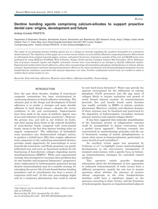
  • 2. Fig. 1 Crystal structure of biogenic HCAp. dentine surfaces might take place in presence of phosphate-containing solutions that closely mimic physiological fluids present in the oral environment. With information sources collated from original scientific papers, reviews, and patent literatures, this paper tracked and arranged the early to recent developments in this field into three sections as follows: I) Pathways in dentinal mineralization; II) Top-down remineralization of hybrid layer collagen matrix; III) Development of dentine bonding systems comprising bioactive particles. PATHWAYS IN DENTINAL MINERALIZATION Biologically mineralized tissues have remarkable hierarchical structures that have evolved over time to achieve great functions in a large variety of organisms. Enormous progress has been made over the last few decades in understanding the mineralization processes in mammalian tissues such as bone, enamel and dentine. Dentine is mainly composed of type-I collagen with associated noncollagenous proteins, to form a highly cross-linked three-dimensional matrix reinforced by mineral crystallites10) . The quality of dentine is dependent upon the total sum of characteristics of the tissue that influence its competence: microstructure, mineral density and composition, along with the particular location of the mineral phase with respect to the organic structures. This mineral-reinforced composite was described by Weiner and Wagner as containing oriented plate-like crystals with their c-axis aligned with the long axis of each collagen fibril11) . Their sizes, ranging in length from 20 Å to 1100 Å12) , is not arbitrary; rather, it seems to give them remarkable physical characteristics13,14) , exposing a very large surface area to the extracellular fluids, which is critically important for rapid ion exchange. Minerals start to nucleate within or immediately adjacent to the holes and pores present in the collagen fibrils15) . This intrafibrillar heterogeneous nucleation is catalyzed by the presence of phosphated esters groups16) and carboxylate groups17) present in the collagen fibrils. Subsequently, the growth, or mineralization, takes place along the collagen fibrils, eventually occupying all of the interstitial extrafibrillar spaces (20 nm). The location of these crystals was demonstrated in a study by Traub et al.18) in which mineralized collagen fibrils had the same banded pattern as negatively stained collagen fibrils, with domains of charged amino acids at the border of the gap and overlap zones acting as nucleation sites. Modelling of collagen fibrils showed that these nanosized, positively charged regions are used for mineral infiltration as well as charge-charge attraction. It was proposed that mineral platelets were arranged in parallel within the gap zones and interstices of the fibril like a stack of cards19) . The principal mineral component is carbonated nanocrystalline hydroxyapatite (HCAp) whose structure is far different from stoichiometric hydroxyapatite (HAp), represented by the formula: Me10(XO4)6Y2 Where Me is a divalent metal (e.g. calcium, Ca2+ ), XO4 is a trivalent anion (e.g. phosphate, PO4 −3 ), and Y is a monovalent anion (e.g. hydroxyl, OH− ). Given the unique mechanisms involved in its formation, biogenic HCAp varies in several ways from the corresponding geologically produced mineral. First, HCAp has a hexagonal lattice structure, having a strong ability to form solid solutions, and to accept numerous substitutions (Fig. 1). These substitutions affect the apatitic lattice parameters: the crystal size is decreased, and thereby the surface area is increased compared to stoichiometric HAp, thus permitting additional adsorption of ions and molecules20) . HCAp contains in fact various trace elements from intrinsic or extrinsic origins, namely significant carbonate substitutions, OH− deficiencies, and imperfections in the crystal lattice21) . This phenomenon provides certain physico-chemical and functional features important in the formation and dissolution of crystals in dental tissues. For example, F− ions are readily incorporated into HCAp, forming fluoroapatite, a less soluble phase of Ca/P as compared to HAp, confering to enamel its low dissolution properties to resist acidic attacks. With respect to dentinal structure, the peculiarity is represented by numerous substitutions (i) by hydrogenophosphate (HPO4 2− ) of XO4 groups and (ii) by carbonate (CO3 2− ) of Y2 and XO4 groups. Lastly, biological minerals tend to attain high crystallinity and a more organized structure on the time scale of days or months rather than years22) . To this end, Lowenstam and Weiner23) used transmission electron microscopy to evaluate the ultrastructure of HCAp crystallites and confirmed that the average length and width of crystallites was 20 nm, with an approximate thickness of 2–3 nm, resembling plate-like structures. TOP-DOWN REMINERALIZATION OF HYBRID LAYER COLLAGEN MATRIX Despite their complex hierarchical structures, the basic building blocks of bones and teeth during both initial 2 Dent Mater J 2014; 33(2): 1–10
  • 3. and later formation stages are nanosized mineral particles. Although extensive investigations of Ca/P crystallization have been performed, many have studied the final structures and morphologies and have not emphasised the need to consider the early contacts between mineral nuclei and organic matrix, or the thermodynamic and kinetic controls imposed by the latter and soluble proteins. Current results and concepts of crystal nucleation and growth at the molecular level, and the role of site-specific interactions, provide possible mechanisms of Ca/P crystallization that are related to the remineralization of dentine13,14) . However, the detailed physical and chemical processes by which nucleation control is established and the thermodynamic and kinetic parameters that define those processes remain largely unknown. The site and the amount of mineral deposition are probably determined by the chemical process (deposition) and by the physical condition (mineral distribution profile and transport mechanism). Organic phases play an initial role in templating the structure of mineralized tissues; therefore, their matrices should be sound as a scaffold for the mineral crystals to grow24,25) . Structurally intact collagen fibrils that retain their banded characteristics and intermolecular crosslinks are considered to be physiologically remineralizable26,27) . Also, non-denatured intact collagen is remineralizable as long as seed crystallites are still present28) . Partial demineralization of the collagen matrix by acids derived from bacteria or dentine bonding procedures leaves remnant seed crystallites and noncollagenous phosphoproteins that can act as heterogeneous nucleation sites29,30) . In nanotechnology terminology, this represents a top-down approach via epitaxial growth31) . Mineral/matrix contact is a key factor. This leads to the deposition of a dense network of pre-nucleation clusters bound by polyanions within any nanosized region of the organic template and their subsequent growth involves various possible precursor phases to the final mineral phase32) . Classic crystallization theory assumes that crystals nucleate and grow from elementary building blocks (ions, molecules) in a supersaturated solution, although phase transformations may also occur at later stages. The association of solution species to form “metastable intermediate precursors”33) , or “growth units”, that subsequently dissolve as the precipitation reactions proceed, is a crucial step34) . The nature of the primary mineral phase remains controversial. Dicalcium-phosphate salts have been implicated as possible precursors to the formation of biogenic HCAp with trace of carbonate and fluoride35) . Moreover, in vivo calcifications have also suggested the involvement of an initial amorphous Ca/P phase (ACP) followed by transformation to the final HCAp product36) . According to Posner, HCAp derives from Ca/P clusters [Ca9 (PO4 )6 ] packing randomly with interfacial water to form ACP precursors37) . This theory is supported by the presence of several Ca/P growth inhibitors such as magnesium that stabilize the amorphous state38,39) . These precursor nanoparticle phases grow rapidly in size via ion-by-ion attachment40) and aggregation, with local loss of solvent, undergoing amorphous- crystalline transformations or phase transformations en route to a thermodynamically stable macro-crystal. Unlike biomimetic bottom-up remineralization strategies that take months of protracted chemical reactions to complete, top-down mineral fabrication of HCAp crystals proceeds fast. Thus, while the mineral constituents is being restored, the region of the exposed collagen fibrils is less susceptible to hydrolytic degradation by MMPs. Furthermore, traditional remineralization by epitaxial growth is a thermodynamically favorable process that overcomes the energy barrier of homogeneous nucleation41) . According to Ostwald’s rule42) , normally the first occurring phase in polymorphism is the least stable and closest in free energy to the mother-phase, followed by other phases in order of increasing stability. The formation, dissolution, and transformation of Ca/P bodies depend on their nature (particle size, crystallographic features, density) and the nature of the solution (composition, pH, temperature). Most Ca/ Ps are sparingly soluble in water, and some are very insoluble, but all dissolve in acids. Their solubility, defined as the amount of dissolved solute contained in a saturated solution when particles of solute are continually passing into solution (dissolving) while other particles are returning to the solid solute phase (growth) at exactly the same rate43) , decreases with increments in temperature and in pH44) . Each Ca/P phase possesses its own thermodynamical solubility. For example, at pH=7 and 37ºC, HCAp is the most stable phase32) . However, these thermodynamical considerations are under equilibrium conditions, and therefore they do not take into account kinetics that dictate the formation of one or the other phase under dynamic conditions. In vivo, the interactions between Ca/Ps and their “biological surroundings” are highly complex due to the non-equilibrium conditions and due to the undefined amount of compounds playing a role in these interactions. The second important factor in the stability of Ca/Ps is the characteristics of the solution in which these salts are formed or placed, namely the solution supersaturation in free Ca2+ and PO4 −3 ions45) . At a given pH and temperature, a free Ca2+ and PO4 −3 ion containing solution can be categorized in three different states: (i) the stable (undersaturated) condition, when crystallization is impossible; (ii) the metastable condition, when spontaneous crystallization of Ca/P salt is improbable, although the concentrations are higher than the ones corresponding to the salt solubility. If a crystal seed were placed in such a metastable solution, growth would occur on the seed; (iii) the unstable condition, when spontaneous crystallization of Ca/P is probable, but not inevitable32) . Extracellular fluids that are supersaturated for Ca2+ and PO4 −3 under normal conditions or, alternatively, by means of external sources with tailored treatments 3Dent Mater J 2014; 33(2): 1–10
  • 4. Fig. 2 Schematic diagram of bioactive micro-fillers conversion within the mineral-depleted resin- dentine interface. It is believed that throughout the course of nano-leakage events HCAp can form from dissolved precursor species on top of a silica nucleation layer. In presence of salivary or dentinal fuid, providing a natural source of Ca2+ and PO4 −3 ions, Bioglass® 45S5 and the silicate phases of PCs undergo a series of physicochemical reactions with subsequent precipitation of an amorphous silica-rich layer “Si-gel” or resulting in the formation of a nanoporous matrix/gel of calcium silicate hydrates “CSH phase”. Either way, silica nucleation layers provide the negatively charged sites for the migration and incorporation of mineral ions (e.g. Ca2+ ), which in turn lead to an over-saturated solution that exceeds the solubility product constants for a number of mineral forms, inducing crystal growth. that deliver the same ions (Fig. 2), may induce the heterogeneous nucleation and growth of new Ca/P crystals32) . The newly formed minerals, in their turn, may act as sites for further nucleation promoting a continuous remineralization over time in presence of environmental mineral ions. DEVELOPMENT OF DENTINE BONDING SYSTEMS COMPRISING BIOACTIVE PARTICLES Many modifications and improvements have been made to dentine bonding agents in the years since their inception. Several “generations” of products have been developed in the last few decades, but the advancements have mainly increased ease of use and reduced technique sensitivity. To date, however, little progress has been made with regard to reducing the propensity of the adhesive towards nano-leakage (Fig. 3), though the achievement of improved bond strengths has significantly reduced gross gaps from occurring46) . One possible modification that may significantly reduce the occurrence and extent of nano-leakage is represented by the incorporation of biologically active agents into the resin-bonding procedure to repair any nanometre-sized void that remains within the HL after polymerization of the adhesive. The development of self-sealing dentine bonding systems with therapeutic ability to create a chemical bond in addition to the micromechanical one, enabling dental restorations to have longer lifetimes, is currently one of the main targets of dental biomaterials research47) . Key objectives in the design of ion-releasing interfacial materials for dental restoration In view of the clinical demand on engineered dental tissue, new biologically active adhesive/primers formulations have to exhibit: Ⅰ. A hydrophilic nature to interact with oral fluids, tolerate moisture and absorb water for the necessary ion movement. Ⅱ. Light-curable chemistry characteristics with controlled water sorption and solubility behaviour of the cross-linked network. Ⅲ. Ability to release mineral ions in a sustained manner (bioavailability) and crystallographic inclusion of foreign ions from aqueous environments that contain Ca2+ and PO4 −3 ions, such as saliva and dentinal fluid. Ⅳ. Appropriate filler size, or fineness, to influence hydration rate: the smaller the size, the greater the surface-area-to-volume ratio, and thus, the more area available for water-particle interaction per unit volume48) . Ⅴ. Potential to completely replace water from the resin-sparse regions of the HL with redeposition of thermodynamically stable, apatitic tooth mineral (bioactivity). Ⅵ. Antibacterial properties at the dentine- restoration interfacial region to reduce the risk of reinfection and secondary caries, in particular when using dental composites lacking any antimicrobial activity. These said features thus form the requisites for the improvement of dentine bonding technology with regard to the hydrolytic enzyme-related loss of HL’s 4 Dent Mater J 2014; 33(2): 1–10
  • 5. Fig. 3 Confocal laser scanning microscopy (CLSM) characterization (single-projection images, fluorescence mode) and nano-leakage (fluorescein dye) of resin-dentine interfaces created with a typical 3-step etch-and-rinse adhesive. The adhesive is labeled with rhodamin B to reveal the extent of resin diffusion within the demineralized (acid-etched) dentine at the conclusion of the bonding procedure (left). Note the presence of intense fluorescein dye uptake within the HL and at bottom of the adhesive layer after the ageing period (right). The presence of gaps is due to HL degradation [Adapted from Profeta et al., Dent Mater 2013; 29: 729-741]. organic components and bond strength. An initial appraisal of the effects of calcium/sodium phosphosilicates on the resin bonded- dentine interface Recently, calcium/sodium phosphosilicates have been successfully used to reduce tooth sensitivity due to their ability to act as biomimetic mineralizers, promoting Ca/P precipitation and subsequent crystallization into carbonated HCAp on the glass/tissue interface without toxicological consequences49) . The chemical composition is significant. The components are basically oxides of calcium, sodium, phosphorus, and silicon at certain weight ratios. Silica maintains the glass structure, while sodium aids in maintenance of physiological ionic balance and pH50) . More than three decades of study have revealed a distinct, though as yet not completely understood, series of chemical reactions that takes place after few minutes of exposure to calcium-phosphate- rich environments, beginning with exchange of cations followed by loss of soluble silica. Both steps are characterized by formation of silanols (SiOH). Polycondensation of SiOH to form a hydrated silica gel precedes the formation of an ACP layer. The reaction is concluded with crystallization into HCAp51) . More in detail, the first phase is characterized by rapid release of alkali or alkaline earth elements (Na+ or K+ ), usually through cation exchange with H+ or H3O+ ions, and de- alkalinization of the glass surface. This ion exchange process leads to an increase in interfacial pH, to values >7.4. Network dissolution occurs concurrently by breaking of -S-O-Si-O-Si- bonds of the glass structure through the action of OH− ions (base catalyzed hydrolysis of -S-O-Si-O-Si- bonds). Breakdown of the network is localized and releases silica into the solution in the form of silicic acid (SiOH4). Last stages involve the development of silica rich and ACP layers respectively. Hydrated silica formed on the glass surface by these reactions undergoes rearrangement by polycondenzation of neighboring SiOH, culminating in a silica-rich gel layer. During the precipitation reaction, Ca2+ and PO4 −3 ions liberated from the glass along with those of the solution combine to create a calcia-phosphate-rich (Ca/P) layer on the silica gel. The Ca/P phase that stratifies in the gel surface is initially amorphous. Within 3–6 h, the ACP will crystallize into HCAp by incorporating carbonate anions from the surroundings51) . It is expected that the forming mineral would bond to specific amino acids within exposed dentine collagen as occurs in bone52) . Evidence has emerged suggesting that bonds to bone are mechanically strong and believed to be chemical in nature; force is required to separate the tissue from the glass53,54) . Confirming the relative success of surface reactive glass-ceramic materials incorporation in dentine bonding procedures required assessment of the potential to reduce nano-leakage, as well as their effect upon the strength of the bond over time. In a study by Profeta et al.35) when 0.05 g of bioactive glass of the well-characterized 45S5 formulation (45 wt% SiO2; 24.5 wt% Na2O and CaO; 6 wt% P2O5) (Sylc; OSspray, London, UK) and of an average particle size of <10 μm were applied directly onto H3PO4-etched wet mid-coronal dentine before bonding, a commercially available adhesive was able to form an apparently 5Dent Mater J 2014; 33(2): 1–10
  • 6. Fig. 4 Interfacial characterization (CLSM single-projection images, fluorescence/reflection mode) and nano-leakage (fluorescein dye) of resin-dentine interfaces created with a 3-step etch-and-rinse adhesive comprising Bioglass® 45S5. The adhesive layer shows a strong fluorescent signal due to added dye (rhodamin B) before application (left). It is also possible to observe intense fluorescein signal from the HL (pointer) located underneath a thick adhesive layer characterized by the presence of mineral micro-fillers. After 6 months of artificial ageing (right), it is possible to note reduced fluorescein diffusion within a HL characterized by a strong reflective signal (pointer), indicating a change in dentine’s mineral content within this zone [Adapted from Profeta et al., Eur J Oral Sci 2012; 120: 353-362]. normal HL. Notably, after 6 months of storage in PO4 −3 buffered saline, the occurrence and extent of nanometre- sized voids within the HLs was reduced due to the chemical nature of the mineral precipitation (dicalcium- phosphate salts) within the resin-dentine interface. Conversely, severe water uptake was observed within the resin-bonded dentine interfaces created with the control resin both after 24 h and 6 months of storage. However, a concomitant decrease in microtensile bond strength over time was also observed. On this account, the crystallized bonded-dentine interfaces might have attained mechanical characteristics comparable to those created by conventional glass-ionomer cements applied onto polyacrylic acid-etched dentine when submitted to tensile tests55) , not reflecting their true adhesive strength. Following this promising start, it was decided to include 30 wt% of Bioglass® 45S5 within the composition of a resin adhesive as tailored micro- filler (filler/resin ratio of 30/70 wt%). This approach produced bioactive/protective effects not only in terms of nano-leakage reduction (Fig. 4) but also preserving high adhesive strength and joint integrity. Analysis of the failure modes displayed a pronounced tendency for these samples to fail cohesively; strong adhesion to tooth substrates would shift the fracture plane from the HL to the dentine substrate or resin composite, while the negative controls tended towards failure at the resin-dentine interface. Another important trend was confirmed in this study: the ultramorphology analysis of the fractured specimens demonstrated the formation of mineral crystals embedded within a well-preserved collagen network over the course of the ageing period. The mechanical characteristics of resin-dentine bonds are a fundamental aspect of restorative procedures. It has been advocated that atomic force microscope (AFM)/nano-indentation analysis performed in hydrated dentine, with force and indenter displacement being recorded simultaneously throughout the indentation and where the modulus of elasticity (Ei) and hardness (Hi) are determined from the load displacement curve, may be a suitable method to evaluate the visco-elasticity of the demineralized dentine and the effective remineralization of the collagen matrix56,57) . In 2012, Sauro and coworkers58) reported that experimental bonding agents formulated on bioactive micro-fillers (Bioglass® 45S5 and polycarboxylated zinc-modified bioactive glass) were able to induce a significant increase of the mechanical nano-properties (Ei and Hi) of definite mineral-depleted areas within the bonded-dentine interface (i.e. HL and bottom of the HL) after prolonged storage in a simulated body fluid solution (SBFS). Presence of well shaped indentations was observed in the crystallized SBFS- aged bonded-dentine interfaces. Conversely, scarcely pronounced marks were detected in the specimens stored in SBFS for 24 h due to the visco-elastic properties of the demineralized collagen which prevented the plastic deformation of the HL56,59) . Although the reincorporation of mineral into the demineralized dentine matrix does not represent a 6 Dent Mater J 2014; 33(2): 1–10
  • 7. Fig. 5 Interfacial characterization (CLSM single-projection images, fluorescence mode) and nano-leakage (fluorescein dye) of resin-dentine interfaces created with a 3-step etch- and-rinse adhesive comprising a calcium-silicate Portland-derived micro-filler. At the end of the bonding procedure, it is possible to observe a clear HL with long resin tags (rhodamin B-labeled adhesive) penetrating the dentinal tubules (left). Intense fluorescein uptake was also observed within the entire resin-dentine interface. Following 6 months of artificial ageing (right), the CLSM analysis reveals absence of gaps and diminished fluorescein penetration inside the HL [Adapted from Profeta et al., Dent Mater 2013; 29: 729-741]. full recovery of its functionality, it still plays a very important role, since the remineralized remnant crystallites may be much more resistant to subsequent acid attacks60) . Early developments in etch-and-rinse adhesives containing calcium-silicate Portland-derived micro-fillers Since it was introduced in 1993, mineral trioxide aggregate (MTA) (ProRoot; Dentsply Tulsa Dental, Tulsa, OK) has proven to be one of the most versatile dental materials of this epoch. Abundant research shows excellent biocompatibility and bioactive properties in a multitude of challenging endodontic clinical situations61) . As stated by the patent, MTA is a derivative of a type I ordinary Portland cement (PC) with 4:1 proportions of bismuth oxide just added for radiopacity62) . PCs are hydraulic materials mainly composed of di- and tri-calcium silicate (2CaO•SiO2 belite and 3CaO•SiO2 alite), tricalcium aluminate (3CaO•Al2O3) and gypsum (CaSO4•2H2O) hydrophilic powders63) . The setting reactions of PC remain in many respects obscure, with much controversy and uncertainty, despite its economic importance. Perhaps not surprisingly, the dental literature contains no satisfactory account, although there are a few superficial remarks and suggestions64) . However, taking note of the respective chemical and condition variations, it is possible to deduce a reaction scheme which is probably sufficiently correct to account for the main features of the process. When hydrated, a solid-liquid interface forms on the silicate phases and ion dissolution occurs almost immediately. A high- pH solution containing Ca2+ , OH− and silicate ions is established65) . A series of physicochemical reactions (hydrolysis of SiO4 4− groups) result in the formation of a nanoporous matrix/gel of calcium silicate hydrates (CSH phase), characterized by Si-OH silanol groups whose deprotonation leads to negatively charged SiO− groups66) , and of a soluble fraction of calcium hydroxide Ca(OH)2 or portlandite61) . While SiO− negative groups sets over time to form a solid network (heterogeneous nucleation of HCAp) by bonding Ca2+ on the CSH surface67) , the release of Ca(OH)2 increases the alkalinity of the surrounding medium68) . The soaring pH and concentration of Ca2+ ions enhance supersaturation of the solution and growth of HCAp crystals63) . The similar composition and mechanism of action suggest that additives used in PC industry might also be used to change the physical properties of MTA. In the same manner, this allows initial research studies to be carried out by using PC, which is advantageous considering the significantly lower cost of PC compared with that of MTA. If favorable results are obtained by using PC, then additional studies can then be performed to determine whether the same results can be obtained by using the more expensive MTA material. Calcium silicates appeared to bond chemically to dentinal walls, possibly via a diffusion-controlled reaction63) , but their inclusion in dentine bonding 7Dent Mater J 2014; 33(2): 1–10
  • 8. Fig. 6 Dye-assisted (xylenol orange) CLSM imaging (single-projection images, fluorescence mode) of resin-dentine interfaces created with a 3-step etch-and-rinse adhesive comprising a calcium-silicate Portland-derived micro-filler. Fluorochrome xylenol orange is taken up at the sites of active calcification, due to its ability to form complexes with divalent Ca2+ ions, thus labeling new mineral tissue formation. It is possible to observe strong presence of Ca2+ -chelator dye inside adhesive, resin tags and dentinal tubules. Note the intense Ca2+ deposition at the bottom and within the HL following 24 h of storage in simulated body fluid solution (left). The data from the biophotonic imaging showed a clearly outlined fluorescence signal within the bonding interface and along the walls of dentinal tubules even after 6 months of artificial ageing (right) [Adapted from Profeta et al., Dent Mater 2013; 29: 729-741]. systems is yet to be attempted. In a recent study small amounts of three derivatives and proprietary formulations based on the composition of an ordinary PC (GB patent application no. 1118138.5) were added to the neat resin of a typical 3-step etch-and-rinse adhesive (30 μm average particle size; 40/60 wt% filler/ resin ratio), and teeth were bonded for the purpose of carrying out bond strength tests, ultramorphology and nano-leakage studies69) . The most favorable results were obtained with the addition of set type-I PC modified using either montmorillonite or hydrotalcite poly-ions silicates. The use of resin bonding agents containing these two tailored micro-fillers promoted a therapeutic mineral deposition mechanism within the HL: nano- leakage did indeed appear to be reduced after soaking in simulated body fluid for 6 months (Fig. 5), and consistent bond strength values were maintained when compared to negative controls. The reduced fluorescent- dye uptake observations, along with a strong Ca2+ chelating fluorophore (xylenol orange) signal from the HL and the dentinal tubules, clearly indicated the remineralization of those areas which were previously detected as mineral-deficient/porous zones (Fig. 6). A further supporting result was found in the scanning electron microscopy: the ultramorphological analysis performed on fractured match-sticks demonstrated the presence of mineral crystals within the resin-dentine matrix subsequent to the storage period. Despite no specific biomimetic analogue agents being used in this study, there may have been a sort of biomimetic activity evoked by the main hydration products of the two silicate-based fillers. It was also hypothesized that the alkaline condition induced during the process may have created unfavorable conditions for MMPs expression within the demineralized collagen matrix and may have played a supplementary role in the remineralization kinetics70) . Addition of a PC-based micro-filler containing titanium oxide to the same adhesive was less successful. Again, it was shown that an apparently normal HL was able to form, and the strength of the bonds formed was comparably high following 24 h of storage in simulated body fluid. However, long-term evaluation of the micro-mechanical strength was less definitive than in the other two etch-and-rinse adhesives doped with calcium silicates. Although nano-leakage was substantially reduced, specimens of this experimental adhesive group showed diminution of bond strength durability due to the high hydrophilicity of titanium oxide. CONCLUSIONS Noteworthy conclusions can be drawn from this review paper. Different studies provided preliminary evidence that the durability of the resin-dentine interfaces may be enhanced by modified bonding agents in a clinically relevant manner. The inclusion of highly reactive silicate compounds, such as commercially available 8 Dent Mater J 2014; 33(2): 1–10
  • 9. bioactive glass and calcium-silicate Portland-derived cements, within the composition of representative etch- and-rinse bonding system conferred attractive basic properties, such as: Ⅰ. Light polymerization ability and tendency to interact with oral fluids and wet tooth structures (hydrophylic nature). Ⅱ. High bioavailability and propensity to deliver various remineralizing species; when hydrated, each micro-filler releases the predominant ions of its composition enhancing mineral delivery within and beneath the HL (mineral enrichment effect). Ⅲ. Potential to displace water from the resin-sparse regions of the HL with redeposition of apatitic tooth minerals (bioactivity). Ⅳ. Alkalinizing activity to buffer the environmental acids as well as to interfere with the action of MMPs, and antibacterial properties. Ⅴ. Predisposition to reduce nano-leakage and the rate of enzymatic degradation, two of the major causes of restoration failure, while producing no negative effects on bond strength over time. Ⅵ. Capacity to preserve the mechanical properties and stability of resin-dentine bonded interfaces. The initial evaluation of bioactive silicate compounds as a possible addition to the resin-dentine bonding process has shown their value; a new generation of biologically active dental materials able to induce physicochemical reactions yielding HCAp formation in demineralized dentine has been obtained as promising adhesives to be tested in future studies and clinical trials. ACKNOWLEDGMENTS The author thanks all the research co-workers at King’s College London Dental Institute, Department of Biomaterials, Biomimetics and Biophotonics (B3) Research Group, for their invaluable assistance and discussions in the preparation of this review paper. REFERENCES 1) Breschi L, Mazzoni A, Ruggeri A, Cadenaro M, Di Lenarda R, Dorigo E. Dental adhesion review: aging and stability of the bonded interface. Dent Mater 2008; 24: 90-101. 2) Marshall GW, Marshall SJ, Kinney JH, Balooch M. The dentin substrate: structure and properties related to bonding. J Dent Res 1997; 25: 441-458. 3) Sadek FT, Braga RR, Muench A, Liu Y, Pashley DH, Tay F. Ethanol wet-bonding challenges current anti-degradation strategy. J Dent Res 2010; 89: 1499-1504. 4) Liu Y, Tjäderhane L, Breschi L, Mazzoni A, Li N, Mao J, Pashley DH, Tay FR. Limitations in bonding to dentin and experimental strategies to prevent bond degradation. J Dent Res 2011; 90: 953-968. 5) Liu Y, Li N, Qi Y, Niu LN, Elshafiy S, Mao J, Breschi L, Pashley DH, Tay FR. The use of sodium trimetaphosphate as a biomimetic analog of matrix phosphoproteins for remineralization of artificial caries-like dentin. Dent Mater 2011; 27: 465-477. 6) Tay FR, Pashley DH. Biomimetic remineralization of resin- bonded acid-etched dentin. J Dent Res 2009; 88: 719-724. 7) Tay FR, Pashley DH. Guided tissue remineralisation of partially demineralised human dentine. Biomaterials 2008; 29: 1127-1137. 8) Zhang X, Neoh KG, Lin CC, Kishen A. Remineralization of partially demineralized dentine substrate based on a biomimetic strategy. J Mater Sci Mater Med 2012; 23: 733- 742. 9) Cochrane NJ, Cai F, Huq NL, Burrow MF, Reynolds EC. New approaches to enhanced remineralization of tooth enamel. J Dent Res 2010; 89: 1187-1197. 10) Kinney JH, Habelitz S, Marshall SJ, Marshall GW. The importance of intrafibrillar mineralization of collagen on the mechanical properties of dentin. J Dent Res 2003; 82: 957- 961. 11) Weiner S, Wagner HD. The material bone: structure- mechanical function relations. Annu Rev Mater Sci 1998; 28: 271-298. 12) Kim HM, Rey C, Glimcher MJ. Isolation of calcium-phosphate crystals of bone by nonaqueous methods at low-temperature. J Bone Miner Res 1995; 10: 1589-1601. 13) Wang LJ, Nancollas GH. Calcium orthophosphates: crystallization and dissolution. Chem Rev 2008; 108: 4628- 4669. 14) Wang LJ, Nancollas GH. Biomineralization. From Nature to Application. Sigel A, Sigel H, Sigel RKO, editors. Metal ions in life sciences series 4. Chichester: John Wiley & Sons; 2008. p. 413-456. 15) Glimcher MJ. The nature of the mineral component of bone and the mechanism of calcification. Instr Course Lect 1987; 36: 49-69. 16) GlimcherMJ,KossivaD,Brickley-ParsonsD.Phosphoproteins of chicken bone matrix. Proof of synthesis in bone tissue. J Biol Chem 1984; 259: 290-293. 17) Rhee SH, Lee JD, Tanaka J. Nucleation of hydroxyapatite crystal through chemical interaction with collagen. J Am Ceram Soc 2000; 83: 2890-2892. 18) Traub W, Arad T, Weiner S. Three-dimensional ordered distribution of crystals in turkey tendon collagen fibers. Proc Natl Acad Sci USA 1989; 86: 9822-9826. 19) Palmer LC, Newcomb CJ, Kaltz SR, Spoerke ED, Stupp SI. Biomimetic systems for hydroxyapatite mineralization inspired by bone and enamel. Chem Rev 2008; 108: 4754- 4783. 20) LeGeros RZ. Calcium phosphates in oral biology and medicine. Myers HM, editor. Monographs in oral science. Basel: Karger; 1991. 21) Boskey AL. Mineralization of bones and teeth. Elements Magazine 2007; 3: 385-391. 22) Verdelis K, Lukashova L, Wright JT, Mendelsohn R, Peterson MGE, Doty S, Boskey AL. Maturational changes in dentin mineral properties. Bone 2007; 40: 1399-1407. 23) Lowenstam HA, Weiner S. On biomineralization. New York: Oxford University Press; 1980. 24) Kusboki Y, Ohgushi K, Fusayama T. Collagen biochemistry of the two layers of carious dentin. J Dent Res 1977; 56: 1233- 1237. 25) Gonzalez-CabezasC.Thechemistryofcaries:remineralization and demineralization events with direct clinical relevance. Dent Clin North Am 2010; 54: 469-478. 26) Landis WJ, Song MJ, Leith A, McEwen L, Mc-Ewen BF. Mineral and organic matrix interaction in normally calcifying tendon visualized in three dimensions by high-voltage electron microscopic tomography and graphic image reconstruction. J Struct Biol 1993; 110: 39-54. 27) Landis WJ. Mineral characterization in calcifying tissues: atomic, molecular and macromolecular perspectives. Connect Tissue Res 1996; 34: 239-246. 28) Koutsoukos PG, Nancollas GH. Crystal growth of calcium phosphates —epitaxial considerations. J Cryst Growth 1981; 9Dent Mater J 2014; 33(2): 1–10
  • 10. 53: 10-19. 29) Clarkson BH, Feagin FF, McCurdy SP, Sheetz JH, Speirs R. Effects of phosphoprotein moieties on the remineralization of human root caries. Caries Res 1991; 25: 166-173. 30) Bertassoni LE, Habelitz S, Marshall SJ, Marshall GW. Mechanical recovery of dentin following remineralization in vitro —an indentation study. J Biomech 2011; 44: 176-181. 31) Wong TS, Brough B, Ho CM. Creation of functional micro/ nano systems through top-down and bottom-up approaches. Mol Cell Biomech 2009; 1: 1-55. 32) Barrère F, van Blitterswijk CA, de Groot K. Bone regeneration: molecular and cellular interactions with calcium phosphate ceramics. Int J Nanomedicine 2006; 1: 317-332. 33) Onuma K, Ito A. Cluster growth model for hydroxyapatite. Chem Mater 1998; 10: 3346-3351. 34) Eanes ED, Gillessen IH, Posner AS. Intermediate states in the precipitation of hydroxyapatite. Nature 1965; 208: 365- 367. 35) Profeta AC, Mannocci F, Foxton RM, Thompson I, Watson TF, Sauro S. Bioactive effects of a calcium/sodium phosphosilicate on the resin-dentine interface: a microtensile bond strength, scanning electron microscopy, and confocal microscopy study. Eur J Oral Sci 2012; 120: 353-362. 36) Mahamid J, Sharir A, Addadi L, Weiner S. Amorphous calcium phosphate is a major component of the forming fin bones of zebrafish: Indications for an amorphous precursor phase. Proc Natl Acad Sci USA 2008; 105: 12748-12753. 37) Posner AS. The mineral of bone. Clin Orthop Relat Res 1985; 200: 87-99. 38) Barrére F, Layrolle P, van Blitterswijk CA, de Groot K. Biomimetic calcium phosphate coatings on Ti6AI4V: a crystal growth study of octacalcium phosphate and inhibition by Mg2+ and HCO3− . Bone 1999; 25: 107S-111S. 39) Root MJ. Inhibition of the amorphous calcium phosphate phase transformation reaction by polyphosphates and metal ions. Calcif Tissue Int 1990; 47: 112-116. 40) Niederberger M, Cölfen H. Oriented attachment and mesocrystals: non-classical crystallization mechanisms based on nanoparticle assembly. Phys Chem Chem Phys 2006; 8: 3271-3287. 41) Jiang H, Liu XY. Principles of mimicking and engineering the self-organized structure of hard tissues. J Biol Chem 2004; 297: 41286-41293. 42) Ostwald WF. Studies on formation and transformation of solid materials. Z Phys Chem 1897; 22: 289-302. 43) Wu WJ, Nancollas GH. The dissolution and growth of sparingly soluble inorganic salts: A kinetics and surface energy approach. Pure Appl Chem 1998; 70: 1867-1872. 44) de Groot K. de Groot K, editor. Ceramics of calcium phosphates: preparation and properties. Bioceramics of calcium phosphate. CRC Press Inc; 1983. p.100-111. 45) Tang RK, Nancollas GH, Orme CA. Mechanism of dissolution of sparingly soluble electrolytes. J Am Chem Soc 2001; 123: 5437-5443. 46) Pashley DH, Tay FR, Breschi L, Tjäderhane L, Carvalho RM, Carrilho M, Tezvergil-Mutluay A. State of the art etch-and- rinse adhesives. Dent Mater 2011; 27: 1-16. 47) Liu Y, Mai S, Li N, Yiu CK, Mao J, Pashley DH, Tay FR. Differences between top-down and bottom-up approaches in mineralizing thick, partially demineralized collagen scaffolds. Acta Biomater 2011; 7: 1742-1751. 48) Hashimoto M, Iijima M, Nagano F, Ohno H, Endo K. Effect of monomer composition on crystal growth by resin containing bioglass. J Biomed Mater Res B Appl Biomater 2010; 94: 127- 133. 49) Bauer TW, Smith ST. Bioactive materials in orthopaedic surgery: overview and regulatory considerations. Clin Orthop Relat Res 2002; 395: 11-22. 50) Hench LL, Splinter RJ, Allen WC, Greenlee TK. Bonding mechanisms at the interface of ceramic prosthetic materials. J Biomed Mater Res 1971; 5: 117-141. 51) Hench LL, Andersson O. Hench LL, Wilson J, eds. Bioactive glasses. Advanced series in ceramics. Vol. 1. An introduction to bioceramics. Singapore: World Scientific; 1993. p. 45-47. 52) Efflandt SE, Magne P, Douglas WH, Francis LF. Interaction between bioactive glasses and human dentin. J Mater Sci Mater Med 2002; 13: 557-565. 53) Hench LL, Paschall HA. Direct chemical bond of bioactive glass-ceramic materials to bone and muscle. J Biomed Mater Res 1973; 7: 25-42. 54) Wilson J, Pigott GH, Schoen FJ, Hench LL. Toxicology and biocompatibility of bioglasses. J Biomed Mater Res 1981; 15: 805-817. 55) Yip HK, Tay FR, Ngo HC, Smales RG, Pashley DH. Bonding of contemporary glass ionomer cements to dentin. Dent Mater 2001; 17: 456-470. 56) Balooch M, Habelitz S, Kinney JH, Marshall SJ, Marshall GW. Mechanical properties of mineralized collagen fibrils as influenced by demineralization. J Struct Biol 2008; 162: 404- 410. 57) Habelitz S, Marshall SJ, Marshall GW Jr, Balooch M. Mechanical properties of human dental enamel on the nanometre scale. Arch Oral Biol 2001; 46: 173-183. 58) Sauro S, Osorio R, Watson TF, Toledano M. Therapeutic effects of novel resin bonding systems containing bioactive glasses on mineral-depleted areas within the bonded-dentine interface. J Mater Sci Mater Med 2012; 23: 1521-1532. 59) Schulze KA, Oliveira SA, Wilson RS, Gansky SA, Marshall GW, Marshall SJ. Effect of hydration variability on hybrid layer properties of a self-etching versus an acid-etching system. Biomaterials 2005; 26: 1011-1018. 60) Featherstone JD. Modeling the caries-inhibitory effects of dental materials. Dent Mater 1996; 12: 194-197. 61) Parirokh M, Torabinejad M. Mineral trioxide aggregate: a comprehensive literature review —part III clinical applications, drawbacks and mechanism of action. J Endod 2010; 36: 400-413. 62) Torabinejad M, Hong CU, McDonald F, Pitt Ford T. Physical and chemical properties of a new root-end filling material. J Endod 1995; 21: 349-353. 63) Sarkar NK, Caicedo R, Ritwik P, Moiseyeva R, I. K. Physicochemical basis of the biologic properties of mineral trioxide aggregate. J Endod 2005; 31: 97-100. 64) Darvell BW, Wu RC. “MTA” —an Hydraulic Silicate Cement: review update and setting reaction. Dent Mater 2011; 27: 407-422. 65) Taylor HFW. Cement chemistry. 2nd ed. London: Thomas Telford Ltd,; 1997. 66) Sanchez F, Zhang L. Molecular dynamics modeling of the interface between surface functionalized graphitic structures and calcium-silicate-hydrate: interaction energies, structure, and dynamics. J Colloid Interf Sci 2008; 323: 349-358. 67) Plassard C, Lesniewska E, Pochard I, Nonat A. Nanoscale experimental investigation of particle interactions at the origin of the cohesion of cement. Langmuir 2005; 21: 7263- 7270. 68) Pellenq RJM, Kushima A, Shahsavari R. A realistic molecular model of cement hydrates. Proc Natl Acad Sci USA 2009; 106: 16102-16107. 69) Profeta AC, Mannocci F, Foxton R, Watson TF, Feitosa VP, De Carlo B, Mongiorgi R, Valdré G, Sauro S. Experimental etch-and-rinse adhesives doped with bioactive calcium silicate-based micro-fillers to generate therapeutic resin- dentin interfaces. Dent Mater 2013; 29: 729-741. 70) Somasundaran P, Ofori Amankonah J, Ananthapadmabhan KP. Mineral—solution equilibria in sparingly soluble mineral systems. Colloids Surf 1985; 15: 309-333. 10 Dent Mater J 2014; 33(2): 1–10