Call Girl Number in Panvel Mumbai📲 9833363713 💞 Full Night Enjoy
Pulmonary Tuberculosis
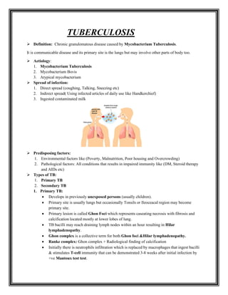
1. TUBERCULOSIS
➢ Definition: Chronic granulomatous disease caused by Mycobacterium Tuberculosis.
It is communicable disease and its primary site is the lungs but may involve other parts of body too.
➢ Aetiology:
1. Mycobacterium Tuberculosis
2. Mycobacterium Bovis
3. Atypical mycobacterium
➢ Spread of infection:
1. Direct spread (coughing, Talking, Sneezing etc)
2. Indirect spread( Using infected articles of daily use like Handkerchief)
3. Ingested contaminated milk
➢ Predisposing factors:
1. Environmental factors like (Poverty, Malnutrition, Poor housing and Overcrowding)
2. Pathological factors: All conditions that results in impaired immunity like (DM, Steroid therapy
and AIDs etc)
➢ Types of TB:
1. Primary TB
2. Secondary TB
1. Primary TB:
• Develops in previously unexposed persons (usually children).
• Primary site is usually lungs but occasionally Tonsils or Ileoceacal region may become
primary site.
• Primary lesion is called Ghon Foci which represents caseating necrosis with fibrosis and
calcification located mostly at lower lobes of lung.
• TB bacilli may reach draining lymph nodes within an hour resulting in Hilar
lymphadenopathy.
• Ghon complex is a collective term for both Ghon foci &Hilar lymphadenopathy.
• Ranke complex: Ghon complex + Radiological finding of calcification
• Initially there is neutrophils infiltration which is replaced by macrophages that ingest bacilli
& stimulates T-cell immunity that can be demonstrated 3-8 weeks after initial infection by
+ve Mantoux test test.
2. • Primary TB is mostly asymptomatic but occasionally may present with low grade fever for
7-14 day and Erythema nodosum formation. (Erythema nodosum also seen in Sarcoidosis)
• Fate of primary TB (Healing, Primary progressive or Secondary TB)
Primary progressive TB
• Seen in children with impaired immunity.
• There is enlargement, caseation and cavitations of primary lesion.
• Lymph nodes also enlarge and may compress bronchi causing obstruction and collapse.
• Infection spread and can also lead to Pleural effusion.
2. Secondary TB:
• Seen in previously exposed persons.
• Results from reactivation or re-infection of primary foci.
• May occur shortly after primary infection but mostly after decades when the immunity is
impaired.
• Pathological feature is cavitations.
• Cavitations is formed when the centre of primary lesion liquefies & discharged into bronchus,
this finding is seen on X ray as area of radiolucent(dark area) surrounded by area of
radioopacity (white area)
• Cavitations are formed mostly at apex of lung where the oxygen tension is high and poor
lymphatic drainage.
➢ Clinical features:
Systemic:
• Low grade fever specially occurs at evening with nigh sweets, weight loss and weakness.
• On examination: Fever, wasting, weight loss, Clubbing, Anaemia. Sometimes it is associated with
cervical lymphadenopathy
Respiratory:
• Cough initially Dry then becomes productive with purulent sputum and in advance stage the
sputum turn into bloody (Haemoptysis).
• On examination: Crepitations may be present during inspiration& there is bronchial breathing,
There are 4 stages of TB lesion, so depending on lesion stages different findings are noted as
given below
1. Consolidation: Dullness on percussion
2. Cavitations: Crepitation and tympanic note on percussion
3. Fibrosis: Apex beat shifted toward the lesion
4. Pleural effusion: Percussion note becomes stony dull
➢ Investigations:
1. X-ray chest: Hilar lymphadenopathy and Nodular opacities or cavitations can be seen especially
on upper lobes.
Milliary TB: This shows multiple, multifocal milliary mottling (also seen in Sarcoidosis and
Staphylococcal pneumonia)
3. 2. Sputum AFB smear: 3 early morning samples are sent for AFB smear.
3. PCR: Rapidly detects mycobacterium DNA in sputum and other body fluids.
4. Culture: sputum or pleural fluid can be cultured.
5. Needle biopsy: Biopsy of pleura, lymph nodes, liver or spinal fluid can be performed that shows
Chronic Granulomatous inflammation.
For taking samples of biopsy or culture from bronchi, bronchial lavage is performed via
Fiberoptic Bronchoscopy
6. Gastric washing: This can be performed if sputum is not produced or Bronchoscopy is not
available. Early morning aspiration of gastric content after overnight fast is gastric washing. This
test is usually limited to paediatric patient to diagnose primary TB.
7. Mantoux test:
• In this test immune reaction against a tuberculin antigen is tested
• For this Purified Protein Derivative (PPD) is injected at flexor surface of arm, 48-72
hours after, the induration is formed which is measured.
• If size of induration is >10mm , the test is +ve
• In AIDs patients even induration of >5mm is considered as +ve.
• Mantoux test may be false negative when there is immunodeficiency state
• Mantoux test may be false positive in persons who are immunized with BCG vaccine.
• Heaf test is similar to Mantoux test but it is Screening test rather than the diagnostic.
➢ Complications of TB:
1. Milliary TB
2. Pleural effusion
3. Pneumothorax
4. Empyema
5. Pyopneumothorax
6. Pericardial effusion
4. 7. Right ventricular failure
8. TB laryngitis, Pharyngitis
9. Blood dissemination
10. Cervical lymphadenopathy
11. TB meningitis
12. Tuberculoma
13. TB enteritis
14. Sub-acute Intestinal Obstruction
15. Ischiorectal abscess
16. Psoas abscess
17. Genito-urinary TB
18. Spinal TB
19. Fungal colonization of cavities
1. Milliary TB
• It is result of acute, diffuse dissemination of Tubercle bacilli.
• Mostly occur in children& young adults as a complication of primary TB.
• Presents with fever, night sweets, weight loss, dyspnoea & productive cough.
• On examination
o CNS: Signs of meningeal irritation
o CVS: Tachycardia
o RS: Crypts bilateral
o Eye: Choroid tubercle
o GIT: Hepatosplenomegally
2. Cryptic TB
• Patient is over 60 y of age
• Presents with low grade fever, weight loss &Hepato-splenomegally
• CXR is normal & TB test is negative
• Bone marrow/ Liver biopsy or trial of ATT helps in diagnosis
3. Pleural TB
• May lead to pleural effusion , empyema & Pyo-pneumothorax
• Discussed in details in respective topics
4. TB lymphadenopathy:
• This is the most common extra-pulmonary TB
• It presents with painless enlargement of lymph nodes especially cervical &
supraclavicular nodes.
• Initially nodes are discrete then become matted. Cold abscess is formed resulting in
fluctuant swelling, ultimately sinus formation occur.
• Diagnosed by FNAC or Surgical biopsy.
5. 5. TB pericarditis:
• Present with fever, chest pain & pericardial rub
• Sometimes exudative or sometimes haemorrhagic effusion present
6. Gastro-intestinal TB
• Results from swallowing of infected sputum
• Ileo-ceacal most common site involved
• May lead to SAIO or TB peritonitis
7. Genito-urinary TB
• Urinary- may present with Haematuria, Dysurea, Flank pain
• Genital- Chronic PID, Infertility, Menstrual abnormalities and Ectopic pregnancy.
8. Spinal TB (Pot’s disease)
• Most commonly lower thoracic or upper lumber are involved.
• Collapse of vertebral bodies leads to Kyphosis & Paraplegia
• A paravertebral cold abscess may also form
• MRI and Biopsy of spine are helpful for diagnosis
9. TB meningitis and Tuberculoma
• TB meningitis presents with features of meningeal irritation e-g Neck rigidity,
kerning’s +ve
• In contrast to bacterial meningitis, The TB meningitis evolves over 1 or 2 weeks.
• Ocular palsy is frequent finding and Hydrocephalus is common complication.
• Diagnosis is made by LP
• Tuberculoma presents with features of space occupying lesion
10. Multi-drug resistant TB (MDR-TB)
TB that is resistant to at least Rifampicin
6. ➢ Treatment:
General:
1. Hydralline / Ventolin expectorant: For Productive cough
2. Panadol: For Fever
3. Steroid: For Pericardial / Pleural effusion & TB meningitis
Specific: ATT which consists of two phase therapy
1. Intensive phase / Bacteriocidal phase
Consists of 4 drugs namely
• Rifampicin
• INH
• Pyrazinamide
• Ethambutol
• Pyridoxin: is indicated for INH neuropathy
2. Continuation phase / Sterilization phase
• Consists of 2 – 3 drugs except Pyrazinamide
1st
line ATT: All 4 1st
line drugs present in market with name of MYRINE-P
1) Rifampicin
2) INH
3) Pyrazinamide
4) Ethambutol
2nd
line ATT:
7. o Ofloxacin
o Ethionamide
o Cycloserine
o PAS
o Amikacin
o Capreomycin
DRUG DOSAGE SIDE EFFECTS
Adult Children
<50kg >50kg
Rifampicin 450mg/day 600mg/day 15mg/kg/day Orange-red colour body
secretions & Hepatitis
INH 200mg/day 300mg/day 15mg/kg/day Peripheral neuropathy, Hepatitis
Pyrazinamide 1500mg/day 2000mg/day 30mg/kg/day GOUT , Hepatitis
Ethambutol 1200mg/day 1600mg /day 15mg/kg/day Optic neuritis
Streptomycin 0.75-1 g/day 8th
nerve palsy, Nephrotoxicity
➢ Monitoring during ATT therapy:
1. AFB smear of sputum: (Clear by 3 months after start of ATT)
2. CXR: Before and after the ATT therapy
3. LFTs: Every month
4. Uric acid level: Every month
5. Visual & Auditory evaluations
6. Urea/Creatinin
➢ DOTS (DIRECTLY OBSERVED THERAPY SHORT COURSE STRATEGY): is the name given to
the tuberculosis (TB) control strategy recommended by the World Health Organization. According to
WHO, "The most cost-effective way to stop the spread of TB in communities with a high incidence is
by curing it. The best curative method for TB is known as DOTS."
DOTS has five main components:
8. 1. Government commitment (including political will at all levels, and establishment of a centralized
and prioritized system of TB monitoring, recording and training)
2. Case detection by sputum smear microscopy
3. Standardized treatment regimen directly of six to nine months observed by a healthcare worker or
community health worker for at least the first two months
4. Drug supply
5. A standardized recording and reporting system that allows assessment of treatment results