DEVELOPMENT OF THE MUSCULOSKELETAL
•
1 like•168 views

The skeletal and muscular systems develop from paraxial mesoderm, lateral plate mesoderm, and neural crest cells. Mesoderm forms somites that differentiate into sclerotome and dermomyotome, with sclerotome cells becoming mesenchyme that migrates and forms cartilage models through endochondral ossification or membraneous bone, while dermomyotome forms myoblasts that fuse into muscle fibers. Limb buds develop from lateral plate mesoderm and rotate as cartilage and bone form through endochondral ossification while surrounding musculature develops from dermomyotome and surrounding nerves

Recommended
More Related Content
What's hot
What's hot (20)
Similar to DEVELOPMENT OF THE MUSCULOSKELETAL
Similar to DEVELOPMENT OF THE MUSCULOSKELETAL (20)
More from Dr. Faiza Munir Ch
More from Dr. Faiza Munir Ch (20)
Recently uploaded
Recently uploaded (20)
DEVELOPMENT OF THE MUSCULOSKELETAL
- 1. DEVELOPMENT OF THE MUSCULOSKELETAL SYSTEM Dr.Faiza Munir Ch Medical Officer (lecturer) dr.faizamunich@gmail.com
- 2. Learning Objectives The students should be able to know: • Different sources of origin of the skeletal and muscular system • Embryonic connective tissue • Development of limb bud and limb musculature. • Intrauterine ossification.
- 3. Development of the Skeletal System Source of Origin: • Paraxial Mesoderm • Lateral plate Mesoderm • Neural Crest Cells • Paraxial mesoderm forms a segmented series of tissue blocks on each side of the neural tube , the ‘Somites’. • These somites differentiate into a; – Sclerotome, (ventromedial part) – Dermomyotome, (dorsolateral part )
- 4. • At the end of 4th week the sclerotome cells become polymorphous and form a loosely organized tissue called mesenchyme or embryonic connective tissue. • It is characteristic of the mesenchymal cell to migrate and to differentiate in many ways.
- 5. The Embryonic connective tissue • Mesenchyme or the embryonic connective tissue is a gelatinous substance with ‘star-shaped’ mesenchymal cells. • The mesenchymal cells migrate & differentiate into many different types of primitive cell lines, such as; – Fibroblasts (adult conn. Tissue forming cells), – Chondroblasts (cartilage forming cells), – Osteoblasts (bone forming cells).
- 6. Origins of the Axial & Appendicular Skeleton • The mesenchyme in the paraxial mesoderm will transform into Osteoblasts that will form the bony elements of the vertebral column (e.g. body, transverse process, spinous process etc). • The mesenchyme in the somatopleuric mesoderm will transform into osteoblasts that will form the Pelvic & Pectoral girdles and also the bones of upper & lower limbs.
- 7. Ossification “The process of bone formation is known as ossification” Bone develops during the intra uterine life through two types of ossifications; • Membranous type, in which the mesenchymal tissue (jelly-like) will directly transform into bone. (e.g. flat bones of skull) • Intra-cartilagenous type, in which the mesenchymal tissue first give rise to a hyaline cartilage model of the bone, then the osteoblasts will convert that model into bone (e.g. long bones, irregular bones like vertebrae etc)
- 8. Intracartilagenous Ossification Mostly seen in the long bones of limbs. • First a hyaline cartilage model is formed in the mesenchyme • Then a primary ossification center appears in the ‘diaphysis’ of the model. • Bone formation/laying starts from the center in both upward & downward direction. • Almost all primary centers of ossification appear before birth. • Most of the secondary centers appear after birth. • The part of a long ossified from Primary center is the ‘Diaphysis’ • The part of a long bone ossified from a secondary center is the ‘Epiphysis’ The fusion between the diaphysis and epiphysis does not occur till puberty
- 12. Development of Skull (Intramembranous Ossification) The skull is divided into two parts; • Neurocranium which forms a protective covering around the brain is derived from the ‘occipital somites’ – Membranous Neurocranium is composed of flat bones of the cranial vault. – Cartilagenous Neurocranium is composed of the irregular bones of the base of skull • Viscerocranium which forms the skeleton of the face is derived from the ‘Neural crest cells’.
- 13. Newborn Skull • At birth the flat bones of the skull are separated from each other by narrow seams of connective tissue the sutures.which are also derived from two sources: neural crest cells (sagittal suture) and paraxial mesoderm (coronal suture) • At point where more than two bones meet ,sutures are wide and are called fontanelles. • Sutures and fontanelles allow the bones of skull to overlap (molding) during birth. Soon after birth, membranous bones move back to their original positions and the skull appears large and round. • Several sutures and fontanelles remain membranous for a considerable time after birth. The bones of the vault continue to grow after birth, mainly because the brain grows. Although a 5- to 7—year-old child has nearly all of his or her cranl capacity. • some sutures remain open until adulthood. In the first few years after birth, palpation of the anterior fontanelle may give valuable information as to whether ossification of the skull is proceeding normally and whether intracranial pressure is normal. In most cases, the anterior fontanelle closes by 18 months of age, and the posterior fontanelle closes by 1 to 2 months of age.
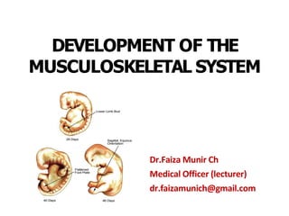
- 15. Development of the Limb Buds • The limb buds become visible as outpocketings from the ventrolateral body wall of the embryo in the beginning of 5th wk. • Initially, they consists of a mesenchymal core derived from parietal layer of lateral plate mesoderm that will form the bones and connective tissues of the limb.
- 16. Development of the Limbs • In the 6th wk, the terminal portion of the limb buds becomes flattened to form hand plates & foot plates and are separated from the proximal segment by a circular constriction (the future wrist crease & ankle crease) later a second constriction divides the proximal portion into two segments. And the main parts of extremities can be recognized. • Fingers and toes are formed when cell death in AER(apical ectodermal ridge)separate this ridge into 5 parts.
- 18. • The mesenchyme in the buds begins to condense, and by the end of 6th wk the first hyaline cartilage models, foreshadowing the bones of extremities can be recognized. • The endochondral ossification of limb bones starts by the end of embryonic period (i.e, after 8th wk).
- 20. Rotation of the Upper & Lower Limbs • During the 7th week, the upper & lower limbs rotate in opposite directions. • The upper limb rotates 90⁰ laterally so that the extensor muscles lie on the posterior surface of the upper limb and the thumbs also come to lie laterally. • The lower limb rotates 90⁰ medially so that the extensor muscles come to lie on the anterior surface of the lower limb and the big toe lies on the medial side.
- 21. Development of the Limb Musculature • The first indication of limb musculature is found in the 7th wk of development as a condensation of mesenchyme near the base of each limb bud. • This mesenchyme is derived from the dermomyotome cells of somites.
- 22. • The upper limb buds lie opposite the lower five cervical and upper two thoracic segments • The lower limb buds lie opposite lower four lumbar and upper two sacral segments
- 23. • As soon as the buds are formed, the neighboring spinal nerves penetrate into the mesenchyme. • At first they enter with isolated dorsal and ventral branches. • Soon these branches unite to form large dorsal and ventral nerves. • Thus in a developing upper limb; – Radial nerve which supplies the extensors (posterior/dorsal muscles), is formed by dorsal segmental branches. – Ulnar and Median nerves, which supply the flexors (anterior/ventral muscles), are formed by ventral segmental branches
- 24. Dermatomal Pattern • The spinal nerves not only play an important role in the differentiation & motor innervation of the limb musculature, but also provide the sensory innervation for the dermatomes. • Although the original dermatomal pattern changes with growth of extremities, an orderly sequence can still be recognized in the adult.
- 25. Development of the Muscular System The entire muscular system of the body develops from the Mesoderm (except for the muscles of Iris, which are derived from the ectoderm of the optic cups). The smooth muscles of gastrointestinal tract develop from the Splanchnic mesoderm surrounding the gut tube . The cardiac muscles of the heart develops from the splanchnic mesoderm surrounding the primitive heart tube.
- 26. Development of the Axial Musculature • The Somites develop from Paraxial mesoderm give rise to the skeletal musculature of axial skeleton, body wall, limbs, and head. • From the occipital region down, somites form and differentiate into the ‘sclerotome and dermomyotome.
- 27. Myogenesis (Development of muscle fibers) Cells of the myotome (Satellite cells) in the body wall and limb regions split and become elongated, spindle-shaped ‘myoblasts’. Multiple myoblasts fuse together and form multinucleated muscle fibers. By the end of 3rd month of IUL, cross striations, a typical feature of skeletal muscles appear
- 28. Formation of Anterior & Posterior Groups of Muscles • At the end of 5th wk, the musculature in the body wall divides into; – a small dorsal portion, the ‘Epimere’, and – a larger ventral portion, the ‘Hypomere’. • The nerve innervating the segmental muscles is also divided into two rami; ‘Dorsal primary ramus’ supplying the Epimere ‘Ventral primary ramus’ supplying the Hypomere
- 29. The muscles of Epimere form the extensors of the vertebral column. The muscles of Hypomere form the ventral and lateral flexors of vertebral column. Formation of Extensors and Flexors
- 30. The Hypomere splits into three layers; In the thorax, these layers are represented by External & Internal intercostals and Transversus thoracic muscles. In the Abdomen, these are represented by External & Internal Obliques and Transversus abdominis muscles.
- 31. Skeletal system Anomaly Achondroplasia: • Caused by a disturbance in the ‘endochondral/intracartilagenous’ ossification in the ‘epiphyseal/growth plates’ of the long bones. • The result is Dwarfism. • Both extremities are short but head is of normal size.
- 33. • Amelia complete absence of one or more extremities • Meromelia partial absence
- 34. Assignment Total marks 15 • Enlist the different sources of origin of the skeletal and muscular system (2) • Define the embryonic connective tissue (3) • Briefly describe the development of limb bud and limb musculature. (5) • Briefly describe the process of ossification. (5)