SlideShare a Scribd company logo
1 of 17
Download to read offline
Anatomical Evidence That the Uninjured Adjacent L4
Nerve Plays a Significant Role in the Development of
Peripheral Neuropathic Pain After L5 Spinal Nerve
Ligation in Rats
Safa Shehab,1
* Mehwish Anwer,1
Divya Galani,1
Afaf Abdulkarim,1
Khuloud Al-Nuaimi,1
Abdullah Al-Baloushi,1
Saeed Tariq,1
Nico Nagelkerke,2
and Milos Ljubisavljevic3
1
Department of Anatomy, College of Medicine and Health Sciences, United Arab Emirates University, Al-Ain, UAE
2
Department of Community Medicine, College of Medicine and Health Sciences, United Arab Emirates University, Al-Ain, UAE
3
Department of Physiology, College of Medicine and Health Sciences, United Arab Emirates University, Al-Ain, UAE
ABSTRACT
Rats develop hyperalgesia and allodynia in the hind
paw after L5 spinal nerve ligation. Phosphorylated
extracellular regulated kinase (pERK) was used as a
pain marker to investigate the potential role of adjacent
uninjured L4 nerve in the development of heat hyperal-
gesia after L5 nerve injury. Left L5 nerve was ligated
and sectioned in rats. Three days later, rats were ran-
domly assigned to five groups; each had both hind
paws immersed in water at different temperatures (no
heat, 37, 42, 47, and 52
C) under sevoflurane anesthe-
sia for 2 minutes. Five minutes after stimulation the
rats were sacrificed and sections of L3–L6 spinal seg-
ments were stained immunocytochemically with pERK
antibody. pERK immunoreactivity, which is not detecta-
ble in the normal spinal cord, was discernible in neu-
rons (not glia) of the superficial dorsal horn after
noxious heat stimuli. pERK-positive neurons clearly
overlapped in laminae I–II with normal unmyelinated
and thin myelinated afferents labeled with calcitonin
gene-related peptide and isolectin B4, and injured
unmyelinated afferents labeled with vasoactive intesti-
nal polypeptide. There was a linear increase in pERK
immunoreactivity on both sides with an increase in tem-
perature. Importantly, the number of positive pERK neu-
rons was significantly higher in the ipsilateral side of L4
spinal segment, which receives innervation from unin-
jured L4 nerve, compared with the contralateral control
side, which receives both uninjured L4 and L5 spinal
nerves. The data demonstrate that the uninjured L4
nerve plays an important role in the development of
heat hyperalgesia at the spinal cord level after L5 nerve
injury. J. Comp. Neurol. 523:1731–1747, 2015.
VC 2015 Wiley Periodicals, Inc.
INDEXING TERMS: L5 nerve injury; heat hyperalgesia; pERK; L4 spinal segment; AB_331768; AB_2315112;
AB_2298772; AB_2224402; AB_2336475; AB_2050884; AB_2314660; AB_2301751; AB_258833
Neuropathic pain is a pathological condition that
develops as a result of injury to the somatosensory
nervous system. Despite being extensively investigated,
the underlying mechanisms of neuropathic pain remain
controversial, with several proposed hypotheses (Yaksh
and Sorkin, 2005; Devor, 2006; Saade and Jabbur,
2008; Sandk€uhler, 2009). However, there is general
consensus that it originates from a lesion of the nerv-
ous system (Campbell and Meyer, 2006) and, therefore,
most of the neuropathic pain models (Campbell and
Meyer, 2006; Ossipov et al., 2006) are based on
peripheral nerve injury (Bennett and Xie, 1988; Seltzer
et al., 1990; Kim and Chung, 1992; Decosterd and
Woolf, 2000). Spinal nerve ligation (SNL) is one of the
most common models of neuropathic pain in rodents
(Kim and Chung, 1992). It is also extensively used to
elucidate the complex neuronal circuitry involved in the
development of sensitization of pain caused by a variety
of nociceptive and noxious stimuli. Typically, in this
Grant sponsor: UAE University; Grant number: 31M070.
*CORRESPONDENCE TO: Professor Safa Shehab, Department of
Anatomy, College of Medicine and Health Sciences, United Arab Emirates
University, Al-Ain, PO BOX 17666, UAE. E-mail: s.shehab@uaeu.ac.ae
Received October 30, 2014; Revised January 15, 2015;
Accepted January 18, 2015.
DOI 10.1002/cne.23750
Published online April 7, 2015 in Wiley Online Library
(wileyonlinelibrary.com)VC 2015 Wiley Periodicals, Inc.
The Journal of Comparative Neurology | Research in Systems Neuroscience 523:1731–1747 (2015) 1731
RESEARCH ARTICLE
model either the fifth and sixth lumbar (L5 and L6)
(Kim and Chung, 1992) or only the L5 spinal nerve are
ligated and cut in rats. Within a few days the rats
develop long-lasting ipsilateral, spontaneous pain,
hyperalgesia, and allodynia in the hind paw similar to
that seen in human neuropathic pain (Bennett and Xie,
1988; Seltzer et al., 1990; Kim and Chung, 1992; Deco-
sterd and Woolf, 2000). Although the primary afferent
injured neurons have been blamed for the spontaneous
pain following peripheral nerve injury (Boucher et al.,
2000; Liu et al., 2000), hyperalgesia and allodynia
require an intact nerve to conduct the noxious informa-
tion from the skin of the plantar surface of the hind
paw of experimental rats to the spinal cord. Since the
skin of the foot of the rat is mainly innervated by L4
and L5 nerves, it can be assumed the L4 nerve would
be involved in the development of abnormal pain sensa-
tion in an SNL model of neuropathic pain (Wu et al.,
2001, 2002; Fukuoka and Noguchi, 2002; Shim et al.,
2007; Meyer and Ringkamp, 2008).
In our previous anatomical study we identified central
terminations of the unmyelinated primary (presumably
nociceptive) afferents of L4 and L5 spinal nerve in rats.
The results showed that the central terminations of the
unmyelinated primary afferents of both nerves are not
restricted to the corresponding spinal segment that
they enter (L4 and L5, respectively) but extend two seg-
ments rostrally and one segment caudally (Shehab
et al., 2008). More important, the central terminations
of unmyelinated primary afferents of the L4 and L5 spi-
nal nerves intermingle with each other in the dorsal
horn at the L3–L5 spinal levels (Shehab et al., 2008).
Consequently, and as predicted, we have shown that
several neurochemical changes which occur after L5
nerve injury have not only taken place at the L5 spinal
segment but also in the rostral (L3 and L4) and the
caudal (L6) spinal segments which receive primary
afferent inputs from the uninjured adjacent nerves
(Shehab, 2014). It was concluded that the neuroplastic
changes in the dorsal horn of the spinal cord in the L4
spinal segment, which receives the central terminations
of the injured (L5) and uninjured (L4) nerves, might
explain the mechanism of hyperalgesia after peripheral
nerve injury (Shehab, 2014). In this study we used
phosphorylation of extracellular regulated kinases
(pERK) as a pain activity marker to provide further evi-
dence for our proposed hypothesis that neuropathic
pain manifestations due to L5 injury might develop
through the involvement of adjacent uninjured L4 nerve
at the level of the dorsal horn of the spinal cord. Previ-
ously, it has been shown that in normal nonstimulated
rats the levels of pERK in L4 and L5 spinal segment are
low or negligible. However, after a few minutes of nox-
ious hind paw stimulation, pERK immunoreactivity was
activated in the dorsal horn of the spinal cord in areas
which are known to receive primary afferent C-fibers of
the stimulated skin (Ji et al., 1999; Polgar et al., 2007).
Only noxious peripheral stimulation including mechani-
cal pinching, heat, and chemically induced painful stim-
uli through the stimulation of C and Ad fibers caused
activation of pERK because innocuous (touch) stimuli or
stimulation of Ab (mechanoreceptor) fibers did not
have any effects (Ji et al., 1999; Wang et al., 2004;
Polgar et al., 2007). The noxious heat threshold of nor-
mal Wistar rats is 43.5
C (B€olcskei et al., 2007). In this
study, therefore, we investigated the effects of temper-
atures below (37, 42
C) and above (47, 52
C) the heat
threshold applied to both hind paws of rats who had
unilateral L5 nerve ligation and transection. The results
were compared with another group of rats without heat
application.
The investigation proceeded in five phases. In the
first phase, we confirmed that heat noxious stimuli
applied to the hind paw caused pERK activation (Ji
et al., 1999; Wang et al., 2004; Polgar et al., 2007). In
the second phase, we used double and triple immuno-
cytochemical techniques and confocal microscopy to
investigate whether the pERK activation in response to
heat noxious stimuli is restricted to neurons or involve
glia as well. In the third phase, three methods were
used to discover the relationship of pERK-activated
cells and the distribution of normal and injured primary
afferents of L5 spinal nerve. In the first method, Ban-
deiraea simplicifolia isolectin (IB4), calcitonin gene-
related peptide (CGRP), vesicular glutamate trans-
porter1 (VGLUT1) immunoreactivities were used as
markers to label normal unmyelinated C, both unmyeli-
nated C and thin myelinated Ad, and myelinated Ab pri-
mary afferents, respectively (Chung et al., 1988; Wang
et al., 1994; Todd et al., 1998; Gerke and Plenderleith,
2004; Hughes et al., 2004; Shehab et al., 2008; Todd,
2010). In the second method, L5 unmyelinated and
myelinated afferents were identified by injection of a
mixture of IB4 (unmyelinated C fiber marker) and chol-
era toxin B subunit (CTb, myelinated nerve fiber marker)
(Robertson and Grant, 1985; LaMotte et al., 1991;
Kitchener et al., 1994; Shehab et al., 2003, 2004,
2008; Shehab, 2009) into L5. In the third method, vaso-
active intestinal polypeptide (VIP) was used as a marker
for injured unmyelinated (and presumably thin myelin-
ated) primary afferents (Shehab and Atkinson, 1986;
Shehab et al., 2003, 2004). In the fourth phase, we
determined the effects of acute and chronic L5 nerve
injury on pERK immunoreactivity in the dorsal horn of
the spinal cord. In the fifth phase, we tested the effects
of different intensities of heat stimulation on pERK
S. Shehab et al.
1732 The Journal of Comparative Neurology | Research in Systems Neuroscience
activation in the dorsal horn of L3–L6 spinal segments
in an SNL model of neuropathic pain.
Part of this study had appeared in a previous
abstract (Shehab et al., 2013).
MATERIALS AND METHODS
All experiments were approved by the Animal Ethics
Committee of the College of Medicine and Health Sci-
ences of the United Arab Emirates University and were
performed in accordance with the guidelines of the
European Communities Council directive of 24 Novem-
ber, 1986 (86/609/EEC).
Spinal nerve ligation and transection
Adult male Wistar rats (240–255 g at the time of sur-
gery) were anesthetized with a mixture of ketamine
(100 mg/kg) and xylazine (20 mg/kg) delivered intra-
peritoneally or intramuscularly. The skin of the back
was incised longitudinally, the transverse processes of
the sixth lumbar vertebra was excised, and the left L5
nerve was ligated with a 6/0 silk suture and sectioned
distally as described before (Shehab, 2014). To deter-
mine the effect of L5 nerve injury on pERK immunore-
activity, animals (n 5 6) were perfused 5–10 minutes
after surgery through the ascending aorta with modified
Zamboni’s fixative (10% formalin containing 15% of satu-
rated picric acid) in 0.1M phosphate buffer (pH 7.4).
The spinal lumbar segments from L3 to L6 were dis-
sected out, postfixed in the same fixative for 3 hours,
and stored in 30% sucrose in phosphate buffer over-
night. In another set of animals (n 5 6) the L5 nerve
was ligated and transected and the rats were perfused
3 days postoperatively without heat stimulation.
In order to determine the effects of heat stimulation
on pERK immunoreactivity in neuropathic animals, 3
days after L5 nerve ligation and transection rats were
randomly assigned to different experimental groups in
which both hind paws (n 5 5–6 per group) were
immersed in a water bath at different temperatures
(37, 42, 47, and 52
C) under sevoflurane anesthesia
for 2 minutes. Five minutes later, enough time to have
pERK activated (Ji et al., 1999; Polgar et al., 2007), the
animals were perfused as mentioned above. As a con-
trol experiment a no heat group was also included in
which rats had only L5 injury without heat exposure.
In order to characterize the type of heat-induced
pERK in the cells of the spinal cord, hind paws of nor-
mal (uninjured) animals (n 5 6) or experimental (injured)
were immersed in 52
C hot water for 2 minutes and
perfused 5 minutes after termination of stimulation.
Sections were incubated with pERK and either nuclear
protein (NeuN, neuronal marker) antibodies or a mixture
of glial fibrillary acidic protein (GFAP, astrocytes
marker) and ionized calcium binding adapter molecule
1 (Iba1, microglia marker) antibodies to find whether
the labeled cells were neurons or glia, respectively.
Sections from the same normal animals were also used
to determine the topographic arrangement and relation-
ship of pERK-positive cells with various types of primary
afferents. Triple immunofluorescent staining of pERK
with any two of the following was performed: IB4 (as a
marker for unmyelinated C fibers), CGRP (as a marker
for unmyelinated C and Ad thin myelinated fibers), and
VGLUT1 (as a marker for myelinated Ab fibers).
Injection of tracers
To label both unmyelinated and myelinated primary
afferents simultaneously, we injected a mixture of IB4 and
CTb, which have been shown to be taken up selectively
and transported transganglionically by these two types of
axons when injected into (uninjured) somatic nerves (She-
hab and Hughes, 2011). The skin of the back was incised
and the left L5 nerve (n5 4) was exposed, ligated, and
transected and injected with 1 ll of a mixture of 1% CTb
(Sigma, St. Louis, MO) and 2% IB4 (Vector, Burlingame,
CA). To ensure that the whole L5 nerve was filled with
the tracer, we used a finely drawn glass micropipette
inserted gently, for a few millimeters, into the nerve in 3–
4 different positions. In addition, the tracer was prepared
in a 0.1% solution of Fast Green. This allowed clear visual-
ization of the injection process, which resulted in colora-
tion of all aspects of the nerve and also served to
confirm that no leakage had taken place during or imme-
diately after the injection. The area was washed with nor-
mal saline after injection and the muscles and the skin
were sutured in layers. Three days after the injections of
the tracer the animals were stimulated with 52
C water
under sevoflurane and perfused as mentioned above.
To investigate the relationship between heat-
activated pERK neurons and injury-induced VIP upregu-
lation, which was used as a marker of injured unmyeli-
nated primary afferents (Shehab and Atkinson, 1986;
Shehab et al., 2003, 2004), the left L5 nerve was
ligated and transected as mentioned above (n 5 4).
After 7 days of nerve injury, a period of time when VIP
is upregulated and neuropathic pain manifestation is
already established (Shehab and Atkinson, 1986; Kim
and Chung, 1992), the hind paws were immersed in
52
C hot water for 2 minutes and the animals were
perfused 5 minutes after the termination of stimulation.
Immunocytochemistry
Transverse sections (50 lm) of L3–L6 spinal seg-
ments of experimental rats were cut in a cryostat and
treated with 50% ethanol to increase antibody
Role of L4 nerve in neuropathic pain
The Journal of Comparative Neurology | Research in Systems Neuroscience 1733
penetration (Llewellyn-Smith and Minson, 1992) and
then incubated overnight in experiment-specific combi-
nations of primary antibodies. Antibodies were diluted
in phosphate-buffered saline (PBS) containing 0.3% Tri-
ton X-100. Details of the primary antibodies used, their
sources, and concentrations are given in Table 1.
For peroxidase staining, sections were incubated
overnight in either mouse anti-pERK (1:10,000; Cell Sig-
naling Technology, Beverly, MA) or rabbit anti-pERK
(1:10,000; Cell Signaling). After rinsing with PBS, the
sections were incubated with biotinylated antimouse or
antirabbit secondary antibody (Jackson ImmunoRe-
search, West Grove, PA) for an hour, followed by extra-
vidin peroxidase conjugate for 1 hour (1:1,000, Sigma-
Aldrich). Finally, the sections were incubated for 5
minutes in a solution of 3,30
-diaminobenzidine (DAB)
solution (25 mg / 50 ml of phosphate buffer, pH 7.4
with 7.5 ll hydrogen peroxide [30%] and 1 ml nickel
chloride [3%] added to it). All sections were mounted
on gelled slides and allowed to air-dry overnight. They
were then washed, dehydrated in graded alcohol,
cleared in xylene, coverslipped, and examined under
light microscope.
For double immunofluorescent staining, sections from
animals treated with heat (52
C without nerve injury)
were incubated overnight with monoclonal rabbit anti-
pERK (diluted 1:1,000) and mouse anti-NeuN (diluted
1:1,000; Millipore, Billerica, MA). After rinsing, the sec-
tions were incubated for 1 hour in species-specific sec-
ondary antibodies which have been raised in donkey
(antirabbit Alexa 488, diluted 1:200 and antimouse Rho-
damine Red, diluted 1:100) supplied by Jackson Immu-
noResearch. Using triple immunofluorescent labeling, a
cocktail of primary antibodies including monoclonal rab-
bit anti-pERK (diluted 1:1,000), goat anti-Iba1 (diluted
1:500; Abcam, Cambridge, UK), and mouse anti-GFAP
(diluted 1:500; Vector, Peterborough, UK) was
employed. This was followed by secondary antibodies
for 1 hour (antirabbit conjugated to Alexa 488, antigoat
conjugated to Rhodamine Red, and antimouse conju-
gated to Cyanine 5 [Jackson ImmunoResearch]).
From rats that were used for investigating the rela-
tionship of primary afferents with pERK-positive cells,
sections were incubated overnight with monoclonal
mouse anti-pERK (diluted 1:1,000), rabbit anti-CGRP
(diluted 1:2,000, BioMol/Enzo Life Science, Lausen,
Switzerland), and goat anti-IB4 (diluted 1:5,000, Vec-
tor). For labeling with IB4, the sections were preincu-
bated with IB4 (Vector) solution (1 lg/ml of PBS in
0.3% triton) for 1 hour. After washing with PBS, sections
were incubated in antimouse Alexa 488 (1:200),
antirabbit Rhodamine Red, and antigoat Cy5 (1:100;
Jackson ImmunoResearch). For another set of triple
TABLE1.
PrimaryAntibodiesUsed,Sources,andConcentrations
Srno.
Nameof
theantibodyStructureoftheimmunogenManufacturer
Catalog
numberRRIDSpecies
Mono/
polyclonalDilutionused
1pERKSyntheticphosphopeptidecorrespondingtoresidues
surroundingThr202/Tyr204ofhumanp44MAPkinase.
CellSignalingTechnology,
Beverly,MA,USA
mAb9106AB_331768MouseMonoclonal1:10k
2pERKSyntheticphosphopeptidecorrespondingtoresiduessurrounding
Thr202/Tyr204ofhumanp44MAPkinase.
CellSignalingTechnology,
Beverly,MA,USA
mAb4370AB_2315112RabbitMonoclonal1:10k
3NeuNPurifiedcellnucleifrommousebrainMillipore,Billerica,MA,USAMAB377AB_2298772MouseMonoclonal1:1k
4Iba1Syntheticpeptidecorrespondingtoaminoacids135–147
(C-TGPPAKKAISELP)ofHumanIba1
Abcam,Cambridge,UKab5076AB_2224402GoatPolyclonal1:500
5GFAPPorcinespinalcordVector,Peterborough,UKVPG805AB_2336475MouseMonoclonal1:500
6CGRPSyntheticpeptidecorrespondingtoaportionofrat
a-calcitoningene-relatedpeptide(CGRP)
BioMol/EnzoLifeScience,
Lausen,Switzerland
CA1134AB_2050884RabbitPolyclonal1:2k
7IB4ByimmunizinggoatswithpurelectinsVector,Peterborough,UKAS2104AB_2314660GoatPolyclonal1:5k
8VGlut1Syntheticpeptidecorrespondingto542–560
(GATHSTVQPPRPPPPVRDY)fromratVGLUT1protein
Chemicon,InternationalInc.
Billerica,MA,UnitedStates
AB5905AB_2301751GuineaPigPolyclonal1:5k
9VIPPurePorcineVIPGiftfromProfJ.AllenRabbit1:5k
10CTbProducedinrabbitusingpurifiedtoxinfromVibrio
choleraeasimmunogen
Sigma-Aldrich,StLouis,
MO,USA
C-3062AB_258833RabbitPolyclonal1:2k
S. Shehab et al.
1734 The Journal of Comparative Neurology | Research in Systems Neuroscience
labeling, primary monoclonal rabbit anti-pERK (diluted
1:1,000), goat anti-IB4 (Vector, diluted 1:5,000) after 1
hour incubation with IB4 and guinea pig anti-VGlut1
(diluted 1:5,000, Chemicon International, Temecula, CA)
were added to sections and left at room temperature
overnight. After PBS rinsing, secondary antirabbit Alexa
488, antigoat Rhodamine Red, and antiguinea-pig Cy5
were added for 1 hour.
For rats that were stimulated with heat 7 days after
injury, sections were incubated with a cocktail of pri-
mary antibodies including rabbit anti-VIP (a gift from J.
Allen, diluted 1:5,000) and mouse anti-pERK (1:1,000).
After rinsing, sections were incubated in mixture of
antirabbit Alexa 488 and antimouse Rhodamine Red.
From those rats that received injection of a mixture
of CTb and IB4 in L5 nerve, transverse sections were
incubated with a cocktail of three primary antibodies:
monoclonal mouse anti-pERK (diluted 1:1,000), goat
anti-IB4 (diluted 1:1,000), and rabbit anti-CTb (diluted
1:2,000, Sigma). After rinsing, the sections were incu-
bated for 1 hour with a mixture of species-specific sec-
ondary antibodies (antimouse Alexa 488, antigoat
Rhodamine Red, and antirabbit Cy5).
Sections were mounted on glass slides with glycerol-
based antifade medium and examined with a Zeiss fluo-
rescent microscope (Carl Zeiss, Germany) equipped
with appropriate filters to reveal Alexa 488 (green) and
Rhodamine Red (red) or a Nikon C1 laser scanning con-
focal microscope to reveal Alexa 488, Rhodamine Red,
and Cy5 (blue) labeling. Representative digital images
were captured using a Zeiss AxioCam HRc Digital cam-
era with AxioVision 3.0 software or a Nikon C1 confocal
microscope. The resulting files were used to generate
figures in Adobe Photoshop software CS6 (San Jose,
CA) in which photomicrographs were adjusted for con-
trast and brightness.
pERK-positive cell count and statistical
analysis
It should be noted that the effects of noxious heat
stimulation were quantified by counting the number of
pERK-positive cells in the ipsilateral and contralateral
side (injured vs. uninjured) of the dorsal horn directly
under the light microscope by two independent observ-
ers. Theoretically, this may have introduced bias as,
undeniably, the dissector method of stereological count-
ing is the most unbiased approach, especially when the
aim is to establish the number of cells in a specific tis-
sue volume. Nevertheless, it has been repeatedly sug-
gested that the conventional method of counting is
sufficiently reliable, particularly when the counted
objects are small relative to the thickness of the sec-
tion (Guillery, 2002; Baquet et al., 2009) and when the
structures being counted do not change in size, shape,
or orientation between the two estimates that are com-
pared (Saper, 1996). Thus, we believe that the conven-
tional counting method used in this study did not affect
the final result by introducing a bias, as all the above
preconditions were met.
Furthermore, to confirm that the morphometric
changes (size) of the neurons did not have biasing effect
on counting, we performed an additional experiment in
which the maximum and minimum rostrocaudal dimen-
sions of pERK-positive neurons (profiles) in the dorsal
horn of the spinal cord in the z-plane were determined.
Experiments were done on three rats that had L5 nerve
ligation and transection 3 days earlier and both hind
paws were exposed to noxious heat stimulation at 52
C.
Sagittal sections of both L4 and L5 segments were
stained with the peroxidase method as mentioned above
and subsequently photographed at 203 using a bright-
field microscope and Axiovision software. The diameters
of 500 cells from either the ipsilateral or contralateral
side were measured. The cells to be counted were cho-
sen randomly and their diameter was measured in the
rostrocaudal axis. On average, the size of the selected
pERK-positive neurons on the injured (ipsilateral) side
was 11.71 lm (60.11 SEM) with minimum and maxi-
mum diameters of 6.71 to 19.88 lm, respectively. Simi-
larly, the average size of the pERK-positive neurons on
the uninjured (contralateral side) side was 11.46 lm
(60.11 SEM) with minimum and the maximum diameters
of 6.37 to 19.49 lm, respectively. The difference was
not statistically significant (t-test: t 5 1.66; df 5 499;
P 5 0.096). This ruled out the possibility that the injury
had induced morphometric changes in size of the
observed neurons.
The chances of bias in profile counting are more
obvious in thin sections because they generate more
split cells than thicker sections (Guillery, 2002). The
thickness of our sample tissue was 50 lm, which is 5
times greater than the observed diameter of neurons
(profile). Based on these observations, a sampling inter-
val of 150 lm between sections to be counted was
determined, with the intention of minimizing the effect of
duplication of particle count in relation to size, shape,
and orientation of the cells. Therefore, six sections per
segment were included from each animal (n 5 5–6 per
group) for counting. The numerical data were analyzed
using SPSS (v.20, Chicago, IL).
Comparison between the five groups (no heat, 37,
42, 47, and 52
C) was done employing the SPSS mixed
procedure and paired sample t-tests. Considering the
unequal variances among groups, the cell counts were
analyzed on the variance stabilizing square root
Role of L4 nerve in neuropathic pain
The Journal of Comparative Neurology | Research in Systems Neuroscience 1735
transformed scale. The model dimensions (variables/fac-
tors) included Rat, Rats*Spinal cord segment, and
Rats*Spinal cord segment*Section as (nested) random
factors (sources of noise/random variation that observa-
tions share and, although not of primary interest, need to
be accounted for). The main/fixed effects (i.e., the
effects that we are primarily interested in) were Spine
segment (L3, L4, L5, L6), Side [Ipsilateral (operated side)
/ Contralateral (control)] and Temperature (No Heat to
52
C). Moreover, in order to demonstrate qualitative dif-
ferences in response at high temperature (52
C),
Heat52*side, Heat52*SpineSegment, and Heat52*Side*
SpineSegment interactions were also included in an addi-
tional model.
In the first analysis, complete data were included in a
mixed linear model to meticulously identify any changed
pattern of variation in pERK-positive cells in various seg-
ments on both sides. As this first Mixed Model analysis,
with interactions, showed that at 52
C the pattern of
variation was more complex than at other temperatures
and would not well fit the Mixed Model without interac-
tions of fixed effects described above, we separately
analyzed temperatures below 52
C (again a Mixed Model
with main effects of Fixed Effects only) and only 52
C.
For analysis of 52
C, we aggregated the data (mean root
cell counts) for each rat, spinal cord segment, section,
and side for this temperature.
Paired t-tests were applied for counts of each seg-
ment separately to identify statistically significant differ-
ences, if any, between the control and treated side of
the section. In order to validate our model we analyzed
the correlation between the predicted values and
square root counts. The correlation was highly signifi-
cant, showing the strength of the predicted values and
the consequent analysis.
Antibody characterization
The VIP, CTb, and the IB4 antibodies were previously
characterized (Shehab et al., 2003; Shehab, 2009). Pre-
treatment of rabbit anti-VIP antibodies with VIP
(M1026
) and rabbit anti-CTb antibodies with CTb (10 lg
/ 1 ml) completely abolished the corresponding immu-
noreactivity. The IB4 antibody was raised in goat
against Griffonia simplicifolia lectin I as an immunogen.
Sections of the spinal cord taken from control rat which
did not have a tracer injection and incubated with CTb
and IB4 antibodies showed no immunoreactivity. In
addition, the specificity of CTb and IB4 antibodies was
shown by the presence of immunostaining in specific
locations in the spinal cord and the lack of immunore-
activity in the contralateral side of the spinal cord.
Pretreatment of rabbit anti-CGRP with CGRP (M1025
,
PolyPeptide Group, San Diego, CA) completely abol-
ished the corresponding immunoreactivity. Furthermore,
no immunoreactivity was detected in sections incubated
with nonimmune rabbit or goat serum.
The pERK antibody detects both ERK1 and ERK2 that
are dually phosphorylated at Thr202 and Tyr204 sites,
and does not crossreact with JNK or p38 MAP kinase
that are phosphorylated at the corresponding residues
(manufacturer’s specification).
The mouse monoclonal antibody NeuN, raised against
cell nuclei from mouse brain, reacts with a neuron-
specific protein (Mullen et al., 1992). In rat spinal cord,
NeuN apparently detects all neurons, but does not label
glial cells (Todd et al., 1998).
Goat Iba1 antibody was a polyclonal IgG. It was immu-
nogen affinity-purified antibody raised against the Iba1
synthetic peptide, corresponding to amino acids 135–
147 of human Iba1 which can be blocked with human
Iba1 peptide (Ab23067, manufacturer’s information).
The monoclonal mouse anti-GFAP antibody (Vector,
Clone GA5) was raised against purified GFAP from por-
cine spinal cord and has been previously characterized
(Debus et al., 1983). This antibody detected the GFAP
in astrocytes in the cerebral cortex, cerebellum, basal
ganglia, hippocampus, and spinal cord (manufacturer’s
information) (Shehab et al., 1990).
RESULTS
Characterization of pERK-positive cells
No pERK immunoreactivity could be detected in the
dorsal horn of L3–L6 spinal segments in normal nonsti-
mulated animals without nerve injury. However, 5
minutes after termination of stimulation of the hind paw
with 52
C hot water, pERK-positive cells were observed
in dorsal horn.
In the next step we investigated whether the acti-
vated pERK-positive cells in response to heat noxious
stimuli or peripheral nerve injury are neurons or glia.
Double immunofluorescent staining of sections from L4
and L5 spinal segments from animals which had heat
stimulation of the hind paw at 52
C showed consistent
colocalization between pERK and NeuN labeled cells
(Fig. 1A–C). With the triple immunofluorescence, none
of the pERK-labeled cells were GFAP- (marker of astro-
cytes) or Iba1- (marker of microglia) positive (Fig. 1D–
G). These results show that, following heat stimulation
at 52
C, the pERK-labeled cells in the dorsal horn of
the spinal cord belong to neurons rather than glia. Simi-
lar results of the presence of colocalization with NeuN
and absence of colocalization with GFAP and Iba1 were
seen in the ipsilateral dorsal horn of animals which had
only L5 nerve injury 5–10 minutes earlier or both L5
nerve injury followed 3 days later with 52
C heat stimu-
lation applied to the ipsilateral hind paw (not shown).
S. Shehab et al.
1736 The Journal of Comparative Neurology | Research in Systems Neuroscience
Relationship of pERK-activated neurons and
the distribution of normal and injured
primary afferents of L5 spinal nerve
In animals which had either L5 spinal nerve injury
alone or followed by heat stimulation of the hind paw 3
days later, pERK immunoreactivity was activated in the
dorsomedial part of the superficial dorsal horn of the
spinal cord. In order to precisely locate the pERK immu-
noreactivity in relation to spinal cord laminae, we first
photographed unstained sections of L4 and L5 spinal
segments in brightfield. The boundaries between lami-
nae I, II, and III were clearly visible when sections were
viewed with brightfield optics, due to the very translu-
cent appearance of lamina II, compared with laminae I
and III. To plot laminar boundaries on confocal images
of pERK, CGRP, IB4, and VGLUT1 labeling in the spinal
dorsal horn, sections were first scanned through a 103
objective lens on a Nikon confocal microscope (Nikon,
Japan) and then brightfield images of each scanned
section were superimposed over the corresponding
Figure 1. Confocal images of spinal cord sections following noxious heat stimulus showing the relationship of activated pERK-positive cells
(green in A and D) to NeuN (a neuronal marker, magenta in B) or Iba 1 (a marker for microglia, red in E), or GFAP (a marker for astro-
cytes, blue in F). Panel C is merged image of and A and B. Panel G is merged image of D–F. pERK-positive cells (arrows in A) were colo-
calized with NeuN (arrows in B,C) but not with Iba1 (E,G) or GFAP (F,G), indicating that noxious heat stimulus activated pERK in the spinal
neurons and not in neuroglia. Images were built from projections of two confocal optical sections at 1 lm z-spacing. Scale bar 5 25 lm.
[Color figure can be viewed in the online issue, which is available at wileyonlinelibrary.com.]
Role of L4 nerve in neuropathic pain
The Journal of Comparative Neurology | Research in Systems Neuroscience 1737
Figure 2. Confocal images of transverse sections of L5 spinal cord segments showing pERK, IB4, CGRP, and VGlut1 labeling following nox-
ious heat stimulus. The sections were photographed first using brightfield objectives to show the different laminae (A,F) and then stained
with antibodies against pERK, IB4, CGRP, and VGlut1. IB4-labeled terminals arborize mainly in the middle of lamina II (C,I), whereas CGRP-
labeled terminals are located in lamina I and outer lamina IIo (H). In comparison, VGlut1-labeled terminals are located in deep lamina IIi
to V (D). (E) Merged image of B–D and panel (J) is merged image of G–I. Note the clear overlapping of pERK (B,G) with CGRP and IB4
and no overlapping with and VGlut1 immunoreactivity. Scale bar 5 200 lm. [Color figure can be viewed in the online issue, which is
available at wileyonlinelibrary.com.]
S. Shehab et al.
1738 The Journal of Comparative Neurology | Research in Systems Neuroscience
projected confocal images using Adobe Photoshop and
Xara designer Pro X software (Gaddesden Place, Great
Gaddesden, Hemel Hempstead, Hertfordshire, UK).
Lamina II was further divided into outer (IIo) and inner
(IIi) layers by a line drawn half way between lamina I/II
and II/III boundaries (Fig. 2).
Upon labeling, pERK-positive neurons were found to be
localized primarily in lamina I and IIo extending partially
to lamina IIi. Very few less brightly labeled neurons were
seen in the ventral laminae (III–VI) of the spinal cord. In
order to find out the relationship between pERK-positive
neurons and different primary afferents we used three
markers (CGRP, IB4, and VGlut1) each for a specific type
of afferent. Triple labeling showed that CGRP labeled
unmyelinated C and thin myelinated Ad nerve terminals
with a distinctive overlap with pERK-positive neurons in
laminae I and II of the dorsal horn (Fig. 2F–J).
As reported previously (Kitchener et al., 1994; Gerke
and Plenderleith, 2004; Shehab and Hughes, 2011), IB4
preferentially labeled unmyelinated primary afferents in
lamina I and II with a dense intensity in the middle of
lamina II. Again, a clear overlapping of IB4 immunoreactiv-
ity with pERK-positive neurons was observed (Fig. 2A–E).
In comparison, VGlut1 labeled primary terminals of
myelinated Ab fibers in the spinal cord which terminate
in the lamina III and deeper laminae showed no overlap
with pERK-positive neurons (Fig. 2A–E).
Relationship of pERK-activated cells and
transganglionically transported CTb and IB4
in central primary afferents of L5 nerve
We used another method to investigate the relation-
ship between activated pERK neurons and central
Figure 3. Images of a transverse section of the spinal cord showing relationship of activated pERK (green) and IB4 (red) and CTb (blue)
labeling in the L4 segment of a rat that had noxious heat stimulus to the hind paw. The rat had a coinjection of IB4 and CTb into ligated
and transected L5 nerve 3 days earlier. Note that the termination of IB4-labeled terminals is confined mainly to the middle of lamina II (C)
with partial overlapping with pERK (A), while the termination of CTb-labeled terminals are located in the deep laminae with no overlapping
with pERK immunoreactivity (B). (D) Merged image of A–C. Scale bar 5 25 lm. [Color figure can be viewed in the online issue, which is
available at wileyonlinelibrary.com.]
Role of L4 nerve in neuropathic pain
The Journal of Comparative Neurology | Research in Systems Neuroscience 1739
termination of primary afferents of L5 in the dorsal
horn. The main purpose of this experiment was to
explore whether the pERK-positive cells in ipsilateral L4
spinal segment were in the territory of the central ter-
mination of injured L5 nerve. Injection of a mixture of
CTb and IB4 into L5 ligated spinal nerve was followed
by 3 days of recovery and then heat stimulation was
applied to the ipsilateral hind paw. In L4 and L5 spinal
segments, pERK-labeled neurons were seen in the lami-
nas I and II corresponding with IB4- and CTb-labeled
terminals mediolaterally and rostrocaudally (Fig. 3).
However, the pERK-labeled neurons showed overlap
with IB4- (in lamina II) but not with CTb- (in lamina III)
labeled nerve terminals. These results also demon-
strated that pERK-positive neurons in the dorsal horn of
L4 were in the territory of the central termination of L5
nerve in L4 segment.
Relationship between noxious heat pERK
activation and VIP upregulation followed by
L5 injury
We have previously shown that both sciatic and L5
nerve injury causes upregulation of VIP in nerve termi-
nals in the lamina I and II of the spinal cord where
these two nerves terminate (Shehab and Atkinson,
1986; Shehab et al., 2003, 2004; Shehab, 2014). In
this study we used this VIP upregulation as a marker
for injured unmyelinated primary afferents in L4 and L5
spinal segments. Seven days after L5 ligation and trans-
ection, noxious heat was applied to the ipsilateral hind
paw. The result showed that pERK-activated neurons
were surrounded by VIP-labeled terminals in lamina I
and II of the dorsal horn of the spinal cord (Fig. 4). In
the contralateral side, only scattered positive VIP nerve
terminals and no pERK cells could be detected. These
results clearly demonstrated that the neuronal pERK
activation in dorsal horn of spinal cord in response to
heat noxious stimuli, which is most likely transmitted
through L4 nerve, were in the same locations of central
primary afferents of injured L5 nerve.
Effects of peripheral nerve injury on pERK
immunoreactivity
We aimed in this study to investigate the effects of
noxious heat stimulation on pERK immunoreactivity in an
animal model of neuropathic pain in rats due to periph-
eral nerve injury. Therefore, it was important first to dis-
cover the effects of peripheral nerve injury itself on pERK
expression without heat stimulation. No pERK immunore-
activity was seen in untreated rats. However, 5–10
minutes after L5 nerve ligation and transection, pERK-
positive cells were detected in the dorsal horn of L3–L6
spinal segments (Fig. 5). The mediolateral and rostrocau-
dal distribution of pERK-positive cells were seen in
exactly the same areas of the dorsal horn where unmyeli-
nated primary afferents of L5 terminate (Shehab et al.,
2008). As expected, no pERK reactivity was observed on
the contralateral side of the spinal cord. Also, as men-
tioned above, the pERK-positive cells were neurons, as
they were also labeled with NeuN and not glia, as there
was no colocalization with GFAP and Iba1 (not shown).
Importantly, this pERK activation following peripheral
nerve injury was temporary, as it disappeared completely
after 3 days. This indicates that the pERK activation in
the dorsal horn, which we observed after noxious heat
stimulation applied to the hind paw 3 days following L5
transection, must be attributed to heat stimulation rather
than nerve injury itself.
Figure 4. Confocal images of a transverse section of spinal cord showing VIP (green in A) and activated pERK neurons (magenta in B) in
the L5 segment following L5 spinal nerve ligation and transection 7 days before dipping hind paws in water at 52
C. (C) A merge of A,B.
The pERK-activated neurons in response to noxious heat stimulation (which are likely due to stimulation of L4 primary afferents of the
hind paw) intermingle with upregulated VIP in the central terminals of injured L5 primary afferent fibers. Images built from projections of
three confocal optical sections at 2 lm z-spacing. Scale bar 5 25 lm. [Color figure can be viewed in the online issue, which is available
at wileyonlinelibrary.com.]
S. Shehab et al.
1740 The Journal of Comparative Neurology | Research in Systems Neuroscience
pERK activation in response to noxious heat
stimulation
Figures 6 and 7 and Table 2 show the effects of the
exposure of the hind paws to heat stimuli at 37–52
C on
the number of pERK-labeled neurons in the dorsal horn.
In this experiment the L5 nerve was ligated and trans-
ected 3 days earlier to induce neuropathic pain in the
ipsilateral hind paw. In a control No heat group of rats
the animals had no heat stimulation. The numbers of
pERK-positive neurons in the ipsilateral side of L3–L6
segment were compared with contralateral uninjured
side. Interestingly, after 3 days of L5 nerve injury the
No heat group of rats showed either few or no pERK-
immunoreactive neurons in the ipsilateral side (Fig. 6A).
This pERK immunoreactivity was not different from that
observed in the contralateral side of the spinal cord
Figure 5. Transverse sections of L3–L6 spinal segments showing the activation of pERK in response to unilateral L5 nerve ligation and trans-
ection 10 minutes before sacrifice. The activated pERK cells are located mainly in laminae I–II of the ipsilateral dorsal horn of L3–L6 (A–D)
in exactly the same areas where the unmyelinated primary afferents of L5 nerve terminate (see fig. 2 in Shehab et al., 2008). Arrows in A–C
indicate that pERK activation would very likely take place in areas of the spinal cord where the dorsal rami of L3–L6 nerves terminate and
were inevitably injured during the surgical operation in which a skin incision of the back was carried out to expose the L5 spinal nerve. Negli-
gible pERK immunoreactivity can be detected in the spinal cord on the contralateral uninjured side (E–H). Scale bar 5 200 lm.
Role of L4 nerve in neuropathic pain
The Journal of Comparative Neurology | Research in Systems Neuroscience 1741
(Fig. 6) or in naive animals. These results indicated that
activation of pERK 5–10 minutes after peripheral nerve
injury was reduced to its normal level 3 days later.
In comparison, upon stimulation with 37
C water, a
few more pERK-positive cells were seen in the ipsilat-
eral dorsal horn which increased in number with the
raise in temperature from 42 to 52
C (Fig. 6B–E), com-
pared with the contralateral side (Fig. 6G–J).
Statistical analysis of pERK-positive cell
counts
In order to quantify and estimate the number of pERK-
positive cells in ipsilateral and contralateral sides of spi-
nal segments (L3–L6) upon heat, a detailed statistical
analysis was carried out. Mixed model analysis revealed
that all fixed effects were statistically highly significant,
including all two-way and three-way interactions
Figure 6. Brightfield images of transverse sections of dorsal horns of L4 spinal segment, showing pERK immunoreactivity on the contralateral
and ipsilateral sides of animals which had left L5 nerve ligation and transection 3 days earlier to induce neuropathic pain in the hind paw. Both
hind paws were either subjected to no heat (A) or immersed in water at 37
C (B), 42
C (C), 47
C (D), and 52
C (E). Note the increase in pERK-
positive neurons in the ipsilateral side (A–E) with increase in temperature compared with contralateral uninjured side (F–J). Scale bar 5 200 lm.
S. Shehab et al.
1742 The Journal of Comparative Neurology | Research in Systems Neuroscience
(P  0.001). In general, it explains that there was signifi-
cant difference in the number of pERK-immunoreactive
neurons on both sides (injured vs. uninjured) of all spinal
cord segments (L3–L6) at all temperatures (including
52
C). The difference between both sides followed a lin-
ear pattern with increasing temperature.
To further elucidate the effects and to simplify inter-
pretation, we divided the data into two groups and ran
separate analysis for 1) temperatures below 52
C
including 37, 42, and 47
C, 2) 52
C only. The same
mixed model was utilized for temperatures below 52
C
with all the terms, except heat52. The results
expressed a highly significant difference between the
injured and control side (P  0.001) with root cell
counts on the treated side 0.45 units higher on average
than on the control side. Also, it was observed that the
L4 spinal segment had the highest number of root cell
difference (0.71), which highlights the increased activa-
tion of pERK in the L4 segment. This observation was
consistent at all temperatures.
When temperature 52
C was analyzed separately, it
was observed that for L3 and L4 the ipsilateral side
was significantly (P  0.001) higher than the contralat-
eral side, in line with temperatures below 52
C. For L5
the pattern was reversed, with the ipsilateral side hav-
ing significantly (P  0.001) lower counts than the con-
tralateral side. The substantial change between both
sides at 52
C was further evaluated by paired samples
t-test which showed statistically significant difference
(P  0.001) between the ipsilateral and contralateral
side of all the spinal segments (L3–L6).
We used the mean cell counts for graphical illustra-
tion of the differences between the injured and unin-
jured side of each spinal cord at various temperatures
(No heat–52
C). The individual mean values for both
sides at all temperatures are provided (Table 2). L4 has
the maximum number of pERK-positive cells, which
increase linearly with temperature (Fig. 7). However, L5
follows the same pattern until it approaches 52
C. At
this temperature the contralateral side (control unin-
jured) has more pERK-positive cells as compared to
ipsilateral side (injured-L5 ligated).
DISCUSSION
In the present study we exploited prior observations
(Ji et al., 1999; Polgar et al., 2007) that application of
heat noxious stimuli to the normal hind paw causes acti-
vation of pERK in the dorsal horn of the spinal cord. The
principal findings of this study were: 1) We showed that
pERK activation in response to both noxious heat stimu-
lation and peripheral nerve injury were in spinal neurons
and not glia. 2) The pERK heat-activated neurons were in
clear overlap with CGRP immunoreactivity at lamina I
and IIo, and IB4 in lamina II with no overlap with
VGLUT1 or CTb transganglionically transported immunor-
eactivities in laminae IIi and III. 3) The pERK heat-
activated neurons in L4 and L5 intermingled with
Figure 7. A histogram showing the comparison of mean of pERK-positive cell counts between contralateral uninjured side (Cont) and ipsi-
lateral injured side (Ipsi) of L3–L6 spinal segments in response to stimulation of both hind paws to either no heat or various degrees of
temperatures at 37, 42, 47, and 52
C. All rats had left L5 nerve ligation and transection 3 days earlier. A linear trend was observed in
the increased number of pERK-positive neurons on injured ipsilateral side from 37–52
C. The number of positive pERK neurons was signifi-
cantly higher in the ipsilateral side of L4 spinal segment, which receives innervation from both uninjured L4 and injured L5 nerves, com-
pared with contralateral control side. Data are expressed as mean 6 SEM. [Color figure can be viewed in the online issue, which is
available at wileyonlinelibrary.com.]
Role of L4 nerve in neuropathic pain
The Journal of Comparative Neurology | Research in Systems Neuroscience 1743
upregulated VIP immunoreactive fibers as a marker of
axotomized unmyelinated afferents of L5 nerve. 4) L5
nerve injury caused temporary activation of pERK in neu-
rons in the L3–L6 spinal segments which disappeared
completely after 3 days. 5) The number of pERK-positive
neurons in the L4 dorsal horn was significantly more on
the injured side compared with the contralateral side
and directly proportional to the increase in the intensity
of heat stimuli in rats that had their L5 nerve ligated and
transected 3 days earlier. The study essentially reports
the hypersensitivity of the uninjured L4 spinal nerve at
the level of the dorsal horn of L4 segment after L5 nerve
injury. These results provide direct anatomical evidence
for the mechanism of the production and maintenance
of heat hyperalgesia in the skin of hind paw in the SNL
model of neuropathic pain in rats.
These results will be discussed first in terms of tech-
nical issues that may influence their interpretation.
They will then be considered in the context of previous
observations and conclude by considering its implica-
tions for the possible mechanisms of neuropathic pain
due to peripheral nerve injury.
Effect of noxious heat stimulation on pERK
immunoreactivity
The activation of pERK along with other mitogen-
activated protein kinases (MAPK) have been shown to
play a significant role in development of neuropathic
pain in different animal models including SNL (Crown,
2012). In agreement with previous reports (Ji et al.,
1999; Polgar et al., 2007), 5 minutes after the immer-
sion of the hind paws in hot water at 52
C, we
observed numerous pERK-immunoreactive neuronal
cells but not glia in the medial part of the ipsilateral
dorsal horn in the L3–L6 spinal cord segment. The loca-
tion of these activated pERK neurons corresponds well
with the termination of the central primary afferents of
the nerves which supply the hind paw in rats. These
neurons were concentrated in a band that occupied
lamina I and II, which clearly overlapped with both
CGRP and IB4 labeled terminals. The location of the
pERK neurons in this study is consistent with previous
reports that showed the densest staining of transient
receptor potential vanilloid 1 (TRPV1, as a receptor for
capsaicin and a transducer of noxious thermal stimuli)
in laminae I and II of the spinal cord (Tominaga et al.,
1998; Guo et al., 1999; Valtschanoff et al., 2001).
Effects of varying degrees of heat
stimulation of the hind paws in neuropathic
pain animals
Having confirmed previous findings (Ji et al., 1999;
Polgar et al., 2007) which showed pERK activation in
TABLE2.
MeanofpERKPositiveNeuronsinContralateral(Uninjured)andIpsilateral(Injured)SideoftheDorsalHornofL3-L6SpinalSegmentsatVariousTemperaturesofRatsWhich
HadL5NerveLigationandTransection3DaysEarlier
NoHeat37
C42
C47
C52
C
ContIpsiContIpsiContIpsiContIpsiContIpsi
L30.6460.101.9160.221.3660.172.8960.352.6860.354.8260.473.9760.657.6060.9821.2062.0030.0062.11
L41.1060.162.0160.221.6960.183.4460.353.2460.365.7560.5212.6361.3324.4861.9051.7662.1566.2262.77
L50.8160.111.5060.171.5060.252.2160.273.5660.334.5760.509.0061.4311.0761.2657.6262.541.8563.5
L61.4960.161.2960.151.5960.221.9760.253.0160.373.1760.312.6660.333.8960.555.4360.454.3260.49
Thedataareexpressedasmean6SEM.
S. Shehab et al.
1744 The Journal of Comparative Neurology | Research in Systems Neuroscience
response to noxious heat stimulation, in the next step
we investigated the effects of different degrees of heat
stimulation in animals which had L5 nerve ligation and
transection 3 days earlier. The reason for leaving the
animals for 3 days was due to previous observations
that rats completely develop neuropathic pain manifes-
tations after this postoperative period of time following
L5 nerve injury (Kim and Chung, 1992). Our prediction
was that noxious heat stimulation would result in an
increase in pERK immunoreactivity in dorsal horn of the
spinal cord on the ipsilateral injured side compared
with the contralateral uninjured side. Indeed, the
expression of pERK in the dorsal horn of L3–L6 spinal
segments on the lesion side, over a range of heat stim-
ulation (42–52
C), always exceeded that on the control
uninjured side except in L5 at 52
C. This exceptionally
amplified pain response in the uninjured side at 52
C is
possibly because of intense peripheral noxious heat
stimulation and its transmission through both intact L4
and L5 nerves, whereas on the injured side there was
no sensory transmission through L5 nerve.
Mechanisms of neuropathic pain
There has been considerable debate about the mech-
anisms of neuropathic pain after nerve injury and many
hypotheses and potential explanations have been pro-
posed (Yaksh and Sorkin, 2005; Campbell and Meyer,
2006; Devor, 2006), but it is more likely that a number
of mechanisms are active in parallel and/or in
sequence (Sandk€uhler, 2009). There is also debate
about whether injured or uninjured primary afferents
are responsible for neuropathic pain (Sheen and Chung,
1993; Gold, 2000; Li et al., 2000; Ringkamp and Meyer,
2005; Campbell and Meyer, 2006; Jang et al., 2007)
and about the role of different classes of primary affer-
ents (Ringkamp and Meyer, 2005; Campbell and Meyer,
2006). However, following the transection of L5 or L5
and L6 spinal nerves in the Chung model of neuropathic
pain (Kim and Chung, 1992), the responsiveness to
hind paw stimulation, including hyperalgesia and allody-
nia, depends on the innervation of the adjacent unin-
jured L4 primary afferents.
In normal rats, L4 and L5 spinal nerves are the major
source of skin innervation of the hind paw. Anatomi-
cally, the unmyelinated primary afferents of L5 nerve
terminate mainly in dorsal horn of L5 and L4 with a
minor projection in L3 and L6 spinal segments (Shehab
et al., 2008). Similarly, the central terminals of nerve
L4 were found in both L4 and L3, again with less label-
ing in L2 and L5 (Shehab et al., 2008). This means that
neurons in dorsal horn of L4 spinal segment would
receive primary afferents of both L4 and L5. Mathemati-
cally, elimination of the central terminals of L5 from L4
spinal segment following L5 nerve injury would result in
a reduction in the activation of spinal neurons in the L4
segment in response to painful stimuli applied to the
hind paw. However, in the current study the reverse
was observed. Noxious heat stimulation with increasing
temperature showed an increase in nociception
response revealed by an increase in pERK activation.
This is consistent with the general belief that central
spinal sensitization, following peripheral nerve injury, as
the main explanation for the development of the neuro-
pathic pain signs and symptoms (Ji et al., 2003; Camp-
bell and Meyer, 2006; Latremoliere and Woolf, 2009).
Additional evidence supporting the hypothesis that
the uninjured primary afferents play a significant role in
the development of neuropathic pain (Fukuoka et al.,
2002; Ringkamp and Meyer, 2005; Meyer and Ring-
kamp 2008) comes from electrophysiological studies
(Wu et al., 2001, 2002; Shim et al., 2007; Meyer and
Ringkamp, 2008). They demonstrated that the conduc-
tion properties of the uninjured unmyelinated fibers in
the L4 spinal nerve of the rat were altered following L5
spinal nerve lesion (Wu et al., 2001, 2002; Shim et al.,
2007; Meyer and Ringkamp, 2008). Changes of L4
nerves may have been caused by Wallerian degenera-
tion of L5 nerves and its direct effects on L4 nerves
probably at the level of the sciatic nerve, where both
nerves run together, or in the skin of the foot, where
their terminations overlap. Their conclusion was that
the interaction between degenerated injured nerves
fibers and intact fibers of the adjacent nerve play a crit-
ical role in the initiation and the maintenance of
mechanical hyperalgesia (Wu et al., 2001, 2002; Shim
et al., 2007; Meyer and Ringkamp, 2008).
Furthermore, Fukuoka et al. (2012) reported that a
brain-derived neurotrophic factor (BDNF) was upregu-
lated in the uninjured neurons in the L4 DRG following
L5 nerve injury. They proposed that the increase in this
neuromodulator might contribute to the hypersensitivity
induced by the L5 SNL. Although the results of this
study do not rule out the possibility of the alteration in
L4 of peripheral origin, previous work (Hammond et al.,
2004) and recent data from our laboratory showed
marked phenotypical changes, including upregulation of
neuropeptide Y (NPY), VIP, and activating transcription
factor 3 (ATF3) and downregulation of substance P
(SP), CGRP, and IB4 binding in the injured neurons in
L5 DRG with minimal changes in the uninjured neurons
in L4 DRG following L5 nerve injury (Shehab, 2014). It
was therefore concluded that these minor changes in
neurons of L4 DRG do not seem to contribute to the
development of neuropathic-like manifestations follow-
ing L5 nerve injury (Hammond et al., 2004; Shehab,
2014). Our findings demonstrated that the phenotypical
Role of L4 nerve in neuropathic pain
The Journal of Comparative Neurology | Research in Systems Neuroscience 1745
changes including upregulation of NPY, VIP, and neuro-
kinin 1 receptor and downregulation of SP, CGRP, and
IB4 binding following L5 nerve injury did not only occur
in the dorsal horn of L5 segment where the injured (L5)
enters, but also extended to two segments rostrally and
one segment caudally (Shehab, 2014) where the unin-
jured primary afferents of L4 nerve terminate (Shehab
et al., 2008). Taken together, the data of the present
study, which showed significantly more pERK activation,
and from our previous work (Shehab, 2014), which
showed neuroplastic changes in the dorsal horn where
primary afferents of adjacent injured and uninjured
nerves terminate, provide evidence for the mechanism
of the initiation and maintenance of hyperalgesia in the
SNL model of neuropathic pain.
These spinal cord neuroplastic changes could be one
of the most probable causes for central sensitization of
the neurons in the denervated region of the dorsal horn
after nerve injury (Ji et al., 2003; Campbell and Meyer,
2006; Latremoliere and Woolf, 2009). Since neurons in
L4 spinal segment receive contacts from both L4 and
L5 nerves (Shehab et al., 2008), their sensitization due
to L5 injury would very likely contribute to exaggerated
responses to noxious or subnoxious stimuli applied to
the skin of the hind paw which is transmitted by the
uninjured L4 nerve resulting in hyperalgesia.
In conclusion, our data demonstrate the role of spinal
neurons that receive primary afferents of both injured
and uninjured nerves in the production of heat hyperal-
gesia in peripheral neuropathic pain.
CONFLICT OF INTEREST
The authors declare no conflict of interest.
ROLE OF AUTHORS
All authors had full access to the data in the study
and read and accepted the final version of the article.
The roles of the authors: Study concept and design: SS.
Acquisition of data: SS, MA, DG, AA, KA, AA-B, ST. Anal-
ysis and interpretation of data: SS, MA, DG, NN. Writing
the article: SS, MA, DG, NN, ML. Statistical analysis:
SS, MA, DG, NN. Obtained funding: SS, ML. Carried out
the experiments: SS, MA, DG, AA, KA, AA-B. Study
supervision: SS.
LITERATURE CITED
Baquet ZC, Williams D, Brody J, Smeyne RJ. 2009. A compari-
son of model-based (2D) and design-based (3D) stereo-
logical methods for estimating cell number in the
substantia nigra pars compacta (SNpc) of the C57BL/6J
mouse. Neuroscience 161:1082–1090.
Bennett GJ, Xie YK. 1988. A peripheral mononeuropathy in rat
that produces disorders of pain sensation like those
seen in man. Pain 33:87–107.
B€olcskei K, Horvath D, Szolcsanyi J, Petho G. 2007. Heat injury-
induced drop of the noxious heat threshold measured with
an increasing-temperature water bath: a novel rat thermal
hyperalgesia model. Eur J Pharmacol 564:80–87.
Boucher TJ, Okuse K, Bennett DL, Munson JB, Wood JN,
McMahon SB. 2000. Potent analgesic effects of GDNF in
neuropathic pain states. Science 290:124–127.
Campbell JN, Meyer RA. 2006. Mechanisms of neuropathic
pain. Neuron 52:77–92.
Chung K, Lee WT, Carlton SM. 1988. The effects of dorsal rhi-
zotomy and spinal cord isolation on calcitonin gene-
related peptide-labeled terminals in the rat lumbar dorsal
horn. Neurosci Lett 90:27–32.
Crown ED. 2012. The role of mitogen activated protein kinase
signaling in microglia and neurons in the initiation and
maintenance of chronic pain. Exp Neurol 234:330–339.
Debus E, Weber K, Osborn M. 1983. Monoclonal antibodies
specific for glial fibrillary acidic (GFA) protein and for
each of the neurofilament triplet polypeptides. Differen-
tiation 25:193–203.
Decosterd I, Woolf CJ. 2000. Spared nerve injury: an animal
model of persistent peripheral neuropathic pain. Pain 87:
149–158.
Devor M. 2006. Response of nerves to injury in relation to
neuropathic pain. In: McMahon SB, Koltzenburg M, edi-
tors. Wall Melzack’s textbook of pain. London: Elsevier. p
905–927.
Fukuoka T, Noguchi K. 2002. Contribution of the spared pri-
mary afferent neurons to the pathomechanisms of neuro-
pathic pain. Mol Neurobiol 26:57–67.
Fukuoka T, Yamanaka H, Kobayashi K, Okubo M, Miyoshi K,
Dai Y, Noguchi K. 2012. Re-evaluation of the phenotypic
changes in L4 dorsal root ganglion neurons after L5 spi-
nal nerve ligation. Pain 153:68–79.
Gerke MB, Plenderleith MB. 2004. Ultrastructural analysis of
the central terminals of primary sensory neurones
labelled by transganglionic transport of Bandeiraea sim-
plicifolia I-isolectin B4. Neuroscience 127:165–175.
Gold MS. 2000. Spinal nerve ligation: what to blame for the
pain and why. Pain 84:117–120.
Guillery RW. 2002. On counting and counting errors. J Comp
Neurol 447:1–7.
Guo A, Vulchanova L, Wang J, Li X, Elde R. 1999. Immunocy-
tochemical localization of the vanilloid receptor 1 (VR1):
relationship to neuropeptides, the P2X3 purinoceptor
and IB4 binding sites. Eur J Neurosci 11:946–958.
Hammond DL, Ackerman L, Holdsworth R, Elzey B. 2004.
Effects of spinal nerve ligation on immunohistochemically
identified neurons in the L4 and L5 dorsal root ganglia
of the rat. J Comp Neurol 475:575–589.
Hughes DI, Polgar E, Shehab SAS, Todd AJ. 2004. Peripheral
axotomy induces depletion of the vesicular glutamate trans-
porter VGLUT1 in central terminals of myelinated afferent
fibres in the rat spinal cord. Brain Res 1017:69–76.
Jang JH, Kim KH, Nam TS, Lee WT, Park KA, Kim D-W, Leem
JW. 2007. The role of uninjured C-afferents and injured
afferents in the generation of mechanical hypersensitivity
after partial peripheral nerve injury in the rat. Exp Neurol
204:288–298.
Ji RR, Baba H, Brenner GJ, Woolf CJ. 1999. Nociceptive-spe-
cific activation of ERK in spinal neurons contributes to
pain hypersensitivity. Nat Neurosci 2:1114–1119.
Ji R-R, Kohno T, Moore KA, Woolf CJ. 2003. Central sensitiza-
tion and LTP: do pain and memory share similar mecha-
nisms? Trends Neurosci 26:696–705.
Kim SH, Chung JM. 1992. An experimental model for periph-
eral neuropathy produced by segmental spinal nerve liga-
tion in the rat. Pain 50:355–363.
S. Shehab et al.
1746 The Journal of Comparative Neurology | Research in Systems Neuroscience
Kitchener PD, Lapiz MD, Wilson P, Snow PJ. 1994. Transgan-
glionic labelling of primary sensory afferents in the rat
lumbar spinal cord: comparison between wheatgerm
agglutinin and the I-B4 isolectin from Bandeiraea simpli-
cifolia. J Neurocytol 23:745–757.
LaMotte CC, Kapadia SE, Shapiro CM. 1991. Central projec-
tions of the sciatic, saphenous, median, and ulnar nerves
of the rat demonstrated by transganglionic transport of
choleragenoid-HRP (B-HRP) and wheat germ agglutinin-
HRP (WGA-HRP). J Comp Neurol 311:546–562.
Latremoliere A, Woolf CJ. 2009. Central sensitization: a gener-
ator of pain hypersensitivity by central neural plasticity.
J Pain 10:895–926.
Li Y, Dorsi MJ, Meyer RA, Belzberg AJ. 2000. Mechanical
hyperalgesia after an L5 spinal nerve lesion in the rat is
not dependent on input from injured nerve fibers. Pain
85:493–502.
Liu X, Eschenfelder S, Blenk KH, J€anig W, H€abler H. 2000.
Spontaneous activity of axotomized afferent neurons
after L5 spinal nerve injury in rats. Pain 84:309–318.
Llewellyn-Smith IJ, Minson JB. 1992. Complete penetration of
antibodies into vibratome sections after glutaraldehyde fix-
ation and ethanol treatment: light and electron microscopy
for neuropeptides. J Histochem Cytochem 40:1741–1749.
Meyer R, Ringkamp M. 2008. A role for uninjured afferents in
neuropathic pain. Acta Physiol Sin 60:605–609.
Mullen RJ, Buck CR, Smith AM. 1992. NeuN, a neuronal spe-
cific nuclear protein in vertebrates. Development 116:
201–211.
Ossipov MH, Lai J, Porreca F. 2006. Mechanisms of experi-
mental neuropathic pain: integration from animal models.
In: McMahon SB, Koltzenburg M, editors. Wall Melzack’s
textbook of pain. London: Elsevier. p 929–946.
Polgar E, Campbell AD, MacIntyre LM, Watanabe M, Todd AJ.
2007. Phosphorylation of ERK in neurokinin 1 receptor-
expressing neurons in laminae III and IV of the rat spinal
dorsal horn following noxious stimulation. Mol Pain 3:4.
Ringkamp M, Meyer RA. 2005. Injured versus uninjured affer-
ents: who is to blame for neuropathic pain? Anesthesiol-
ogy 103:221–223.
Robertson B, Grant G. 1985. A comparison between wheat
germ agglutinin-and choleragenoid-horseradish peroxidase
as anterogradely transported markers in central branches
of primary sensory neurones in the rat with some observa-
tions in the cat. Neuroscience 14:895–905.
Saade NE, Jabbur SJ. 2008. Nociceptive behavior in animal
models for peripheral neuropathy: spinal and supraspinal
mechanisms. Prog Neurobiol 86:22–47.
Sandk€uhler J. 2009. Models and mechanisms of hyperalgesia
and allodynia. Physiol Rev 89:707–758.
Saper CB. 1996. Any way you cut it: a new journal policy for the
use of unbiased counting methods. J Comp Neurol 364:5.
Seltzer Z, Dubner R, Shir Y. 1990. A novel behavioral model
of neuropathic pain disorders produced in rats by partial
sciatic nerve injury. Pain 43:205–218.
Sheen K, Chung JM. 1993. Signs of neuropathic pain depend
on signals from injured nerve fibers in a rat model. Brain
Res 610:62–68.
Shehab SAS. 2009. Acute and chronic sectioning of fifth lum-
bar spinal nerve has equivalent effects on the primary
afferents of sciatic nerve in rat spinal cord. J Comp Neu-
rol 517:481–492.
Shehab SAS. 2014. Fifth lumbar spinal nerve injury causes
neurochemical changes in corresponding as well as adja-
cent spinal segments: a possible mechanism underlying
neuropathic pain. J Chem Neuroanat 55:38–50.
Shehab SA, Atkinson ME. 1986. Vasoactive intestinal polypep-
tide (VIP) increases in the spinal cord after peripheral
axotomy of the sciatic nerve originate from primary affer-
ent neurons. Brain Res 372:37–44.
Shehab SAS, Hughes DI. 2011. Simultaneous identification of
unmyelinated and myelinated primary somatic afferents
by co-injection of isolectin B4 and cholera toxin subunit
B into the sciatic nerve of the rat. J Neurosci Methods
198:213–221.
Shehab SA, Cronly-Dillon JR, Nona SN, Stafford CA. 1990.
Preferential histochemical staining of protoplasmic and
fibrous astrocytes in rat CNS with GFAP antibodies using
different fixatives. Brain Res 518:347–352.
Shehab SA, Spike R, Todd A. 2003. Evidence against cholera
toxin B subunit as a reliable tracer for sprouting of pri-
mary afferents following peripheral nerve injury. Brain
Res 964:218–227.
Shehab SAS, Spike RC, Todd AJ. 2004. Do central terminals
of intact myelinated primary afferents sprout into the
superficial dorsal horn of rat spinal cord after injury to a
neighboring peripheral nerve? J Comp Neurol 474:427–
437.
Shehab SAS, Al-Marashda K, Al-Zahmi A, Abdul-Kareem A, Al-
Sultan MAH. 2008. Unmyelinated primary afferents from
adjacent spinal nerves intermingle in the spinal dorsal
horn: a possible mechanism contributing to neuropathic
pain. Brain Res 1208:111–119.
Shehab S, Anwer M, Galani D, AbdulKarim A, Al-Nuaimi K, Al-
Baloushi A, Ljubisavljevic M. 2013. A possible role of
uninjured adjacent L4 nerve in the development of
peripheral neuropathic pain after L5 spinal nerve ligation
in rats. Reg Anesth Pain Med 38(Suppl PP)E1–E259.
Shim B, Ringkamp M, Lambrinos GL, Hartke T V, Griffin JW,
Meyer RA. 2007. Activity-dependent slowing of conduc-
tion velocity in uninjured L4 C fibers increases after an
L5 spinal nerve injury in the rat. Pain 128:40–51.
Todd AJ. 2010. Neuronal circuitry for pain processing in the
dorsal horn. Nat Rev Neurosci 11:823–836.
Todd AJ, Spike RC, Polgar E. 1998. A quantitative study of
neurons which express neurokinin-1 or somatostatin
sst2a receptor in rat spinal dorsal horn. Neuroscience
85:459–473.
Tominaga M, Caterina MJ, Malmberg AB, Rosen TA, Gilbert H,
Skinner K, Raumann BE, Basbaum AI, Julius D. 1998. The
cloned capsaicin receptor integrates multiple pain-
producing stimuli. Neuron 21:531–543.
Valtschanoff JG, Rustioni A, Guo A, Hwang SJ. 2001. Vanilloid
receptor VR1 is both presynaptic and postsynaptic in the
superficial laminae of the rat dorsal horn. J Comp Neurol
436:225–235.
Wang H, Rivero-Melian C, Robertson B, Grant G. 1994. Trans-
ganglionic transport and binding of the isolectin B4 from
Griffonia simplicifolia I in rat primary sensory neurons.
Neuroscience 62:539–551.
Wang H, Dai Y, Fukuoka T, Yamanaka H, Obata K, Tokunaga
A, Noguchi K. 2004. Enhancement of stimulation-induced
ERK activation in the spinal dorsal horn and gracile
nucleus neurons in rats with peripheral nerve injury. Eur
J Neurosci 19:884–890.
Wu G, Ringkamp M, Hartke TV, Murinson BB, Campbell JN,
Griffin JW, Meyer RA. 2001. Early onset of spontaneous
activity in uninjured C-fiber nociceptors after injury to
neighboring nerve fibers. J Neurosci 21:RC140.
Wu G, Ringkamp M, Murinson BB, Pogatzki EM, Hartke TV,
Weerahandi HM, Campbell JN, Griffin JW, Meyer RA.
2002. Degeneration of myelinated efferent fibers induces
spontaneous activity in uninjured C-fiber afferents.
J Neurosci 22:7746–7753.
Yaksh T, Sorkin L. 2005. mechanisms of neuropathic pain.
Curr Med Chem-Central Nerv Syst Agents 129–140.
Role of L4 nerve in neuropathic pain
The Journal of Comparative Neurology | Research in Systems Neuroscience 1747

More Related Content

Viewers also liked

Viewers also liked (12)

Інформаційні аспекти творчості
Інформаційні аспекти творчостіІнформаційні аспекти творчості
Інформаційні аспекти творчості
 
P6 choques
P6 choquesP6 choques
P6 choques
 
Daniela
DanielaDaniela
Daniela
 
Rciu2
Rciu2Rciu2
Rciu2
 
Lumminesce - Reparador da Noite
Lumminesce - Reparador da NoiteLumminesce - Reparador da Noite
Lumminesce - Reparador da Noite
 
Album Entrenamiento Silueta Xtream
Album Entrenamiento Silueta XtreamAlbum Entrenamiento Silueta Xtream
Album Entrenamiento Silueta Xtream
 
V13 016
V13 016V13 016
V13 016
 
Problema derivadas
Problema  derivadasProblema  derivadas
Problema derivadas
 
Causal Analysis w b tie
Causal Analysis w b tieCausal Analysis w b tie
Causal Analysis w b tie
 
Karl zahn's presentation for 2011YDW (Miaoli)
Karl zahn's presentation for 2011YDW (Miaoli)Karl zahn's presentation for 2011YDW (Miaoli)
Karl zahn's presentation for 2011YDW (Miaoli)
 
Cyber Security Challenges and Strategies
Cyber Security Challenges and StrategiesCyber Security Challenges and Strategies
Cyber Security Challenges and Strategies
 
Ayudandote
AyudandoteAyudandote
Ayudandote
 

Similar to Shehab_et_al-2015-Journal_of_Comparative_Neurology

NZSSD - Conference 2012 Poster - ESWT and DPN
NZSSD - Conference 2012 Poster - ESWT and DPNNZSSD - Conference 2012 Poster - ESWT and DPN
NZSSD - Conference 2012 Poster - ESWT and DPNKenneth Craig
 
Senior thesis(2015)_Ganesh
Senior thesis(2015)_GaneshSenior thesis(2015)_Ganesh
Senior thesis(2015)_GaneshArjun Ganesh
 
Pain Management by Acupuncture
Pain Management by AcupuncturePain Management by Acupuncture
Pain Management by Acupunctureyusiye
 
neurodynamic-testing-and-neural-mobilization.pdf
neurodynamic-testing-and-neural-mobilization.pdfneurodynamic-testing-and-neural-mobilization.pdf
neurodynamic-testing-and-neural-mobilization.pdfmupt77
 
The Acute Effects Of Vibroacoustically-Induced Microvibrations On EEG Activit...
The Acute Effects Of Vibroacoustically-Induced Microvibrations On EEG Activit...The Acute Effects Of Vibroacoustically-Induced Microvibrations On EEG Activit...
The Acute Effects Of Vibroacoustically-Induced Microvibrations On EEG Activit...researchinventy
 
2 Brain activation and exhaustion - Kilty et al 2011-annotated.pdf
2 Brain activation and exhaustion - Kilty et al 2011-annotated.pdf2 Brain activation and exhaustion - Kilty et al 2011-annotated.pdf
2 Brain activation and exhaustion - Kilty et al 2011-annotated.pdfJorgeSilva638591
 
Hip muscle exercise JOSPT 2013
Hip muscle exercise  JOSPT 2013Hip muscle exercise  JOSPT 2013
Hip muscle exercise JOSPT 2013Victor Olivares
 
ITS 833 – INFORMATION GOVERNANCEChapter 7Dr. Omar Mohamed.docx
ITS 833 – INFORMATION GOVERNANCEChapter 7Dr. Omar Mohamed.docxITS 833 – INFORMATION GOVERNANCEChapter 7Dr. Omar Mohamed.docx
ITS 833 – INFORMATION GOVERNANCEChapter 7Dr. Omar Mohamed.docxdonnajames55
 
Umn lmn research article
Umn lmn research articleUmn lmn research article
Umn lmn research articleitsnotmee
 
A Critical Role of Erythropoietin Receptor in Neurogenesis and Post-Stroke Re...
A Critical Role of Erythropoietin Receptor in Neurogenesis and Post-Stroke Re...A Critical Role of Erythropoietin Receptor in Neurogenesis and Post-Stroke Re...
A Critical Role of Erythropoietin Receptor in Neurogenesis and Post-Stroke Re...johnohab
 
Muscle senses reflex organization
Muscle senses reflex organizationMuscle senses reflex organization
Muscle senses reflex organizationCsilla Egri
 
Hypothermia Poster 071312bmX3
Hypothermia Poster 071312bmX3Hypothermia Poster 071312bmX3
Hypothermia Poster 071312bmX3Arielle Dennis
 
ESP block - future direction and remaining questions
ESP block - future direction and remaining questionsESP block - future direction and remaining questions
ESP block - future direction and remaining questionsAmit Pawa
 
Anatomy and Related Etiology of Groin Pain
Anatomy and Related Etiology of Groin PainAnatomy and Related Etiology of Groin Pain
Anatomy and Related Etiology of Groin PainGeorge S. Ferzli
 
Cpc Icd-9-Cm Coding Essay
Cpc Icd-9-Cm Coding EssayCpc Icd-9-Cm Coding Essay
Cpc Icd-9-Cm Coding EssayDivya Watson
 

Similar to Shehab_et_al-2015-Journal_of_Comparative_Neurology (20)

NZSSD - Conference 2012 Poster - ESWT and DPN
NZSSD - Conference 2012 Poster - ESWT and DPNNZSSD - Conference 2012 Poster - ESWT and DPN
NZSSD - Conference 2012 Poster - ESWT and DPN
 
Senior thesis(2015)_Ganesh
Senior thesis(2015)_GaneshSenior thesis(2015)_Ganesh
Senior thesis(2015)_Ganesh
 
Pain Management by Acupuncture
Pain Management by AcupuncturePain Management by Acupuncture
Pain Management by Acupuncture
 
neurodynamic-testing-and-neural-mobilization.pdf
neurodynamic-testing-and-neural-mobilization.pdfneurodynamic-testing-and-neural-mobilization.pdf
neurodynamic-testing-and-neural-mobilization.pdf
 
The Acute Effects Of Vibroacoustically-Induced Microvibrations On EEG Activit...
The Acute Effects Of Vibroacoustically-Induced Microvibrations On EEG Activit...The Acute Effects Of Vibroacoustically-Induced Microvibrations On EEG Activit...
The Acute Effects Of Vibroacoustically-Induced Microvibrations On EEG Activit...
 
Pan1 3rd Brian Litt
Pan1 3rd Brian LittPan1 3rd Brian Litt
Pan1 3rd Brian Litt
 
van Leeuwen et al., 2017
van Leeuwen et al., 2017van Leeuwen et al., 2017
van Leeuwen et al., 2017
 
2 Brain activation and exhaustion - Kilty et al 2011-annotated.pdf
2 Brain activation and exhaustion - Kilty et al 2011-annotated.pdf2 Brain activation and exhaustion - Kilty et al 2011-annotated.pdf
2 Brain activation and exhaustion - Kilty et al 2011-annotated.pdf
 
Edgardo Arroyo CV
Edgardo Arroyo CVEdgardo Arroyo CV
Edgardo Arroyo CV
 
Hip muscle exercise JOSPT 2013
Hip muscle exercise  JOSPT 2013Hip muscle exercise  JOSPT 2013
Hip muscle exercise JOSPT 2013
 
Gbs
GbsGbs
Gbs
 
ITS 833 – INFORMATION GOVERNANCEChapter 7Dr. Omar Mohamed.docx
ITS 833 – INFORMATION GOVERNANCEChapter 7Dr. Omar Mohamed.docxITS 833 – INFORMATION GOVERNANCEChapter 7Dr. Omar Mohamed.docx
ITS 833 – INFORMATION GOVERNANCEChapter 7Dr. Omar Mohamed.docx
 
Umn lmn research article
Umn lmn research articleUmn lmn research article
Umn lmn research article
 
A Critical Role of Erythropoietin Receptor in Neurogenesis and Post-Stroke Re...
A Critical Role of Erythropoietin Receptor in Neurogenesis and Post-Stroke Re...A Critical Role of Erythropoietin Receptor in Neurogenesis and Post-Stroke Re...
A Critical Role of Erythropoietin Receptor in Neurogenesis and Post-Stroke Re...
 
Muscle senses reflex organization
Muscle senses reflex organizationMuscle senses reflex organization
Muscle senses reflex organization
 
Hypothermia Poster 071312bmX3
Hypothermia Poster 071312bmX3Hypothermia Poster 071312bmX3
Hypothermia Poster 071312bmX3
 
ESP block - future direction and remaining questions
ESP block - future direction and remaining questionsESP block - future direction and remaining questions
ESP block - future direction and remaining questions
 
Neurodynamics
NeurodynamicsNeurodynamics
Neurodynamics
 
Anatomy and Related Etiology of Groin Pain
Anatomy and Related Etiology of Groin PainAnatomy and Related Etiology of Groin Pain
Anatomy and Related Etiology of Groin Pain
 
Cpc Icd-9-Cm Coding Essay
Cpc Icd-9-Cm Coding EssayCpc Icd-9-Cm Coding Essay
Cpc Icd-9-Cm Coding Essay
 

Shehab_et_al-2015-Journal_of_Comparative_Neurology

  • 1. Anatomical Evidence That the Uninjured Adjacent L4 Nerve Plays a Significant Role in the Development of Peripheral Neuropathic Pain After L5 Spinal Nerve Ligation in Rats Safa Shehab,1 * Mehwish Anwer,1 Divya Galani,1 Afaf Abdulkarim,1 Khuloud Al-Nuaimi,1 Abdullah Al-Baloushi,1 Saeed Tariq,1 Nico Nagelkerke,2 and Milos Ljubisavljevic3 1 Department of Anatomy, College of Medicine and Health Sciences, United Arab Emirates University, Al-Ain, UAE 2 Department of Community Medicine, College of Medicine and Health Sciences, United Arab Emirates University, Al-Ain, UAE 3 Department of Physiology, College of Medicine and Health Sciences, United Arab Emirates University, Al-Ain, UAE ABSTRACT Rats develop hyperalgesia and allodynia in the hind paw after L5 spinal nerve ligation. Phosphorylated extracellular regulated kinase (pERK) was used as a pain marker to investigate the potential role of adjacent uninjured L4 nerve in the development of heat hyperal- gesia after L5 nerve injury. Left L5 nerve was ligated and sectioned in rats. Three days later, rats were ran- domly assigned to five groups; each had both hind paws immersed in water at different temperatures (no heat, 37, 42, 47, and 52 C) under sevoflurane anesthe- sia for 2 minutes. Five minutes after stimulation the rats were sacrificed and sections of L3–L6 spinal seg- ments were stained immunocytochemically with pERK antibody. pERK immunoreactivity, which is not detecta- ble in the normal spinal cord, was discernible in neu- rons (not glia) of the superficial dorsal horn after noxious heat stimuli. pERK-positive neurons clearly overlapped in laminae I–II with normal unmyelinated and thin myelinated afferents labeled with calcitonin gene-related peptide and isolectin B4, and injured unmyelinated afferents labeled with vasoactive intesti- nal polypeptide. There was a linear increase in pERK immunoreactivity on both sides with an increase in tem- perature. Importantly, the number of positive pERK neu- rons was significantly higher in the ipsilateral side of L4 spinal segment, which receives innervation from unin- jured L4 nerve, compared with the contralateral control side, which receives both uninjured L4 and L5 spinal nerves. The data demonstrate that the uninjured L4 nerve plays an important role in the development of heat hyperalgesia at the spinal cord level after L5 nerve injury. J. Comp. Neurol. 523:1731–1747, 2015. VC 2015 Wiley Periodicals, Inc. INDEXING TERMS: L5 nerve injury; heat hyperalgesia; pERK; L4 spinal segment; AB_331768; AB_2315112; AB_2298772; AB_2224402; AB_2336475; AB_2050884; AB_2314660; AB_2301751; AB_258833 Neuropathic pain is a pathological condition that develops as a result of injury to the somatosensory nervous system. Despite being extensively investigated, the underlying mechanisms of neuropathic pain remain controversial, with several proposed hypotheses (Yaksh and Sorkin, 2005; Devor, 2006; Saade and Jabbur, 2008; Sandk€uhler, 2009). However, there is general consensus that it originates from a lesion of the nerv- ous system (Campbell and Meyer, 2006) and, therefore, most of the neuropathic pain models (Campbell and Meyer, 2006; Ossipov et al., 2006) are based on peripheral nerve injury (Bennett and Xie, 1988; Seltzer et al., 1990; Kim and Chung, 1992; Decosterd and Woolf, 2000). Spinal nerve ligation (SNL) is one of the most common models of neuropathic pain in rodents (Kim and Chung, 1992). It is also extensively used to elucidate the complex neuronal circuitry involved in the development of sensitization of pain caused by a variety of nociceptive and noxious stimuli. Typically, in this Grant sponsor: UAE University; Grant number: 31M070. *CORRESPONDENCE TO: Professor Safa Shehab, Department of Anatomy, College of Medicine and Health Sciences, United Arab Emirates University, Al-Ain, PO BOX 17666, UAE. E-mail: s.shehab@uaeu.ac.ae Received October 30, 2014; Revised January 15, 2015; Accepted January 18, 2015. DOI 10.1002/cne.23750 Published online April 7, 2015 in Wiley Online Library (wileyonlinelibrary.com)VC 2015 Wiley Periodicals, Inc. The Journal of Comparative Neurology | Research in Systems Neuroscience 523:1731–1747 (2015) 1731 RESEARCH ARTICLE
  • 2. model either the fifth and sixth lumbar (L5 and L6) (Kim and Chung, 1992) or only the L5 spinal nerve are ligated and cut in rats. Within a few days the rats develop long-lasting ipsilateral, spontaneous pain, hyperalgesia, and allodynia in the hind paw similar to that seen in human neuropathic pain (Bennett and Xie, 1988; Seltzer et al., 1990; Kim and Chung, 1992; Deco- sterd and Woolf, 2000). Although the primary afferent injured neurons have been blamed for the spontaneous pain following peripheral nerve injury (Boucher et al., 2000; Liu et al., 2000), hyperalgesia and allodynia require an intact nerve to conduct the noxious informa- tion from the skin of the plantar surface of the hind paw of experimental rats to the spinal cord. Since the skin of the foot of the rat is mainly innervated by L4 and L5 nerves, it can be assumed the L4 nerve would be involved in the development of abnormal pain sensa- tion in an SNL model of neuropathic pain (Wu et al., 2001, 2002; Fukuoka and Noguchi, 2002; Shim et al., 2007; Meyer and Ringkamp, 2008). In our previous anatomical study we identified central terminations of the unmyelinated primary (presumably nociceptive) afferents of L4 and L5 spinal nerve in rats. The results showed that the central terminations of the unmyelinated primary afferents of both nerves are not restricted to the corresponding spinal segment that they enter (L4 and L5, respectively) but extend two seg- ments rostrally and one segment caudally (Shehab et al., 2008). More important, the central terminations of unmyelinated primary afferents of the L4 and L5 spi- nal nerves intermingle with each other in the dorsal horn at the L3–L5 spinal levels (Shehab et al., 2008). Consequently, and as predicted, we have shown that several neurochemical changes which occur after L5 nerve injury have not only taken place at the L5 spinal segment but also in the rostral (L3 and L4) and the caudal (L6) spinal segments which receive primary afferent inputs from the uninjured adjacent nerves (Shehab, 2014). It was concluded that the neuroplastic changes in the dorsal horn of the spinal cord in the L4 spinal segment, which receives the central terminations of the injured (L5) and uninjured (L4) nerves, might explain the mechanism of hyperalgesia after peripheral nerve injury (Shehab, 2014). In this study we used phosphorylation of extracellular regulated kinases (pERK) as a pain activity marker to provide further evi- dence for our proposed hypothesis that neuropathic pain manifestations due to L5 injury might develop through the involvement of adjacent uninjured L4 nerve at the level of the dorsal horn of the spinal cord. Previ- ously, it has been shown that in normal nonstimulated rats the levels of pERK in L4 and L5 spinal segment are low or negligible. However, after a few minutes of nox- ious hind paw stimulation, pERK immunoreactivity was activated in the dorsal horn of the spinal cord in areas which are known to receive primary afferent C-fibers of the stimulated skin (Ji et al., 1999; Polgar et al., 2007). Only noxious peripheral stimulation including mechani- cal pinching, heat, and chemically induced painful stim- uli through the stimulation of C and Ad fibers caused activation of pERK because innocuous (touch) stimuli or stimulation of Ab (mechanoreceptor) fibers did not have any effects (Ji et al., 1999; Wang et al., 2004; Polgar et al., 2007). The noxious heat threshold of nor- mal Wistar rats is 43.5 C (B€olcskei et al., 2007). In this study, therefore, we investigated the effects of temper- atures below (37, 42 C) and above (47, 52 C) the heat threshold applied to both hind paws of rats who had unilateral L5 nerve ligation and transection. The results were compared with another group of rats without heat application. The investigation proceeded in five phases. In the first phase, we confirmed that heat noxious stimuli applied to the hind paw caused pERK activation (Ji et al., 1999; Wang et al., 2004; Polgar et al., 2007). In the second phase, we used double and triple immuno- cytochemical techniques and confocal microscopy to investigate whether the pERK activation in response to heat noxious stimuli is restricted to neurons or involve glia as well. In the third phase, three methods were used to discover the relationship of pERK-activated cells and the distribution of normal and injured primary afferents of L5 spinal nerve. In the first method, Ban- deiraea simplicifolia isolectin (IB4), calcitonin gene- related peptide (CGRP), vesicular glutamate trans- porter1 (VGLUT1) immunoreactivities were used as markers to label normal unmyelinated C, both unmyeli- nated C and thin myelinated Ad, and myelinated Ab pri- mary afferents, respectively (Chung et al., 1988; Wang et al., 1994; Todd et al., 1998; Gerke and Plenderleith, 2004; Hughes et al., 2004; Shehab et al., 2008; Todd, 2010). In the second method, L5 unmyelinated and myelinated afferents were identified by injection of a mixture of IB4 (unmyelinated C fiber marker) and chol- era toxin B subunit (CTb, myelinated nerve fiber marker) (Robertson and Grant, 1985; LaMotte et al., 1991; Kitchener et al., 1994; Shehab et al., 2003, 2004, 2008; Shehab, 2009) into L5. In the third method, vaso- active intestinal polypeptide (VIP) was used as a marker for injured unmyelinated (and presumably thin myelin- ated) primary afferents (Shehab and Atkinson, 1986; Shehab et al., 2003, 2004). In the fourth phase, we determined the effects of acute and chronic L5 nerve injury on pERK immunoreactivity in the dorsal horn of the spinal cord. In the fifth phase, we tested the effects of different intensities of heat stimulation on pERK S. Shehab et al. 1732 The Journal of Comparative Neurology | Research in Systems Neuroscience
  • 3. activation in the dorsal horn of L3–L6 spinal segments in an SNL model of neuropathic pain. Part of this study had appeared in a previous abstract (Shehab et al., 2013). MATERIALS AND METHODS All experiments were approved by the Animal Ethics Committee of the College of Medicine and Health Sci- ences of the United Arab Emirates University and were performed in accordance with the guidelines of the European Communities Council directive of 24 Novem- ber, 1986 (86/609/EEC). Spinal nerve ligation and transection Adult male Wistar rats (240–255 g at the time of sur- gery) were anesthetized with a mixture of ketamine (100 mg/kg) and xylazine (20 mg/kg) delivered intra- peritoneally or intramuscularly. The skin of the back was incised longitudinally, the transverse processes of the sixth lumbar vertebra was excised, and the left L5 nerve was ligated with a 6/0 silk suture and sectioned distally as described before (Shehab, 2014). To deter- mine the effect of L5 nerve injury on pERK immunore- activity, animals (n 5 6) were perfused 5–10 minutes after surgery through the ascending aorta with modified Zamboni’s fixative (10% formalin containing 15% of satu- rated picric acid) in 0.1M phosphate buffer (pH 7.4). The spinal lumbar segments from L3 to L6 were dis- sected out, postfixed in the same fixative for 3 hours, and stored in 30% sucrose in phosphate buffer over- night. In another set of animals (n 5 6) the L5 nerve was ligated and transected and the rats were perfused 3 days postoperatively without heat stimulation. In order to determine the effects of heat stimulation on pERK immunoreactivity in neuropathic animals, 3 days after L5 nerve ligation and transection rats were randomly assigned to different experimental groups in which both hind paws (n 5 5–6 per group) were immersed in a water bath at different temperatures (37, 42, 47, and 52 C) under sevoflurane anesthesia for 2 minutes. Five minutes later, enough time to have pERK activated (Ji et al., 1999; Polgar et al., 2007), the animals were perfused as mentioned above. As a con- trol experiment a no heat group was also included in which rats had only L5 injury without heat exposure. In order to characterize the type of heat-induced pERK in the cells of the spinal cord, hind paws of nor- mal (uninjured) animals (n 5 6) or experimental (injured) were immersed in 52 C hot water for 2 minutes and perfused 5 minutes after termination of stimulation. Sections were incubated with pERK and either nuclear protein (NeuN, neuronal marker) antibodies or a mixture of glial fibrillary acidic protein (GFAP, astrocytes marker) and ionized calcium binding adapter molecule 1 (Iba1, microglia marker) antibodies to find whether the labeled cells were neurons or glia, respectively. Sections from the same normal animals were also used to determine the topographic arrangement and relation- ship of pERK-positive cells with various types of primary afferents. Triple immunofluorescent staining of pERK with any two of the following was performed: IB4 (as a marker for unmyelinated C fibers), CGRP (as a marker for unmyelinated C and Ad thin myelinated fibers), and VGLUT1 (as a marker for myelinated Ab fibers). Injection of tracers To label both unmyelinated and myelinated primary afferents simultaneously, we injected a mixture of IB4 and CTb, which have been shown to be taken up selectively and transported transganglionically by these two types of axons when injected into (uninjured) somatic nerves (She- hab and Hughes, 2011). The skin of the back was incised and the left L5 nerve (n5 4) was exposed, ligated, and transected and injected with 1 ll of a mixture of 1% CTb (Sigma, St. Louis, MO) and 2% IB4 (Vector, Burlingame, CA). To ensure that the whole L5 nerve was filled with the tracer, we used a finely drawn glass micropipette inserted gently, for a few millimeters, into the nerve in 3– 4 different positions. In addition, the tracer was prepared in a 0.1% solution of Fast Green. This allowed clear visual- ization of the injection process, which resulted in colora- tion of all aspects of the nerve and also served to confirm that no leakage had taken place during or imme- diately after the injection. The area was washed with nor- mal saline after injection and the muscles and the skin were sutured in layers. Three days after the injections of the tracer the animals were stimulated with 52 C water under sevoflurane and perfused as mentioned above. To investigate the relationship between heat- activated pERK neurons and injury-induced VIP upregu- lation, which was used as a marker of injured unmyeli- nated primary afferents (Shehab and Atkinson, 1986; Shehab et al., 2003, 2004), the left L5 nerve was ligated and transected as mentioned above (n 5 4). After 7 days of nerve injury, a period of time when VIP is upregulated and neuropathic pain manifestation is already established (Shehab and Atkinson, 1986; Kim and Chung, 1992), the hind paws were immersed in 52 C hot water for 2 minutes and the animals were perfused 5 minutes after the termination of stimulation. Immunocytochemistry Transverse sections (50 lm) of L3–L6 spinal seg- ments of experimental rats were cut in a cryostat and treated with 50% ethanol to increase antibody Role of L4 nerve in neuropathic pain The Journal of Comparative Neurology | Research in Systems Neuroscience 1733
  • 4. penetration (Llewellyn-Smith and Minson, 1992) and then incubated overnight in experiment-specific combi- nations of primary antibodies. Antibodies were diluted in phosphate-buffered saline (PBS) containing 0.3% Tri- ton X-100. Details of the primary antibodies used, their sources, and concentrations are given in Table 1. For peroxidase staining, sections were incubated overnight in either mouse anti-pERK (1:10,000; Cell Sig- naling Technology, Beverly, MA) or rabbit anti-pERK (1:10,000; Cell Signaling). After rinsing with PBS, the sections were incubated with biotinylated antimouse or antirabbit secondary antibody (Jackson ImmunoRe- search, West Grove, PA) for an hour, followed by extra- vidin peroxidase conjugate for 1 hour (1:1,000, Sigma- Aldrich). Finally, the sections were incubated for 5 minutes in a solution of 3,30 -diaminobenzidine (DAB) solution (25 mg / 50 ml of phosphate buffer, pH 7.4 with 7.5 ll hydrogen peroxide [30%] and 1 ml nickel chloride [3%] added to it). All sections were mounted on gelled slides and allowed to air-dry overnight. They were then washed, dehydrated in graded alcohol, cleared in xylene, coverslipped, and examined under light microscope. For double immunofluorescent staining, sections from animals treated with heat (52 C without nerve injury) were incubated overnight with monoclonal rabbit anti- pERK (diluted 1:1,000) and mouse anti-NeuN (diluted 1:1,000; Millipore, Billerica, MA). After rinsing, the sec- tions were incubated for 1 hour in species-specific sec- ondary antibodies which have been raised in donkey (antirabbit Alexa 488, diluted 1:200 and antimouse Rho- damine Red, diluted 1:100) supplied by Jackson Immu- noResearch. Using triple immunofluorescent labeling, a cocktail of primary antibodies including monoclonal rab- bit anti-pERK (diluted 1:1,000), goat anti-Iba1 (diluted 1:500; Abcam, Cambridge, UK), and mouse anti-GFAP (diluted 1:500; Vector, Peterborough, UK) was employed. This was followed by secondary antibodies for 1 hour (antirabbit conjugated to Alexa 488, antigoat conjugated to Rhodamine Red, and antimouse conju- gated to Cyanine 5 [Jackson ImmunoResearch]). From rats that were used for investigating the rela- tionship of primary afferents with pERK-positive cells, sections were incubated overnight with monoclonal mouse anti-pERK (diluted 1:1,000), rabbit anti-CGRP (diluted 1:2,000, BioMol/Enzo Life Science, Lausen, Switzerland), and goat anti-IB4 (diluted 1:5,000, Vec- tor). For labeling with IB4, the sections were preincu- bated with IB4 (Vector) solution (1 lg/ml of PBS in 0.3% triton) for 1 hour. After washing with PBS, sections were incubated in antimouse Alexa 488 (1:200), antirabbit Rhodamine Red, and antigoat Cy5 (1:100; Jackson ImmunoResearch). For another set of triple TABLE1. PrimaryAntibodiesUsed,Sources,andConcentrations Srno. Nameof theantibodyStructureoftheimmunogenManufacturer Catalog numberRRIDSpecies Mono/ polyclonalDilutionused 1pERKSyntheticphosphopeptidecorrespondingtoresidues surroundingThr202/Tyr204ofhumanp44MAPkinase. CellSignalingTechnology, Beverly,MA,USA mAb9106AB_331768MouseMonoclonal1:10k 2pERKSyntheticphosphopeptidecorrespondingtoresiduessurrounding Thr202/Tyr204ofhumanp44MAPkinase. CellSignalingTechnology, Beverly,MA,USA mAb4370AB_2315112RabbitMonoclonal1:10k 3NeuNPurifiedcellnucleifrommousebrainMillipore,Billerica,MA,USAMAB377AB_2298772MouseMonoclonal1:1k 4Iba1Syntheticpeptidecorrespondingtoaminoacids135–147 (C-TGPPAKKAISELP)ofHumanIba1 Abcam,Cambridge,UKab5076AB_2224402GoatPolyclonal1:500 5GFAPPorcinespinalcordVector,Peterborough,UKVPG805AB_2336475MouseMonoclonal1:500 6CGRPSyntheticpeptidecorrespondingtoaportionofrat a-calcitoningene-relatedpeptide(CGRP) BioMol/EnzoLifeScience, Lausen,Switzerland CA1134AB_2050884RabbitPolyclonal1:2k 7IB4ByimmunizinggoatswithpurelectinsVector,Peterborough,UKAS2104AB_2314660GoatPolyclonal1:5k 8VGlut1Syntheticpeptidecorrespondingto542–560 (GATHSTVQPPRPPPPVRDY)fromratVGLUT1protein Chemicon,InternationalInc. Billerica,MA,UnitedStates AB5905AB_2301751GuineaPigPolyclonal1:5k 9VIPPurePorcineVIPGiftfromProfJ.AllenRabbit1:5k 10CTbProducedinrabbitusingpurifiedtoxinfromVibrio choleraeasimmunogen Sigma-Aldrich,StLouis, MO,USA C-3062AB_258833RabbitPolyclonal1:2k S. Shehab et al. 1734 The Journal of Comparative Neurology | Research in Systems Neuroscience
  • 5. labeling, primary monoclonal rabbit anti-pERK (diluted 1:1,000), goat anti-IB4 (Vector, diluted 1:5,000) after 1 hour incubation with IB4 and guinea pig anti-VGlut1 (diluted 1:5,000, Chemicon International, Temecula, CA) were added to sections and left at room temperature overnight. After PBS rinsing, secondary antirabbit Alexa 488, antigoat Rhodamine Red, and antiguinea-pig Cy5 were added for 1 hour. For rats that were stimulated with heat 7 days after injury, sections were incubated with a cocktail of pri- mary antibodies including rabbit anti-VIP (a gift from J. Allen, diluted 1:5,000) and mouse anti-pERK (1:1,000). After rinsing, sections were incubated in mixture of antirabbit Alexa 488 and antimouse Rhodamine Red. From those rats that received injection of a mixture of CTb and IB4 in L5 nerve, transverse sections were incubated with a cocktail of three primary antibodies: monoclonal mouse anti-pERK (diluted 1:1,000), goat anti-IB4 (diluted 1:1,000), and rabbit anti-CTb (diluted 1:2,000, Sigma). After rinsing, the sections were incu- bated for 1 hour with a mixture of species-specific sec- ondary antibodies (antimouse Alexa 488, antigoat Rhodamine Red, and antirabbit Cy5). Sections were mounted on glass slides with glycerol- based antifade medium and examined with a Zeiss fluo- rescent microscope (Carl Zeiss, Germany) equipped with appropriate filters to reveal Alexa 488 (green) and Rhodamine Red (red) or a Nikon C1 laser scanning con- focal microscope to reveal Alexa 488, Rhodamine Red, and Cy5 (blue) labeling. Representative digital images were captured using a Zeiss AxioCam HRc Digital cam- era with AxioVision 3.0 software or a Nikon C1 confocal microscope. The resulting files were used to generate figures in Adobe Photoshop software CS6 (San Jose, CA) in which photomicrographs were adjusted for con- trast and brightness. pERK-positive cell count and statistical analysis It should be noted that the effects of noxious heat stimulation were quantified by counting the number of pERK-positive cells in the ipsilateral and contralateral side (injured vs. uninjured) of the dorsal horn directly under the light microscope by two independent observ- ers. Theoretically, this may have introduced bias as, undeniably, the dissector method of stereological count- ing is the most unbiased approach, especially when the aim is to establish the number of cells in a specific tis- sue volume. Nevertheless, it has been repeatedly sug- gested that the conventional method of counting is sufficiently reliable, particularly when the counted objects are small relative to the thickness of the sec- tion (Guillery, 2002; Baquet et al., 2009) and when the structures being counted do not change in size, shape, or orientation between the two estimates that are com- pared (Saper, 1996). Thus, we believe that the conven- tional counting method used in this study did not affect the final result by introducing a bias, as all the above preconditions were met. Furthermore, to confirm that the morphometric changes (size) of the neurons did not have biasing effect on counting, we performed an additional experiment in which the maximum and minimum rostrocaudal dimen- sions of pERK-positive neurons (profiles) in the dorsal horn of the spinal cord in the z-plane were determined. Experiments were done on three rats that had L5 nerve ligation and transection 3 days earlier and both hind paws were exposed to noxious heat stimulation at 52 C. Sagittal sections of both L4 and L5 segments were stained with the peroxidase method as mentioned above and subsequently photographed at 203 using a bright- field microscope and Axiovision software. The diameters of 500 cells from either the ipsilateral or contralateral side were measured. The cells to be counted were cho- sen randomly and their diameter was measured in the rostrocaudal axis. On average, the size of the selected pERK-positive neurons on the injured (ipsilateral) side was 11.71 lm (60.11 SEM) with minimum and maxi- mum diameters of 6.71 to 19.88 lm, respectively. Simi- larly, the average size of the pERK-positive neurons on the uninjured (contralateral side) side was 11.46 lm (60.11 SEM) with minimum and the maximum diameters of 6.37 to 19.49 lm, respectively. The difference was not statistically significant (t-test: t 5 1.66; df 5 499; P 5 0.096). This ruled out the possibility that the injury had induced morphometric changes in size of the observed neurons. The chances of bias in profile counting are more obvious in thin sections because they generate more split cells than thicker sections (Guillery, 2002). The thickness of our sample tissue was 50 lm, which is 5 times greater than the observed diameter of neurons (profile). Based on these observations, a sampling inter- val of 150 lm between sections to be counted was determined, with the intention of minimizing the effect of duplication of particle count in relation to size, shape, and orientation of the cells. Therefore, six sections per segment were included from each animal (n 5 5–6 per group) for counting. The numerical data were analyzed using SPSS (v.20, Chicago, IL). Comparison between the five groups (no heat, 37, 42, 47, and 52 C) was done employing the SPSS mixed procedure and paired sample t-tests. Considering the unequal variances among groups, the cell counts were analyzed on the variance stabilizing square root Role of L4 nerve in neuropathic pain The Journal of Comparative Neurology | Research in Systems Neuroscience 1735
  • 6. transformed scale. The model dimensions (variables/fac- tors) included Rat, Rats*Spinal cord segment, and Rats*Spinal cord segment*Section as (nested) random factors (sources of noise/random variation that observa- tions share and, although not of primary interest, need to be accounted for). The main/fixed effects (i.e., the effects that we are primarily interested in) were Spine segment (L3, L4, L5, L6), Side [Ipsilateral (operated side) / Contralateral (control)] and Temperature (No Heat to 52 C). Moreover, in order to demonstrate qualitative dif- ferences in response at high temperature (52 C), Heat52*side, Heat52*SpineSegment, and Heat52*Side* SpineSegment interactions were also included in an addi- tional model. In the first analysis, complete data were included in a mixed linear model to meticulously identify any changed pattern of variation in pERK-positive cells in various seg- ments on both sides. As this first Mixed Model analysis, with interactions, showed that at 52 C the pattern of variation was more complex than at other temperatures and would not well fit the Mixed Model without interac- tions of fixed effects described above, we separately analyzed temperatures below 52 C (again a Mixed Model with main effects of Fixed Effects only) and only 52 C. For analysis of 52 C, we aggregated the data (mean root cell counts) for each rat, spinal cord segment, section, and side for this temperature. Paired t-tests were applied for counts of each seg- ment separately to identify statistically significant differ- ences, if any, between the control and treated side of the section. In order to validate our model we analyzed the correlation between the predicted values and square root counts. The correlation was highly signifi- cant, showing the strength of the predicted values and the consequent analysis. Antibody characterization The VIP, CTb, and the IB4 antibodies were previously characterized (Shehab et al., 2003; Shehab, 2009). Pre- treatment of rabbit anti-VIP antibodies with VIP (M1026 ) and rabbit anti-CTb antibodies with CTb (10 lg / 1 ml) completely abolished the corresponding immu- noreactivity. The IB4 antibody was raised in goat against Griffonia simplicifolia lectin I as an immunogen. Sections of the spinal cord taken from control rat which did not have a tracer injection and incubated with CTb and IB4 antibodies showed no immunoreactivity. In addition, the specificity of CTb and IB4 antibodies was shown by the presence of immunostaining in specific locations in the spinal cord and the lack of immunore- activity in the contralateral side of the spinal cord. Pretreatment of rabbit anti-CGRP with CGRP (M1025 , PolyPeptide Group, San Diego, CA) completely abol- ished the corresponding immunoreactivity. Furthermore, no immunoreactivity was detected in sections incubated with nonimmune rabbit or goat serum. The pERK antibody detects both ERK1 and ERK2 that are dually phosphorylated at Thr202 and Tyr204 sites, and does not crossreact with JNK or p38 MAP kinase that are phosphorylated at the corresponding residues (manufacturer’s specification). The mouse monoclonal antibody NeuN, raised against cell nuclei from mouse brain, reacts with a neuron- specific protein (Mullen et al., 1992). In rat spinal cord, NeuN apparently detects all neurons, but does not label glial cells (Todd et al., 1998). Goat Iba1 antibody was a polyclonal IgG. It was immu- nogen affinity-purified antibody raised against the Iba1 synthetic peptide, corresponding to amino acids 135– 147 of human Iba1 which can be blocked with human Iba1 peptide (Ab23067, manufacturer’s information). The monoclonal mouse anti-GFAP antibody (Vector, Clone GA5) was raised against purified GFAP from por- cine spinal cord and has been previously characterized (Debus et al., 1983). This antibody detected the GFAP in astrocytes in the cerebral cortex, cerebellum, basal ganglia, hippocampus, and spinal cord (manufacturer’s information) (Shehab et al., 1990). RESULTS Characterization of pERK-positive cells No pERK immunoreactivity could be detected in the dorsal horn of L3–L6 spinal segments in normal nonsti- mulated animals without nerve injury. However, 5 minutes after termination of stimulation of the hind paw with 52 C hot water, pERK-positive cells were observed in dorsal horn. In the next step we investigated whether the acti- vated pERK-positive cells in response to heat noxious stimuli or peripheral nerve injury are neurons or glia. Double immunofluorescent staining of sections from L4 and L5 spinal segments from animals which had heat stimulation of the hind paw at 52 C showed consistent colocalization between pERK and NeuN labeled cells (Fig. 1A–C). With the triple immunofluorescence, none of the pERK-labeled cells were GFAP- (marker of astro- cytes) or Iba1- (marker of microglia) positive (Fig. 1D– G). These results show that, following heat stimulation at 52 C, the pERK-labeled cells in the dorsal horn of the spinal cord belong to neurons rather than glia. Simi- lar results of the presence of colocalization with NeuN and absence of colocalization with GFAP and Iba1 were seen in the ipsilateral dorsal horn of animals which had only L5 nerve injury 5–10 minutes earlier or both L5 nerve injury followed 3 days later with 52 C heat stimu- lation applied to the ipsilateral hind paw (not shown). S. Shehab et al. 1736 The Journal of Comparative Neurology | Research in Systems Neuroscience
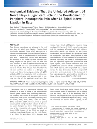
  • 7. Relationship of pERK-activated neurons and the distribution of normal and injured primary afferents of L5 spinal nerve In animals which had either L5 spinal nerve injury alone or followed by heat stimulation of the hind paw 3 days later, pERK immunoreactivity was activated in the dorsomedial part of the superficial dorsal horn of the spinal cord. In order to precisely locate the pERK immu- noreactivity in relation to spinal cord laminae, we first photographed unstained sections of L4 and L5 spinal segments in brightfield. The boundaries between lami- nae I, II, and III were clearly visible when sections were viewed with brightfield optics, due to the very translu- cent appearance of lamina II, compared with laminae I and III. To plot laminar boundaries on confocal images of pERK, CGRP, IB4, and VGLUT1 labeling in the spinal dorsal horn, sections were first scanned through a 103 objective lens on a Nikon confocal microscope (Nikon, Japan) and then brightfield images of each scanned section were superimposed over the corresponding Figure 1. Confocal images of spinal cord sections following noxious heat stimulus showing the relationship of activated pERK-positive cells (green in A and D) to NeuN (a neuronal marker, magenta in B) or Iba 1 (a marker for microglia, red in E), or GFAP (a marker for astro- cytes, blue in F). Panel C is merged image of and A and B. Panel G is merged image of D–F. pERK-positive cells (arrows in A) were colo- calized with NeuN (arrows in B,C) but not with Iba1 (E,G) or GFAP (F,G), indicating that noxious heat stimulus activated pERK in the spinal neurons and not in neuroglia. Images were built from projections of two confocal optical sections at 1 lm z-spacing. Scale bar 5 25 lm. [Color figure can be viewed in the online issue, which is available at wileyonlinelibrary.com.] Role of L4 nerve in neuropathic pain The Journal of Comparative Neurology | Research in Systems Neuroscience 1737
  • 8. Figure 2. Confocal images of transverse sections of L5 spinal cord segments showing pERK, IB4, CGRP, and VGlut1 labeling following nox- ious heat stimulus. The sections were photographed first using brightfield objectives to show the different laminae (A,F) and then stained with antibodies against pERK, IB4, CGRP, and VGlut1. IB4-labeled terminals arborize mainly in the middle of lamina II (C,I), whereas CGRP- labeled terminals are located in lamina I and outer lamina IIo (H). In comparison, VGlut1-labeled terminals are located in deep lamina IIi to V (D). (E) Merged image of B–D and panel (J) is merged image of G–I. Note the clear overlapping of pERK (B,G) with CGRP and IB4 and no overlapping with and VGlut1 immunoreactivity. Scale bar 5 200 lm. [Color figure can be viewed in the online issue, which is available at wileyonlinelibrary.com.] S. Shehab et al. 1738 The Journal of Comparative Neurology | Research in Systems Neuroscience
  • 9. projected confocal images using Adobe Photoshop and Xara designer Pro X software (Gaddesden Place, Great Gaddesden, Hemel Hempstead, Hertfordshire, UK). Lamina II was further divided into outer (IIo) and inner (IIi) layers by a line drawn half way between lamina I/II and II/III boundaries (Fig. 2). Upon labeling, pERK-positive neurons were found to be localized primarily in lamina I and IIo extending partially to lamina IIi. Very few less brightly labeled neurons were seen in the ventral laminae (III–VI) of the spinal cord. In order to find out the relationship between pERK-positive neurons and different primary afferents we used three markers (CGRP, IB4, and VGlut1) each for a specific type of afferent. Triple labeling showed that CGRP labeled unmyelinated C and thin myelinated Ad nerve terminals with a distinctive overlap with pERK-positive neurons in laminae I and II of the dorsal horn (Fig. 2F–J). As reported previously (Kitchener et al., 1994; Gerke and Plenderleith, 2004; Shehab and Hughes, 2011), IB4 preferentially labeled unmyelinated primary afferents in lamina I and II with a dense intensity in the middle of lamina II. Again, a clear overlapping of IB4 immunoreactiv- ity with pERK-positive neurons was observed (Fig. 2A–E). In comparison, VGlut1 labeled primary terminals of myelinated Ab fibers in the spinal cord which terminate in the lamina III and deeper laminae showed no overlap with pERK-positive neurons (Fig. 2A–E). Relationship of pERK-activated cells and transganglionically transported CTb and IB4 in central primary afferents of L5 nerve We used another method to investigate the relation- ship between activated pERK neurons and central Figure 3. Images of a transverse section of the spinal cord showing relationship of activated pERK (green) and IB4 (red) and CTb (blue) labeling in the L4 segment of a rat that had noxious heat stimulus to the hind paw. The rat had a coinjection of IB4 and CTb into ligated and transected L5 nerve 3 days earlier. Note that the termination of IB4-labeled terminals is confined mainly to the middle of lamina II (C) with partial overlapping with pERK (A), while the termination of CTb-labeled terminals are located in the deep laminae with no overlapping with pERK immunoreactivity (B). (D) Merged image of A–C. Scale bar 5 25 lm. [Color figure can be viewed in the online issue, which is available at wileyonlinelibrary.com.] Role of L4 nerve in neuropathic pain The Journal of Comparative Neurology | Research in Systems Neuroscience 1739
  • 10. termination of primary afferents of L5 in the dorsal horn. The main purpose of this experiment was to explore whether the pERK-positive cells in ipsilateral L4 spinal segment were in the territory of the central ter- mination of injured L5 nerve. Injection of a mixture of CTb and IB4 into L5 ligated spinal nerve was followed by 3 days of recovery and then heat stimulation was applied to the ipsilateral hind paw. In L4 and L5 spinal segments, pERK-labeled neurons were seen in the lami- nas I and II corresponding with IB4- and CTb-labeled terminals mediolaterally and rostrocaudally (Fig. 3). However, the pERK-labeled neurons showed overlap with IB4- (in lamina II) but not with CTb- (in lamina III) labeled nerve terminals. These results also demon- strated that pERK-positive neurons in the dorsal horn of L4 were in the territory of the central termination of L5 nerve in L4 segment. Relationship between noxious heat pERK activation and VIP upregulation followed by L5 injury We have previously shown that both sciatic and L5 nerve injury causes upregulation of VIP in nerve termi- nals in the lamina I and II of the spinal cord where these two nerves terminate (Shehab and Atkinson, 1986; Shehab et al., 2003, 2004; Shehab, 2014). In this study we used this VIP upregulation as a marker for injured unmyelinated primary afferents in L4 and L5 spinal segments. Seven days after L5 ligation and trans- ection, noxious heat was applied to the ipsilateral hind paw. The result showed that pERK-activated neurons were surrounded by VIP-labeled terminals in lamina I and II of the dorsal horn of the spinal cord (Fig. 4). In the contralateral side, only scattered positive VIP nerve terminals and no pERK cells could be detected. These results clearly demonstrated that the neuronal pERK activation in dorsal horn of spinal cord in response to heat noxious stimuli, which is most likely transmitted through L4 nerve, were in the same locations of central primary afferents of injured L5 nerve. Effects of peripheral nerve injury on pERK immunoreactivity We aimed in this study to investigate the effects of noxious heat stimulation on pERK immunoreactivity in an animal model of neuropathic pain in rats due to periph- eral nerve injury. Therefore, it was important first to dis- cover the effects of peripheral nerve injury itself on pERK expression without heat stimulation. No pERK immunore- activity was seen in untreated rats. However, 5–10 minutes after L5 nerve ligation and transection, pERK- positive cells were detected in the dorsal horn of L3–L6 spinal segments (Fig. 5). The mediolateral and rostrocau- dal distribution of pERK-positive cells were seen in exactly the same areas of the dorsal horn where unmyeli- nated primary afferents of L5 terminate (Shehab et al., 2008). As expected, no pERK reactivity was observed on the contralateral side of the spinal cord. Also, as men- tioned above, the pERK-positive cells were neurons, as they were also labeled with NeuN and not glia, as there was no colocalization with GFAP and Iba1 (not shown). Importantly, this pERK activation following peripheral nerve injury was temporary, as it disappeared completely after 3 days. This indicates that the pERK activation in the dorsal horn, which we observed after noxious heat stimulation applied to the hind paw 3 days following L5 transection, must be attributed to heat stimulation rather than nerve injury itself. Figure 4. Confocal images of a transverse section of spinal cord showing VIP (green in A) and activated pERK neurons (magenta in B) in the L5 segment following L5 spinal nerve ligation and transection 7 days before dipping hind paws in water at 52 C. (C) A merge of A,B. The pERK-activated neurons in response to noxious heat stimulation (which are likely due to stimulation of L4 primary afferents of the hind paw) intermingle with upregulated VIP in the central terminals of injured L5 primary afferent fibers. Images built from projections of three confocal optical sections at 2 lm z-spacing. Scale bar 5 25 lm. [Color figure can be viewed in the online issue, which is available at wileyonlinelibrary.com.] S. Shehab et al. 1740 The Journal of Comparative Neurology | Research in Systems Neuroscience
  • 11. pERK activation in response to noxious heat stimulation Figures 6 and 7 and Table 2 show the effects of the exposure of the hind paws to heat stimuli at 37–52 C on the number of pERK-labeled neurons in the dorsal horn. In this experiment the L5 nerve was ligated and trans- ected 3 days earlier to induce neuropathic pain in the ipsilateral hind paw. In a control No heat group of rats the animals had no heat stimulation. The numbers of pERK-positive neurons in the ipsilateral side of L3–L6 segment were compared with contralateral uninjured side. Interestingly, after 3 days of L5 nerve injury the No heat group of rats showed either few or no pERK- immunoreactive neurons in the ipsilateral side (Fig. 6A). This pERK immunoreactivity was not different from that observed in the contralateral side of the spinal cord Figure 5. Transverse sections of L3–L6 spinal segments showing the activation of pERK in response to unilateral L5 nerve ligation and trans- ection 10 minutes before sacrifice. The activated pERK cells are located mainly in laminae I–II of the ipsilateral dorsal horn of L3–L6 (A–D) in exactly the same areas where the unmyelinated primary afferents of L5 nerve terminate (see fig. 2 in Shehab et al., 2008). Arrows in A–C indicate that pERK activation would very likely take place in areas of the spinal cord where the dorsal rami of L3–L6 nerves terminate and were inevitably injured during the surgical operation in which a skin incision of the back was carried out to expose the L5 spinal nerve. Negli- gible pERK immunoreactivity can be detected in the spinal cord on the contralateral uninjured side (E–H). Scale bar 5 200 lm. Role of L4 nerve in neuropathic pain The Journal of Comparative Neurology | Research in Systems Neuroscience 1741
  • 12. (Fig. 6) or in naive animals. These results indicated that activation of pERK 5–10 minutes after peripheral nerve injury was reduced to its normal level 3 days later. In comparison, upon stimulation with 37 C water, a few more pERK-positive cells were seen in the ipsilat- eral dorsal horn which increased in number with the raise in temperature from 42 to 52 C (Fig. 6B–E), com- pared with the contralateral side (Fig. 6G–J). Statistical analysis of pERK-positive cell counts In order to quantify and estimate the number of pERK- positive cells in ipsilateral and contralateral sides of spi- nal segments (L3–L6) upon heat, a detailed statistical analysis was carried out. Mixed model analysis revealed that all fixed effects were statistically highly significant, including all two-way and three-way interactions Figure 6. Brightfield images of transverse sections of dorsal horns of L4 spinal segment, showing pERK immunoreactivity on the contralateral and ipsilateral sides of animals which had left L5 nerve ligation and transection 3 days earlier to induce neuropathic pain in the hind paw. Both hind paws were either subjected to no heat (A) or immersed in water at 37 C (B), 42 C (C), 47 C (D), and 52 C (E). Note the increase in pERK- positive neurons in the ipsilateral side (A–E) with increase in temperature compared with contralateral uninjured side (F–J). Scale bar 5 200 lm. S. Shehab et al. 1742 The Journal of Comparative Neurology | Research in Systems Neuroscience
  • 13. (P 0.001). In general, it explains that there was signifi- cant difference in the number of pERK-immunoreactive neurons on both sides (injured vs. uninjured) of all spinal cord segments (L3–L6) at all temperatures (including 52 C). The difference between both sides followed a lin- ear pattern with increasing temperature. To further elucidate the effects and to simplify inter- pretation, we divided the data into two groups and ran separate analysis for 1) temperatures below 52 C including 37, 42, and 47 C, 2) 52 C only. The same mixed model was utilized for temperatures below 52 C with all the terms, except heat52. The results expressed a highly significant difference between the injured and control side (P 0.001) with root cell counts on the treated side 0.45 units higher on average than on the control side. Also, it was observed that the L4 spinal segment had the highest number of root cell difference (0.71), which highlights the increased activa- tion of pERK in the L4 segment. This observation was consistent at all temperatures. When temperature 52 C was analyzed separately, it was observed that for L3 and L4 the ipsilateral side was significantly (P 0.001) higher than the contralat- eral side, in line with temperatures below 52 C. For L5 the pattern was reversed, with the ipsilateral side hav- ing significantly (P 0.001) lower counts than the con- tralateral side. The substantial change between both sides at 52 C was further evaluated by paired samples t-test which showed statistically significant difference (P 0.001) between the ipsilateral and contralateral side of all the spinal segments (L3–L6). We used the mean cell counts for graphical illustra- tion of the differences between the injured and unin- jured side of each spinal cord at various temperatures (No heat–52 C). The individual mean values for both sides at all temperatures are provided (Table 2). L4 has the maximum number of pERK-positive cells, which increase linearly with temperature (Fig. 7). However, L5 follows the same pattern until it approaches 52 C. At this temperature the contralateral side (control unin- jured) has more pERK-positive cells as compared to ipsilateral side (injured-L5 ligated). DISCUSSION In the present study we exploited prior observations (Ji et al., 1999; Polgar et al., 2007) that application of heat noxious stimuli to the normal hind paw causes acti- vation of pERK in the dorsal horn of the spinal cord. The principal findings of this study were: 1) We showed that pERK activation in response to both noxious heat stimu- lation and peripheral nerve injury were in spinal neurons and not glia. 2) The pERK heat-activated neurons were in clear overlap with CGRP immunoreactivity at lamina I and IIo, and IB4 in lamina II with no overlap with VGLUT1 or CTb transganglionically transported immunor- eactivities in laminae IIi and III. 3) The pERK heat- activated neurons in L4 and L5 intermingled with Figure 7. A histogram showing the comparison of mean of pERK-positive cell counts between contralateral uninjured side (Cont) and ipsi- lateral injured side (Ipsi) of L3–L6 spinal segments in response to stimulation of both hind paws to either no heat or various degrees of temperatures at 37, 42, 47, and 52 C. All rats had left L5 nerve ligation and transection 3 days earlier. A linear trend was observed in the increased number of pERK-positive neurons on injured ipsilateral side from 37–52 C. The number of positive pERK neurons was signifi- cantly higher in the ipsilateral side of L4 spinal segment, which receives innervation from both uninjured L4 and injured L5 nerves, com- pared with contralateral control side. Data are expressed as mean 6 SEM. [Color figure can be viewed in the online issue, which is available at wileyonlinelibrary.com.] Role of L4 nerve in neuropathic pain The Journal of Comparative Neurology | Research in Systems Neuroscience 1743
  • 14. upregulated VIP immunoreactive fibers as a marker of axotomized unmyelinated afferents of L5 nerve. 4) L5 nerve injury caused temporary activation of pERK in neu- rons in the L3–L6 spinal segments which disappeared completely after 3 days. 5) The number of pERK-positive neurons in the L4 dorsal horn was significantly more on the injured side compared with the contralateral side and directly proportional to the increase in the intensity of heat stimuli in rats that had their L5 nerve ligated and transected 3 days earlier. The study essentially reports the hypersensitivity of the uninjured L4 spinal nerve at the level of the dorsal horn of L4 segment after L5 nerve injury. These results provide direct anatomical evidence for the mechanism of the production and maintenance of heat hyperalgesia in the skin of hind paw in the SNL model of neuropathic pain in rats. These results will be discussed first in terms of tech- nical issues that may influence their interpretation. They will then be considered in the context of previous observations and conclude by considering its implica- tions for the possible mechanisms of neuropathic pain due to peripheral nerve injury. Effect of noxious heat stimulation on pERK immunoreactivity The activation of pERK along with other mitogen- activated protein kinases (MAPK) have been shown to play a significant role in development of neuropathic pain in different animal models including SNL (Crown, 2012). In agreement with previous reports (Ji et al., 1999; Polgar et al., 2007), 5 minutes after the immer- sion of the hind paws in hot water at 52 C, we observed numerous pERK-immunoreactive neuronal cells but not glia in the medial part of the ipsilateral dorsal horn in the L3–L6 spinal cord segment. The loca- tion of these activated pERK neurons corresponds well with the termination of the central primary afferents of the nerves which supply the hind paw in rats. These neurons were concentrated in a band that occupied lamina I and II, which clearly overlapped with both CGRP and IB4 labeled terminals. The location of the pERK neurons in this study is consistent with previous reports that showed the densest staining of transient receptor potential vanilloid 1 (TRPV1, as a receptor for capsaicin and a transducer of noxious thermal stimuli) in laminae I and II of the spinal cord (Tominaga et al., 1998; Guo et al., 1999; Valtschanoff et al., 2001). Effects of varying degrees of heat stimulation of the hind paws in neuropathic pain animals Having confirmed previous findings (Ji et al., 1999; Polgar et al., 2007) which showed pERK activation in TABLE2. MeanofpERKPositiveNeuronsinContralateral(Uninjured)andIpsilateral(Injured)SideoftheDorsalHornofL3-L6SpinalSegmentsatVariousTemperaturesofRatsWhich HadL5NerveLigationandTransection3DaysEarlier NoHeat37 C42 C47 C52 C ContIpsiContIpsiContIpsiContIpsiContIpsi L30.6460.101.9160.221.3660.172.8960.352.6860.354.8260.473.9760.657.6060.9821.2062.0030.0062.11 L41.1060.162.0160.221.6960.183.4460.353.2460.365.7560.5212.6361.3324.4861.9051.7662.1566.2262.77 L50.8160.111.5060.171.5060.252.2160.273.5660.334.5760.509.0061.4311.0761.2657.6262.541.8563.5 L61.4960.161.2960.151.5960.221.9760.253.0160.373.1760.312.6660.333.8960.555.4360.454.3260.49 Thedataareexpressedasmean6SEM. S. Shehab et al. 1744 The Journal of Comparative Neurology | Research in Systems Neuroscience
  • 15. response to noxious heat stimulation, in the next step we investigated the effects of different degrees of heat stimulation in animals which had L5 nerve ligation and transection 3 days earlier. The reason for leaving the animals for 3 days was due to previous observations that rats completely develop neuropathic pain manifes- tations after this postoperative period of time following L5 nerve injury (Kim and Chung, 1992). Our prediction was that noxious heat stimulation would result in an increase in pERK immunoreactivity in dorsal horn of the spinal cord on the ipsilateral injured side compared with the contralateral uninjured side. Indeed, the expression of pERK in the dorsal horn of L3–L6 spinal segments on the lesion side, over a range of heat stim- ulation (42–52 C), always exceeded that on the control uninjured side except in L5 at 52 C. This exceptionally amplified pain response in the uninjured side at 52 C is possibly because of intense peripheral noxious heat stimulation and its transmission through both intact L4 and L5 nerves, whereas on the injured side there was no sensory transmission through L5 nerve. Mechanisms of neuropathic pain There has been considerable debate about the mech- anisms of neuropathic pain after nerve injury and many hypotheses and potential explanations have been pro- posed (Yaksh and Sorkin, 2005; Campbell and Meyer, 2006; Devor, 2006), but it is more likely that a number of mechanisms are active in parallel and/or in sequence (Sandk€uhler, 2009). There is also debate about whether injured or uninjured primary afferents are responsible for neuropathic pain (Sheen and Chung, 1993; Gold, 2000; Li et al., 2000; Ringkamp and Meyer, 2005; Campbell and Meyer, 2006; Jang et al., 2007) and about the role of different classes of primary affer- ents (Ringkamp and Meyer, 2005; Campbell and Meyer, 2006). However, following the transection of L5 or L5 and L6 spinal nerves in the Chung model of neuropathic pain (Kim and Chung, 1992), the responsiveness to hind paw stimulation, including hyperalgesia and allody- nia, depends on the innervation of the adjacent unin- jured L4 primary afferents. In normal rats, L4 and L5 spinal nerves are the major source of skin innervation of the hind paw. Anatomi- cally, the unmyelinated primary afferents of L5 nerve terminate mainly in dorsal horn of L5 and L4 with a minor projection in L3 and L6 spinal segments (Shehab et al., 2008). Similarly, the central terminals of nerve L4 were found in both L4 and L3, again with less label- ing in L2 and L5 (Shehab et al., 2008). This means that neurons in dorsal horn of L4 spinal segment would receive primary afferents of both L4 and L5. Mathemati- cally, elimination of the central terminals of L5 from L4 spinal segment following L5 nerve injury would result in a reduction in the activation of spinal neurons in the L4 segment in response to painful stimuli applied to the hind paw. However, in the current study the reverse was observed. Noxious heat stimulation with increasing temperature showed an increase in nociception response revealed by an increase in pERK activation. This is consistent with the general belief that central spinal sensitization, following peripheral nerve injury, as the main explanation for the development of the neuro- pathic pain signs and symptoms (Ji et al., 2003; Camp- bell and Meyer, 2006; Latremoliere and Woolf, 2009). Additional evidence supporting the hypothesis that the uninjured primary afferents play a significant role in the development of neuropathic pain (Fukuoka et al., 2002; Ringkamp and Meyer, 2005; Meyer and Ring- kamp 2008) comes from electrophysiological studies (Wu et al., 2001, 2002; Shim et al., 2007; Meyer and Ringkamp, 2008). They demonstrated that the conduc- tion properties of the uninjured unmyelinated fibers in the L4 spinal nerve of the rat were altered following L5 spinal nerve lesion (Wu et al., 2001, 2002; Shim et al., 2007; Meyer and Ringkamp, 2008). Changes of L4 nerves may have been caused by Wallerian degenera- tion of L5 nerves and its direct effects on L4 nerves probably at the level of the sciatic nerve, where both nerves run together, or in the skin of the foot, where their terminations overlap. Their conclusion was that the interaction between degenerated injured nerves fibers and intact fibers of the adjacent nerve play a crit- ical role in the initiation and the maintenance of mechanical hyperalgesia (Wu et al., 2001, 2002; Shim et al., 2007; Meyer and Ringkamp, 2008). Furthermore, Fukuoka et al. (2012) reported that a brain-derived neurotrophic factor (BDNF) was upregu- lated in the uninjured neurons in the L4 DRG following L5 nerve injury. They proposed that the increase in this neuromodulator might contribute to the hypersensitivity induced by the L5 SNL. Although the results of this study do not rule out the possibility of the alteration in L4 of peripheral origin, previous work (Hammond et al., 2004) and recent data from our laboratory showed marked phenotypical changes, including upregulation of neuropeptide Y (NPY), VIP, and activating transcription factor 3 (ATF3) and downregulation of substance P (SP), CGRP, and IB4 binding in the injured neurons in L5 DRG with minimal changes in the uninjured neurons in L4 DRG following L5 nerve injury (Shehab, 2014). It was therefore concluded that these minor changes in neurons of L4 DRG do not seem to contribute to the development of neuropathic-like manifestations follow- ing L5 nerve injury (Hammond et al., 2004; Shehab, 2014). Our findings demonstrated that the phenotypical Role of L4 nerve in neuropathic pain The Journal of Comparative Neurology | Research in Systems Neuroscience 1745
  • 16. changes including upregulation of NPY, VIP, and neuro- kinin 1 receptor and downregulation of SP, CGRP, and IB4 binding following L5 nerve injury did not only occur in the dorsal horn of L5 segment where the injured (L5) enters, but also extended to two segments rostrally and one segment caudally (Shehab, 2014) where the unin- jured primary afferents of L4 nerve terminate (Shehab et al., 2008). Taken together, the data of the present study, which showed significantly more pERK activation, and from our previous work (Shehab, 2014), which showed neuroplastic changes in the dorsal horn where primary afferents of adjacent injured and uninjured nerves terminate, provide evidence for the mechanism of the initiation and maintenance of hyperalgesia in the SNL model of neuropathic pain. These spinal cord neuroplastic changes could be one of the most probable causes for central sensitization of the neurons in the denervated region of the dorsal horn after nerve injury (Ji et al., 2003; Campbell and Meyer, 2006; Latremoliere and Woolf, 2009). Since neurons in L4 spinal segment receive contacts from both L4 and L5 nerves (Shehab et al., 2008), their sensitization due to L5 injury would very likely contribute to exaggerated responses to noxious or subnoxious stimuli applied to the skin of the hind paw which is transmitted by the uninjured L4 nerve resulting in hyperalgesia. In conclusion, our data demonstrate the role of spinal neurons that receive primary afferents of both injured and uninjured nerves in the production of heat hyperal- gesia in peripheral neuropathic pain. CONFLICT OF INTEREST The authors declare no conflict of interest. ROLE OF AUTHORS All authors had full access to the data in the study and read and accepted the final version of the article. The roles of the authors: Study concept and design: SS. Acquisition of data: SS, MA, DG, AA, KA, AA-B, ST. Anal- ysis and interpretation of data: SS, MA, DG, NN. Writing the article: SS, MA, DG, NN, ML. Statistical analysis: SS, MA, DG, NN. Obtained funding: SS, ML. Carried out the experiments: SS, MA, DG, AA, KA, AA-B. Study supervision: SS. LITERATURE CITED Baquet ZC, Williams D, Brody J, Smeyne RJ. 2009. A compari- son of model-based (2D) and design-based (3D) stereo- logical methods for estimating cell number in the substantia nigra pars compacta (SNpc) of the C57BL/6J mouse. Neuroscience 161:1082–1090. Bennett GJ, Xie YK. 1988. A peripheral mononeuropathy in rat that produces disorders of pain sensation like those seen in man. Pain 33:87–107. B€olcskei K, Horvath D, Szolcsanyi J, Petho G. 2007. Heat injury- induced drop of the noxious heat threshold measured with an increasing-temperature water bath: a novel rat thermal hyperalgesia model. Eur J Pharmacol 564:80–87. Boucher TJ, Okuse K, Bennett DL, Munson JB, Wood JN, McMahon SB. 2000. Potent analgesic effects of GDNF in neuropathic pain states. Science 290:124–127. Campbell JN, Meyer RA. 2006. Mechanisms of neuropathic pain. Neuron 52:77–92. Chung K, Lee WT, Carlton SM. 1988. The effects of dorsal rhi- zotomy and spinal cord isolation on calcitonin gene- related peptide-labeled terminals in the rat lumbar dorsal horn. Neurosci Lett 90:27–32. Crown ED. 2012. The role of mitogen activated protein kinase signaling in microglia and neurons in the initiation and maintenance of chronic pain. Exp Neurol 234:330–339. Debus E, Weber K, Osborn M. 1983. Monoclonal antibodies specific for glial fibrillary acidic (GFA) protein and for each of the neurofilament triplet polypeptides. Differen- tiation 25:193–203. Decosterd I, Woolf CJ. 2000. Spared nerve injury: an animal model of persistent peripheral neuropathic pain. Pain 87: 149–158. Devor M. 2006. Response of nerves to injury in relation to neuropathic pain. In: McMahon SB, Koltzenburg M, edi- tors. Wall Melzack’s textbook of pain. London: Elsevier. p 905–927. Fukuoka T, Noguchi K. 2002. Contribution of the spared pri- mary afferent neurons to the pathomechanisms of neuro- pathic pain. Mol Neurobiol 26:57–67. Fukuoka T, Yamanaka H, Kobayashi K, Okubo M, Miyoshi K, Dai Y, Noguchi K. 2012. Re-evaluation of the phenotypic changes in L4 dorsal root ganglion neurons after L5 spi- nal nerve ligation. Pain 153:68–79. Gerke MB, Plenderleith MB. 2004. Ultrastructural analysis of the central terminals of primary sensory neurones labelled by transganglionic transport of Bandeiraea sim- plicifolia I-isolectin B4. Neuroscience 127:165–175. Gold MS. 2000. Spinal nerve ligation: what to blame for the pain and why. Pain 84:117–120. Guillery RW. 2002. On counting and counting errors. J Comp Neurol 447:1–7. Guo A, Vulchanova L, Wang J, Li X, Elde R. 1999. Immunocy- tochemical localization of the vanilloid receptor 1 (VR1): relationship to neuropeptides, the P2X3 purinoceptor and IB4 binding sites. Eur J Neurosci 11:946–958. Hammond DL, Ackerman L, Holdsworth R, Elzey B. 2004. Effects of spinal nerve ligation on immunohistochemically identified neurons in the L4 and L5 dorsal root ganglia of the rat. J Comp Neurol 475:575–589. Hughes DI, Polgar E, Shehab SAS, Todd AJ. 2004. Peripheral axotomy induces depletion of the vesicular glutamate trans- porter VGLUT1 in central terminals of myelinated afferent fibres in the rat spinal cord. Brain Res 1017:69–76. Jang JH, Kim KH, Nam TS, Lee WT, Park KA, Kim D-W, Leem JW. 2007. The role of uninjured C-afferents and injured afferents in the generation of mechanical hypersensitivity after partial peripheral nerve injury in the rat. Exp Neurol 204:288–298. Ji RR, Baba H, Brenner GJ, Woolf CJ. 1999. Nociceptive-spe- cific activation of ERK in spinal neurons contributes to pain hypersensitivity. Nat Neurosci 2:1114–1119. Ji R-R, Kohno T, Moore KA, Woolf CJ. 2003. Central sensitiza- tion and LTP: do pain and memory share similar mecha- nisms? Trends Neurosci 26:696–705. Kim SH, Chung JM. 1992. An experimental model for periph- eral neuropathy produced by segmental spinal nerve liga- tion in the rat. Pain 50:355–363. S. Shehab et al. 1746 The Journal of Comparative Neurology | Research in Systems Neuroscience
  • 17. Kitchener PD, Lapiz MD, Wilson P, Snow PJ. 1994. Transgan- glionic labelling of primary sensory afferents in the rat lumbar spinal cord: comparison between wheatgerm agglutinin and the I-B4 isolectin from Bandeiraea simpli- cifolia. J Neurocytol 23:745–757. LaMotte CC, Kapadia SE, Shapiro CM. 1991. Central projec- tions of the sciatic, saphenous, median, and ulnar nerves of the rat demonstrated by transganglionic transport of choleragenoid-HRP (B-HRP) and wheat germ agglutinin- HRP (WGA-HRP). J Comp Neurol 311:546–562. Latremoliere A, Woolf CJ. 2009. Central sensitization: a gener- ator of pain hypersensitivity by central neural plasticity. J Pain 10:895–926. Li Y, Dorsi MJ, Meyer RA, Belzberg AJ. 2000. Mechanical hyperalgesia after an L5 spinal nerve lesion in the rat is not dependent on input from injured nerve fibers. Pain 85:493–502. Liu X, Eschenfelder S, Blenk KH, J€anig W, H€abler H. 2000. Spontaneous activity of axotomized afferent neurons after L5 spinal nerve injury in rats. Pain 84:309–318. Llewellyn-Smith IJ, Minson JB. 1992. Complete penetration of antibodies into vibratome sections after glutaraldehyde fix- ation and ethanol treatment: light and electron microscopy for neuropeptides. J Histochem Cytochem 40:1741–1749. Meyer R, Ringkamp M. 2008. A role for uninjured afferents in neuropathic pain. Acta Physiol Sin 60:605–609. Mullen RJ, Buck CR, Smith AM. 1992. NeuN, a neuronal spe- cific nuclear protein in vertebrates. Development 116: 201–211. Ossipov MH, Lai J, Porreca F. 2006. Mechanisms of experi- mental neuropathic pain: integration from animal models. In: McMahon SB, Koltzenburg M, editors. Wall Melzack’s textbook of pain. London: Elsevier. p 929–946. Polgar E, Campbell AD, MacIntyre LM, Watanabe M, Todd AJ. 2007. Phosphorylation of ERK in neurokinin 1 receptor- expressing neurons in laminae III and IV of the rat spinal dorsal horn following noxious stimulation. Mol Pain 3:4. Ringkamp M, Meyer RA. 2005. Injured versus uninjured affer- ents: who is to blame for neuropathic pain? Anesthesiol- ogy 103:221–223. Robertson B, Grant G. 1985. A comparison between wheat germ agglutinin-and choleragenoid-horseradish peroxidase as anterogradely transported markers in central branches of primary sensory neurones in the rat with some observa- tions in the cat. Neuroscience 14:895–905. Saade NE, Jabbur SJ. 2008. Nociceptive behavior in animal models for peripheral neuropathy: spinal and supraspinal mechanisms. Prog Neurobiol 86:22–47. Sandk€uhler J. 2009. Models and mechanisms of hyperalgesia and allodynia. Physiol Rev 89:707–758. Saper CB. 1996. Any way you cut it: a new journal policy for the use of unbiased counting methods. J Comp Neurol 364:5. Seltzer Z, Dubner R, Shir Y. 1990. A novel behavioral model of neuropathic pain disorders produced in rats by partial sciatic nerve injury. Pain 43:205–218. Sheen K, Chung JM. 1993. Signs of neuropathic pain depend on signals from injured nerve fibers in a rat model. Brain Res 610:62–68. Shehab SAS. 2009. Acute and chronic sectioning of fifth lum- bar spinal nerve has equivalent effects on the primary afferents of sciatic nerve in rat spinal cord. J Comp Neu- rol 517:481–492. Shehab SAS. 2014. Fifth lumbar spinal nerve injury causes neurochemical changes in corresponding as well as adja- cent spinal segments: a possible mechanism underlying neuropathic pain. J Chem Neuroanat 55:38–50. Shehab SA, Atkinson ME. 1986. Vasoactive intestinal polypep- tide (VIP) increases in the spinal cord after peripheral axotomy of the sciatic nerve originate from primary affer- ent neurons. Brain Res 372:37–44. Shehab SAS, Hughes DI. 2011. Simultaneous identification of unmyelinated and myelinated primary somatic afferents by co-injection of isolectin B4 and cholera toxin subunit B into the sciatic nerve of the rat. J Neurosci Methods 198:213–221. Shehab SA, Cronly-Dillon JR, Nona SN, Stafford CA. 1990. Preferential histochemical staining of protoplasmic and fibrous astrocytes in rat CNS with GFAP antibodies using different fixatives. Brain Res 518:347–352. Shehab SA, Spike R, Todd A. 2003. Evidence against cholera toxin B subunit as a reliable tracer for sprouting of pri- mary afferents following peripheral nerve injury. Brain Res 964:218–227. Shehab SAS, Spike RC, Todd AJ. 2004. Do central terminals of intact myelinated primary afferents sprout into the superficial dorsal horn of rat spinal cord after injury to a neighboring peripheral nerve? J Comp Neurol 474:427– 437. Shehab SAS, Al-Marashda K, Al-Zahmi A, Abdul-Kareem A, Al- Sultan MAH. 2008. Unmyelinated primary afferents from adjacent spinal nerves intermingle in the spinal dorsal horn: a possible mechanism contributing to neuropathic pain. Brain Res 1208:111–119. Shehab S, Anwer M, Galani D, AbdulKarim A, Al-Nuaimi K, Al- Baloushi A, Ljubisavljevic M. 2013. A possible role of uninjured adjacent L4 nerve in the development of peripheral neuropathic pain after L5 spinal nerve ligation in rats. Reg Anesth Pain Med 38(Suppl PP)E1–E259. Shim B, Ringkamp M, Lambrinos GL, Hartke T V, Griffin JW, Meyer RA. 2007. Activity-dependent slowing of conduc- tion velocity in uninjured L4 C fibers increases after an L5 spinal nerve injury in the rat. Pain 128:40–51. Todd AJ. 2010. Neuronal circuitry for pain processing in the dorsal horn. Nat Rev Neurosci 11:823–836. Todd AJ, Spike RC, Polgar E. 1998. A quantitative study of neurons which express neurokinin-1 or somatostatin sst2a receptor in rat spinal dorsal horn. Neuroscience 85:459–473. Tominaga M, Caterina MJ, Malmberg AB, Rosen TA, Gilbert H, Skinner K, Raumann BE, Basbaum AI, Julius D. 1998. The cloned capsaicin receptor integrates multiple pain- producing stimuli. Neuron 21:531–543. Valtschanoff JG, Rustioni A, Guo A, Hwang SJ. 2001. Vanilloid receptor VR1 is both presynaptic and postsynaptic in the superficial laminae of the rat dorsal horn. J Comp Neurol 436:225–235. Wang H, Rivero-Melian C, Robertson B, Grant G. 1994. Trans- ganglionic transport and binding of the isolectin B4 from Griffonia simplicifolia I in rat primary sensory neurons. Neuroscience 62:539–551. Wang H, Dai Y, Fukuoka T, Yamanaka H, Obata K, Tokunaga A, Noguchi K. 2004. Enhancement of stimulation-induced ERK activation in the spinal dorsal horn and gracile nucleus neurons in rats with peripheral nerve injury. Eur J Neurosci 19:884–890. Wu G, Ringkamp M, Hartke TV, Murinson BB, Campbell JN, Griffin JW, Meyer RA. 2001. Early onset of spontaneous activity in uninjured C-fiber nociceptors after injury to neighboring nerve fibers. J Neurosci 21:RC140. Wu G, Ringkamp M, Murinson BB, Pogatzki EM, Hartke TV, Weerahandi HM, Campbell JN, Griffin JW, Meyer RA. 2002. Degeneration of myelinated efferent fibers induces spontaneous activity in uninjured C-fiber afferents. J Neurosci 22:7746–7753. Yaksh T, Sorkin L. 2005. mechanisms of neuropathic pain. Curr Med Chem-Central Nerv Syst Agents 129–140. Role of L4 nerve in neuropathic pain The Journal of Comparative Neurology | Research in Systems Neuroscience 1747