SlideShare a Scribd company logo
1 of 107
Trauma From Occlusion
DR.SWAPNA EDIGA
By-
DR.SWAPNA.E
Contents
 Introduction
 Definition & Terms
 Classification
 Stages of tissue response to
increased occlusal forces
 Role of occlusion in pathogenesis
of Periodontal disease
 Human studies & Clinical trials
 Animal experiments
 TFO & Plaque Associated
Periodontal Disease
 Diagnosis
 Treatment
 Conclusion
 References
Introduction
• To function occlusal harmony-
masticatory apparatus- teeth &
supporting tissues, TMJ & associated
neuromuscular skeletal structures
must operate in an integrated &
dynamic manner
• Loss of integrated function &
homeostasis in response to functional
demand may lead to exacerbation of
existing periodontal condition.
Angle defined occlusion as the normal relation of the occlusal inclined
planes of the teeth when the jaws are closed
Teeth transmit occlusal forces- mastication &
swallowing
Magnitude of occlusal forces controlled by
muscles of mastication.
Majority of occlusal forces are directed axially.
The periodontium is designed to resist axial
forces but some lateral forces can be tolerated
Definitions- Trauma from Occlusion
• A condition where injury results to the supporting structures of the
teeth by the act of bringing the jaws into closed position- (Stillman
1917 )
• When the occlusal forces exceed the adaptive capacity of the
periodontal tissues, tissue injury results. The resultant injury is termed
as trauma from occlusion.
(Orban and Glickman 1928,1933 )
• Damage in the periodontium caused by stress on the teeth produced
directly or indirectly by teeth of opposing jaw”
(WHO 1978)
• “An injury to the attachment apparatus as a result of excessive occlusal
forces”
(Glossary of Periodontic terms AAP 2001)
Traumatogenic Occlusion
• Any occlusion (e.g malocclusion) that produces forces that cause an
injury to the attachment apparatus.
• Other terms:
▫ Traumatic occlusion
▫ Occlusal disharmony
▫ Functional imbalance
▫ Occlusal dystrophy
Occlusion Traumatism
• The overall process by which a traumatogenic occlusion produces
injury in the periodontal attachment apparatus.
• Traumatizing Occlusion may act on individual tooth/groups of teeth
• In premature contact relationship
• In conjunction with parafunction- clenching, bruxism
• In conjunction with loss of migration of premolars & molar teeth with
gradually developing spread of anterior of teeth of maxilla
Adaptive capacity of Periodontium to
occlusal forces
• Varies in different persons & in same person at different times
• Influenced by
Magnitude : when magnitude of occlusal forces
is increased ,there is widening of PDL space,
increase in No.& width of PDL fibers, increase
in density of alveolar bone.
Direction: changing direction of occlusal forces
s re-orientation of stresses & strains within
periodontium(Glickman 1970). Principal fibers
accommodate occlusal forces along long axis of
tooth. Lateral(horizontal) forces &
torque(rotational) forces are more likely to
injure periodontium.
Duration : constant pressure on bone is more
injurious than intermittent forces.
Frequency: more frequent intermittent forces
more injurious .
Physiologically normal occlusal forces in chewing and swallowing :
• Positive stimulus in maintaining the periodontium and the
• Alveolar bone in a healthy and functional condition.
Impact forces : mainly high & short duration periodontium can sustain
high forces for short period
• Exceeding the viscoelastic buffer capacities of PDL results  in loss of
tooth and bone.
Continuous forces :Very low forces (eg, orthodontic forces),
• continuous in one direction  displacing tooth by remodeling
alveolus.
Jiggling forces : Intermittent forces in 2 directions (premature contacts
eg, crowns, fillings)
result in widening of the alveolus and increased mobility.
TYPES OF OCCLUSAL FORCES
Forces in one direction cause tipping of the tooth in the opposite direction or tooth displacement
parallel to the force resulting in a ‘bodily movement . The clinical result is a (temporary) increased
mobility.
However, there are no changes in the supracrestal fibres, no loss of periodontal attachment, or an
increased probing pocket depth. The increased tooth mobility is functional adaptation to the forces
exerted on that tooth.
In the compression zone, pressure stimulates osteoclasts in the adjacent bone and the alveolar wall
is resorbed until a new connection is formed with the hyalinised bone (‘undermining resorption ’).
In the tension zone, bone apposition and rupture of the collagen fibres occur. After removal of the
force the periodontal ligament is reorganised and after some time develops a normal histological
appearance.
Forces too high (above the adaptation level),
aseptic necrosis of the PDL
(hyalinisation)
root resorption occur
resulting in shorter roots.`
Jiggling forces
different and opposite directions.
Therotically same events occurs. However, they are
not clearly seperated.
Histologically :
Aposition & resorption on either sides of PDL 
widening of PDL space ( observed on radiographs)
This phenomenon
the increased mobility
without pocket formation,
migration and tipping.
Hypermobility
as long as the forces are exerted on the tooth
not adaptation.
not a sign of an ongoing process
but result of a previous jiggling force.
Reasons of deflective occlusal
forces.
Patterns of mastication,
Loss of teeth,
Loss of periodontal support,
Dental caries,
Faulty restorations
Flawed orthodontics,
Defective occlusal adjustment,
Occlusal habits,
Inadequate form and position of the
teeth.
EFFECTS OF OCCLUSAL DISHARMONY
Discomfort in the region of the TMJ
Food impaction
Abnormal habits
Pain in and around teeth with improper restorations
Pulpal disturbances
Facial pain of obscure origin
Periodontal abscess
Cheek biting
Sensitivity of occlusal/incisal surfaces of teeth due to their abnormal
wear
Mobility of teeth
Gingival enlargement, Gingival recession
Bruxism; due to faulty occlusion, Unilateral mastication
Restricted excursion of the mandible resulting in insufficient wear
Extreme excursions of the mandible resulting in excessive wear.
Migration of upper and lower teeth
Terminologies
• Occlusal trauma can be divided into 3 general categories:
Occlusal Trauma: An injury to the attachment apparatus
as a result of excessive occlusal force . Occlusal trauma is
the tissue injury, and not the occlusal force.
• Injury resulting from excessive occlusal forces
applied to a tooth or teeth with normal support .
Examples include high restorations, bruxism,
drifting or extrusion into edentulous spaces, and
orthodontic movement.
Primary Occlusal
Trauma:
• Injury resulting from normal occlusal forces applied
to a tooth or teeth with inadequate support .
Secondary Occlusal
Trauma:
• Injury from an excessive occlusal force on a
diseased periodontium . In this case, there is
gingival inflammation, some pocket formation, and
the excessive occlusal forces are generally from
parafunctional movements.
Combined Occlusal
Trauma:
• Traumatogenic Occlusion: Any occlusion that produces forces that cause
an injury to the attachment apparatus.
• Occlusal Traumatism: The overall process by which a traumatogenic
occlusion produces injury in the periodontal attachment apparatus.
TYPES
Trauma from
occlusion
Acute
External force
Chronic
Primary
Secondary
ACUTE TRAUMA
• Acute TFO develops from
• An abrupt occlusal impact biting on a hard
object.
• Restorations, prosthetic appliances that interfere
with or alter the direction of occlusal force
• Clinical features
• Tooth pain, sensitivity to percussion, and increased
tooth mobility.
• Outcome –
• Injury heals and the symptoms subside if the force
is dissipated 1) shift in the position of the tooth or
2) wearing away or correction of the restoration.
• Injury may worsen and develops into necrosis
accompanied by periodontal abscess formation, or
• Persist as a symptom-free chronic condition
• Result in cemental tears.
CHRONIC TRAUMA
• More common and is of greater clinical significance
• Develops from-
• Gradual changes in occlusion produced by
• 1) Tooth wear,
• 2) Drifting ,
• 3) Extrusion ,
• 4) With parafunctional habits such as bruxism and
clenching, rather than as a sequel of acute TFO.
PRIMARY TFO
• When TFO is the result of alterations in occlusal
forces
• Insertion of a “high filling”
• Insertion of a prosthetic replacement that creates
excessive forces on abutment and antagonist teeth.
• Orthodontic movement of teeth into functionally
unacceptable positions.
• Drifting movement or extension of teeth into spaces
created by unreplaced missing teeth, OR
• Changes produced
• Do not alter the level of connective tissue
attachment and do not initiate pocket formation.
• Probably because the supracrestal gingival fibers
are not affected and therefore prevent apical
migration of the junctional epithelium.
SECONDARY TFO
•Occurs when the adaptive capacity of the tissues to withstand occlusal forces is impaired by
bone loss resulting from marginal inflammation.
• This reduces the periodontal attachment area and alters the leverage on the remaining
tissues.
•The periodontium becomes more vulnerable to injury and previously well-tolerated
occlusal forces become traumatic.
Stages of Tissue response to increased
occlusal forces
• 3 stages:
• Stage I: injury
• Stage II: repair
• Stage III: adaptive remodeling of periodontium
Stage I: Injury- increase in areas of
resorption, decrease in bone formation
Injury- excess occlusal forces
Repair- forces are diminished.
Furcation- most
susceptible to
injury
Histological:
temporary
depression in
mitotic activity &
rate of proliferation
& differentiation of
fibroblasts in
collagen formation
& bone formation.
Slightly excessive pressure: PDL widening,
bone resorption.
Slightly excessive tension: elongation of PDL
fibers, Apposition of alveolar bone
Severe pressure: pressure forces root to bone-
undermining resorption: necrosis of PDL &
bone.
Severe tension:PDL widening,
thrombosis,hemorrhage, PDL tearing,bone
resorption.
• Increased pressure areas: blood vessels numerous & reduced in size.
• Areas of increased tension: enlarged blood vessels(Zaki 1963).
• Greater pressure:
▫ compression of PDL fibers, areas of hyalinization(Rygh 1974).
▫ injury to fibroblasts & connective tissue –necrosis of ligament.
▫ Vascular changes:-within 30 mins-impairment & stasis of blood flow,
▫ 2-3 hours blood vessels packed with erythrocytes fragment ,
▫ 1- 7 days disintegration of blood vessels & release contents into surrounding tissue.
▫ Increased resorption of alveolar bone & tooth surface occur.
Histoligicaly
Stage II : Repair-decreased resorption,
increased bone formation
• Damaged cells are removed & new CT cells & fibers , bone , Cementum
are formed in an attempt to restore injured periodontium
• Buttressing bone formation:
• Central: occurs within jaw. Endosteal cells deposit new bone, restore the
bony trabeculae, reduces the size of marrow spaces.
• Peripheral: on bone surface.On facial & lingual bony plates.Lipping-
Shelf like thickening of alveolar margin
Stage III: Adaptive remodeling of periodontium:
resorption & formation return to normal
• If repair process does not keep pace with destruction caused by occlusion,
periodontium is remodeled in an effort to create structural relationship
• Thickened PDL- funnel shaped at crest, angular defects in bone with no
pocket formation. Tooth is loose. Increased vascularization
Role of occlusion in pathogenesis of
Periodontal Disease
• Over 100 years occlusal trauma is associated with PD
• 1901 Karolyi: Association excessive occlusal forces & Periodontal
destruction
• 1917-1926:Stillman- excessive occlusal force was primary cause of PD
• End of 1930: Excessive occlusal forces were a causative factor for PD &
Occlusal discrepancies should be prophylactically treated to prevent PD
• 1933- Orban & Weinman , 1941- Weinmann- effect of excessive occlusal
forces on periodontium and Concluded -: no relationship & cause was
gingival inflammation
• 1950’s- 1960’s( Stahl 1962,Weinmann 1954,Ramjford 1971,) animal
research- rats ,monkeys, dogs- effect of occlusal forces on periodontium:
results : no relation
Glickman & co workers 1960
• 1960 Glickman & co workers : animal models & human Autopsy material :
dogs & monkeys high restorations : no association
• Rhesus monkeys & Human autopsy models: phenomenon :- “Altered
pathway of destruction” when occlusal forces were present.
• Altered pathway of destruction : change in orientation of periodontal &
gingival fibers which occurred in presence of excessive gingival
inflammation to extend along PDL
• This pathway caused vertical bony defects
Glickman & co workers conclusion
• Excessive forces in presence of plaque associated inflammation caused a
change in the alignment of periodontal ligament ,allowing a altered
pathway of inflammation/ destruction resulting in vertical bony defects
• Process was termed “ Co destructive effect”
• Zone of irritation: marginal & interdental
gingiva. gingival inflammation cannot be
induced by TFO but is result of irritation from
microbial plaque. First involvement of alveolar
bone & later PDL. Progression : an even
(horizontal)bone destruction
• Zone of co-destruction:
• PDL, Cementum, Alveolar bone. Coronally
demarcated by trans-septal (interdental) &
dento-alveolar collagen fiber bundles.the
tissue in this zone becomes a lesion of TFO
• Fiber bundles which separate zone of co
destruction affect from 2 directions:
1. Inflammatory lesion maintained by plaque in
zone of irritation
2. Trauma induced changes in zone of co-
destruction
Glickmans Concept (1965,1967)
Periodontal structures divided into 2 zones
Waerhaug(1979) concept
• “Plaque front”- was always in close approximation to epithelial attachment
level & always followed morphology of bony defect.
• Also found excesssive occlusal forces bore no relationship to underlying
bony defect & that vertical bony defects were found equally around
traumatized & non traumatized teeth
• Conclusion: bone loss was always associated with down growth of plaque
& there was no relationship between excessive occlusal forces & vertical
bone loss.
2 extensive studies- Polson & Lindhe( 1970)
Polson (1982)& co workers Lindhe (1982)& co workers
• squirrel monkeys-
• mesiodistal compression forces
comparable to orthodontic forces
• beagle dogs,
• buccolingual forces using a high
occlusal contact and a finger
spring
Effect of plaque & excessive occlusal forces in animal models
Results: similar results
Conclusion: excessive occlusal forces in absence of plaque cause loss of bone
density & mobility of affected tooth but no evidence was found that occlusal
force alone could cause attachment loss
Conclude: excessive force alone does not cause loss of attachment, with plaque it
causes.
Human studies & clinical trials
• World workshop in Periodontics “prospective studies on effect of occlusal
forces on progression of Periodontics are not ethically acceptable in
humans” Gher 1996
• Human studies are limited to retrospective & observational research.
Rosling et al 1976 Advanced PD, multiple
vertical defects, mobile teeth
subjected to antimicrobial
therapy
Infrabony pocket at Hyper
mobile teeth exhibited same
degree of healing as firm
teeth
Fleszar et al 1980 Tooth mobility on healing
following periodontal therapy
Pockets of clinically mobile
teeth do not respond well to
those of firm teeth exhibiting
same degree of disease
severity.
Philstrom et al 1986 TFO & periodontitis Teeth with increase mobility
had deeper pockets,
attachment loss, bone loss
compared to teeth without.
Clinical trials
• Wang & Ramjford 1994-molars with furcation involvement & mobility
have greater probing depths compared to molars that are clinically
immobile.
• Ramjford & Burgett 1992: patients who received occlusal adjustment as
part of their periodontal treatment had greater attachment gain than patients
who did not.
• These studies suggest that occlusal adjustment should be performed where
indicated as part of periodontal treatment
• Mc Guire & Nunn 1996: mobility & para functional habits that are not
treated with biteguard are associated with increased attachment loss,
worsening prognosis & tooth loss.
• Cortelleni & Tonetti 2001- mobile teeth treated with regenerative surgery
did not respond compared to non mobile teeth.
• Harrel & Nunn 2001: occlusal discrepancies appeared to be a significant
risk factor that contribute to more rapid periodontal destruction & that
treatment of occlusal discrepancies seemed to slow periodontal destruction.
Animal Experiments
Orthodontic type trauma
Muhlemann 1961, Waerhaug 1966, karring 1982
Teeth exposed to unilateral forces of magnitude, frequency/duration
that they are unable to withstand
Certain well defined reactions develop in PDL leading to bone
resorption.
Jiggling type trauma
• Healthy periodontium with normal height
Supralaveolar CT was not influenced by occlusal
forces:
reason: tissue compartment is
bordered by hard tissue only on one side- gingiva
was non
inflamed at start of experiment remained non
inflamed
& not aggravated by jiggling forces.
Healthy periodontium
Reduced height
Within certain limits a healthy
periodontium with reduced height has
capacity similar to periodontium of
normal height to adapt to altered
functional demands
Plaque associated Periodontal disease
Occlusal forces which allow adaptive
alterations to develop in the
pressure/tension zone of PDL will not
aggravate a plaque associated PD
Lindhe & Svanberg
1974
If magnitude & direction of occlusal
forces are not adapted by tissues-injury in
Co-destruction is permanent. Angular
bone loss continues Progressive mobility
Plaque associated lesion in area of
irritation & inflammatory lesion in area
of co- destruction merged, the epithelium
proliferated in an apical direction & PD
aggravated.
Effects of insufficient occlusal forces
• May also be injurious
• Insufficient stimulation causes thinning of PDL
• Atrophy of fibers
• Osteoporosis of alveolar bone
• Reduction in bone height
• Hypo function causes:
▫ Open bite relationship
▫ Absence of functional antagonists
▫ Unilateral chewing habits
Reversibility of traumatic lesions
TFO is reversible
The injurious force must
be relived for repair to
occur(Glickman 1961,
Polson 1976).
Presence of inflammation
due to plaque
accumulation in
periodontium may impair
reversibility of traumatic
lesions
Effects of excessive occlusal forces on
pulp
Disappearance of pulpal
symptoms after correction
of excessive occlusal
forces
Animal subjects(Cooper
1971, Landay 1970):
pulpal reactions to
increased occlusal forces.
No response to minimum
or short periods.
Diagnosis Clinical
symptoms
Clinical signs
Examination
Increased tooth
mobility is not
always indicative
of trauma from
occlusion.
It is important,
however, that
hypermobility
which does occur
as a result of
trauma from
occlusion is
detected in
patients with
reduced
periodontal
attachment.
The reason for
this is that trauma
from occlusion
may accelerate
further reduction
in attachment in a
patient with
active
periodontitis.
A clinical
diagnosis of
occlusal trauma
can only be
confirmed where
progressive
mobility can be
identified
through a series
of repeated
measurements
over an extended
period.
This means that
simple but
reliable
monitoring needs
to be undertaken.
A simple
monitoring
protocol is
needed
• SIGNS
MOBLITY + FREMITUS
CHANGE IN
PERCUSSION
MIGRATION WEAR PATTERNS PDLABCESS
GINGIVAL
CHANGES
HYPERTONICITY
OF MUSCLES
Radiographic features
1. Widening PDL space, thickening of lamina dura in lateral aspect of
root, in apical regions & bifurcation areas.
2. Vertical rather than horizontal destruction of interdental septum
3. Radiolucency & condensation of alveolar bone.
4. Root resorption
Detection of occlusal trauma clinically
• Analysis of patients occlusal relationship- Angle’s classification.
• The relationship between cusps is the most important factor in transmittal
of occlusal forces to the periodontal structures.
• Initial contact between teeth is detected by gentle manipulating patient's
mandible into retruded position
• Retruded position: both right & left condyles are firmly placed in fossa of
TMJ
• Patient is asked to close until patient feels first contact between teeth.
• Centric relation: initial contact in retruded position.
• Patient is asked to continue to close the jaws together until maximum
contact between the teeth is achieved.
• Centric occlusion: jaw position of maximum tooth contact
Treatment
• Occlusal adjustment
• Management of parafunctional habits
• Temporary, provisional or long term stabilization of mobile teeth with
appliances
• Orthodontic tooth movement
• Occlusal reconstruction
• Extraction of selected teeth
Occlusal adjustment (Selective Grinding)
• Non reversible. Reshaping of occlusal surfaces to create harmonious
contact relationships between the upper and lower teeth.
• Thorough occlusal analysis must be embarked:
• Clinical assessment of occlusion:- comprehensive assessment of teeth
• Interarch relationships, jaws & associated muscles TMJ & their
movements,
• examination of articulate study casts: semi adjustable articulator with
models mounted in retruded position
Occlusal adjustment
Indications Contraindications
• To reduce traumatic forces to
teeth
• To reduce damage from
parafunctional habits
• To reshape teeth: reduce soft
tissue injury
• To reduce food impaction
• Occlusal adjustment without
▫ pre-treatment study & patient
education
▫ Signs & symptoms of occlusal trauma
▫ Microbial induced inflammatory PD
treatment.
• Bruxism Rx based on patient history
without evidence of damage,
pathosis/pain
• When patients emotion precludes
satisfactory result.
• Treatment for severe extrusion, mobility/
malposition teeth that would not respond
to occlusal adjustment alone.
Equilibrating mobile teeth
• In a patient with mobile teeth, it may be necessary to temporarily
stabilise those teeth before equilibration is possible.
• If a tooth is mobile, it is very difficult if not impossible to
effectively modify its shape with the aim of reducing the occlusal
forces acting upon it (equilibration).
• Whether the inflammatory periodontitis has been treated
successfully.
• If there is an inflammatory periodontal process this should
be treated initially.
• Subsequently when the periodontal condition is stable,
occlusal therapy may be necessary for some patients and
could involve either occlusal equilibration or splinting.
• The radiographic appearance of the periodontal support.
occlusal equilibration is indicated will depend upon:
• Occlusal equilibration is considered an effective form of therapy for
teeth with increased mobility which has developed together with an
increase in the width of the periodontal ligament (PDL).
• Reducing the occlusal interference on a tooth with normal bone support
will normalize the width and height of the PDL.
• Eliminating any occlusal interferences for a tooth which has a reduced
bone height as a result of periodontal disease will result in bone
formation and remodelling of the alveolus only to the pre-trauma level
• It is generally accepted that occlusal adjustment directed solely at
establishing an ideal conceptualized pattern is contraindicated.
• Rather, it should only be performed when the objective is to facilitate
treatment or intercept actively destructive forces.
• When occlusal therapy is planned as part of periodontal treatment, it
is usually deferred until initial therapy aimed at minimizing
inflammation throughout the periodontium has been completed.
• This is based upon the fact that inflammation alone can contribute
significantly to a tooth’s mobility.
Splinting
1) Stabilize teeth with increasing
mobility that have not responded to
occlusal adjustment and periodontal
treatment.
2) Stabilize teeth with advanced
mobility that have not responded to
occlusal adjustment and treatment
when there is interference with normal
function and patient comfort.
3) Facilitate treatment of extremely
mobile teeth by splinting them prior to
periodontal instrumentation and
occlusal adjustment procedures.
4) Prevent tipping or drifting of teeth
and extrusion of unopposed teeth.
5) Stabilize teeth, when indicated,
following orthodontic movement.
6) Create adequate occlusal stability
when replacing missing teeth.
7) Splint teeth so that a root can be
removed and the crown retained in its
place.
8) Stabilize teeth following acute
trauma.
1) When the treatment of
inflammatory periodontal
disease has not been
addressed.
2) When occlusal
adjustment to reduce
trauma and /or
interferences has not
been previously
addressed.
3) When the sole
objective of splinting is
to reduce tooth mobility
following the removal of
the splint.
Contra-indications
This means that if periodontal treatment
results in a stable periodontal condition
which is comfortable, splinting is not
needed.
OCCLUSAL PARAFUNCTION: BRUXISM
• Bruxism : clenching or grinding of the teeth when the individual is
not chewing or swallowing – Ramfjord and Ash - 1966
Bruxism can occur as
brief, rhythmic strong
contractions of the jaw
muscles during
eccentric lateral jaw
movements or in
maximum
intercuspation, which is
called clenching.
Bruxism may also take
the form of tapping.
Bruxism often occurs
without any neurologic
disorders or defects and
can be viewed as a
phenomenon present in
healthy individuals.
Bruxism may lead to
tooth wear, fractures of
the teeth or dental
restorations, or
uncosmetic muscle
hypertrophy.
 Presence of tooth wear in patients is not necessarily the cause for signs and
symptoms of TM disorders.
 Data from Rugh indicate that 83% of a group of bruxers, performed bilateral
muscle contraction, whereas 17% performed unilateral contractions.
Nocturnal bruxism & Daytime (diurnal) bruxism.
 Most people are not aware of a bruxism habit until it is brought to their
attention.
 When active tooth gnashing occurs, the enamel rods are fractured and
become highly reflective to light.
Thus shiny, bright facets
TREATMENT OF BRUXISM
1. Behavioural modality – explanation and arousal of the patients awareness
of the habit.
• If pain and stiffness are associated, physical therapy.
• Anti-anxiety drugs..
• Ware – 1982 – advocated use of antidepressants as a means of inhibiting
the REM sleep.
2. Night guard appliance (max stabilization splint).
• Aim is to protect the tooth surface and to dissipate forces.
• More practical for treating nocturnal bruxism..
• Splints are more significant in the management of the destructive effects of
bruxism.
• Patient instructed to wear the splint during sleep.
• Should be adjusted again in 2-3 weeks..
• Should be observed for bruxofacets.
• If present should be burnished away.
• Balancing should be completed before patient dismissal
3. Coronoplasty – recently placed dental restorations or other occlusal
treatments.
• Occlusal adjustment, reconstruction, or orthodontic
treatment are contraindicated as a means of
controlling bruxism.
SELECTIVE
GRINDING
TERMINOLOGY
Intercuspal position (ICP): Synonyms: centric occlusion
(CO), habitual occlusion, acquired centric, habitual centric,
maximum intercuspation.
Median occlusal position (MOP): a dynamic contact position
of the teeth that may be obtained on command by a snap jaw
closure foll moderate jaw opening
Retruded position (RP): Any position of the mandible on the
terminal hinge path. Synonyms: centric relation (CR),
Terminal hinge position.
Retruded contact position (RCP): The end point of terminal
hinge closure Synonym: centric relation contact (CRC).
Laterotrusion : Synonym: working movement.
Mediotrusion: Synonyms: non-working side movement,
balancing movement.
Laterotrusive side: Synonyms: working side, functional side.
Mediotrusive side: Synonyms: nonworking side, balancing
side, nonfunctional side, idling side.
 According to Ramfjord, “centric occlusion is a tooth to tooth and jaw to
jaw relationship, in which the teeth are in ideal intercuspation and all
components of masticatory system – the TMJ, the neuromuscular elements
and occlusal surfaces are in harmonious relationship.”
Centric Relation
• The most orthopedically stable joint position is when the condyles are in
their most superoanterior position in the articular fossae, resting against the
posterior slopes of the articular with the discs propoerly interposed .
• All elevator muscles activated … no occlusal influences
• Musculoskeletal stable position
PATIENT COUNSELING
• PROPER DIAGNOSIS
• Point out loose teeth
• Relate wear problems
• Study the occlusal relationship
• Demonstrate on the mounted casts
• Tell the patient to expect further adjustments.
Never start coronoplasty unless both the dentist and the patient
are committed to complete it.
TIMING IN RELATION TO SURGERY
Preliminary
grinding
Definitive
grinding
Check
grinding
PRELIMINARY GRINDING
• First step in treatment
• Done when the patient complains of pain or discomfort
or when normal function is prevented by excessive
mobility.
• Spot grinding
DEFINITIVE GRINDING
• Progressive tooth mobility.
• Radiographic signs of trauma.
• Performed after eliminating the inflammation in
periodontal tissues.
CHECK GRINDING
• Performed as a final measure.
• Usually a month after surgery, the mouth is checked
for trauma that may have resulted from a slight
shifting of the teeth.
FOR LOCATING CR:
Begin with locating the
musculoskeletally stable
position – CR
if for any reason the
condylar position is in
question selective
grinding shouldn’t be
carried out until a stable
reproducible position has
been achieved
Kinematic
face bow
Semi or
fully
adjustable
articulator
Gothic
arch &
extraoral
tracer
STEPS IN OCCLUSAL
ADJUSTMENT
I. Initial grinding
II. Harmonization in terminal hinge occlusion,
III. Harmonization in protrusive position and
movement,
IV. Harmonization in lateral occlusal position and
lateral excursion,
V. Reestablishment of physiologic occlusal anatomy
and careful polishing of all ground surfaces.
INITIAL GRINDING
1. Narrowing of bucco lingual diameters
2. Shortening of extruded teeth
3. Improvement of esthetics
4. Correction of marginal ridge relationships
5.Reduction of plunger cusps
6. Correction of rotated, malposed, or tilted teeth
7. Correction of facets and abraded teeth
ABNORMAL WEAR
• The occlusal table is made smaller
• Occlusal forces will become
centered over the tooth and will
tend to be transmitted along the
long axis of the tooth
• This step is indicated only when
such narrowing would neither
disturb vertical dimension (by
removing cusp tips that contact in
centric) nor induce cheek biting.
EXTRUSION
• Unesthetic appearance
• May be the premature tooth in
many movements.
• The plane of occlusion is
disturbed
ESTHETICS
• When individual anterior teeth are
disproportionately longer than the
same teeth on the other side of the
mouth, they may be ground to a more
symmetric & regular form.
UNEVEN MARGINAL RIDGES
• Marginal ridges maybe
▫ unequal in height;
▫ may not meet at the contact
area
• Correct them by grinding or
restorative dentistry
PLUNGER CUSP
• The elongated distobuccal cusp of
the maxillary molar wedges the
lower molars and forces them apart.
• Food impaction occurs during
mastication
• Shortening & rounding without
taking the tooth out of CO
ROTATED,MALPOSED OR TILTED
TEETH
• Esthetics a problem
• Careful grinding to improve the crown form
WEAR FACETS AND ABRADED
TEETH
• Teeth subject to masticatory and
parafunctional activity tend to wear.
• Abraded teeth need more force in
mastication so it need to be reduced
SHARP EDGES
• Can cause irritation to the tongue and cheek .
• Sharp edges of restorations protruding beyond
the enamel surface.
• Undermining or chipping of enamel exposing sharp
edges .
ANTERIOR OPEN BITE
Never grind posterior teeth to bring teeth in
contact.
Developing an acceptable centric relation
contact position
The goal of this step is to create desirable tooth contacts when the condyles are in
their musculoskeletally stable position.
In many patients an unstable occlusal condition exists in CR and creates a slide to
the more stable ICP.
A major goal of SG is to develop a stable intercuspal contact position when the
condyles are in the CR position
A slide of the mandible is created by the instability of the contacts between the
opposite tooth inclines. When the cusp tip contacts a flat surface in and force is
applied by the elevator muscles, no shift occurs.
Thus the goal in achieving acceptable contacts in ICP is to alter or reshape all
inclines into either cusp tips or flat surfaces.
Cusp tip-to-flat surface contacts are desirable as they effectively direct occlusal
forces through the long axes of the teeth.
Achieving the centric contact
relation
The patient reclines in the dental chair, and the CR is bimanually
located. The teeth are lightly brought together and the patient
identifies the tooth that is felt to contact first.
The mouth is then opened, and the teeth are are thoroughy dried
with an air syringe or a cotton roll
Thin articulating paper is then held with forceps on the side
identified as the first contact.
The mandible is again guided to CR and the teeth contact,
lightly tapping on the paper.
The contact areas are located for the maxillary and mandibular
teeth.
With light pressure,
encourage small hinge
movements.
Exert more pressure as the
jaw falls downwards and
backwards to the retruded
position, try to seek
ligamentous resistance of
the TMJs.
If not try again.
Talk in low tones, using
repetitive phrases – “just
let it go”
A. In CR, a mesial incline of the maxillary tooth contacts a distal incline
(arrow) of the mandibular tooth
B. the contact closest to the cusp tip is located on the mandibular tooth. This
incline is elimainated allowing only the cusp tip to contact
C. during the next closure this mandibular cusp tip contacts mesial
inclines(arrow) of the maxillary cusp.
D. This incline is reshaped into a flat surface (i.e. hollow grinding)
E. On the next closure the mandibular cusp tip can be seen to contact the
maxillary flat surface and the treatment goals for this pair of contact is
achieved
Mesial
view
• A. the mandibular buccal cusp prematurely contacts, preventing the contact
of the maxillary lingual cusp.
• B. no contact during the laterotrusive movements (large arrows)
• C. no contact during mediotrusive movement (large arrows)
• D. the fossa area opposing the mandibular buccal cusp is reduced
• E. this reduction allows contact of the maxillary lingual cusp tip.
In case there is a contact during the laterotrusive and
mediotrusive contacts  the mandibular buccal cusp
is shortened.
Static occlusion
Static occlusion is described as
occlusion which occurs in
intercuspal position. It is an
occlusion in centric relation with
maximum intercuspation.
Dynamic Occlusion
Dynamic occlusion refers to the occlusal contacts
that are made when the mandible is moving
relative to maxilla.
The mandible is moved due to muscles of
mastication and the pathway along which it moves
is determined not only by muscles of mastication
but by two other guiding systems.
Posterior guidance system is provided by the
temperomandibular joint.
Anterior guidance system by teeth.
Canine guidance
Group function
Ideal Occlusion
In ideal static occlusion, occlusion
occurs in centric relation and there
is provision for free centric
movements.
In protrusive movement there is
contact in canines and disclusion in
the remaining posterior
teeth(Canine Guidance)
In lateral movement there are multiple
contacts on working side i.e. premolars
and molars, and disclusion on the non
working side (group function).
Developing an acceptable lateral and
protrusive guidance
• Technique:
• After the CR contacts are established they should never be altered. All
adjustments for the eccentric contacts occur around the CR contacts
without altering them.
• The patient closes in CR, and the relationship of the anterior teeth is
visualized. It is then determined whether immediate canine guidance is
possible or a group function guidance is necessary
• When a group function is indicated, the teeth that can assist
in the guidance must be selected. The patient moves the
mandible through the various lateral and protrusive
excursions to reveal the most desirable contacts.
• In some instances gross mediotrusive contacts will actually
disocclude to visualize the best guidance e.g during a right
laterotrusive movement there is significant mediotrusive
contact on the left third molars.
• This disoccludes the right side and must be eliminated
before the type of laterotrusive guidance on the right side
can be evaluated.
When the desirable
guidance contacts been
determined, they are
refined and the remaining
eccentric contacts
eliminated.
To ensure that the already
established CR contacts
are not altered, two
different marking paper
are used.
The teeth are dried, and
blue marking paper is
placed between them.
The patient closes and
taps on the posterior
teeth.
Then from CR position a
right excursion is made
with return to centric.
Finally, a straight
protrusive movement is
made with return to
centric.
The mouth is then opened, the
blue paper is removed and
replaced with red paper and
patient closes and taps on the
CR contacts.
The red paper is removed, and
the contacts are inspected.
All eccentric contacts are now
marked in blue, and the CR
contacts are marked in red.
The blue eccentric points are
adjusted to meet the
determined guidance
condition without altering any
red CR contacts.
L
T
C
R
MT
• A red dot with a blue streak extending from it is typically seen.
• This type of marking reveals that the red centric cusp tip contacts an
opposing tooth incline during a particular eccentric movement.
Procedure for canine guidance:
when the anterior tooth relationship provides for canine guidance, all
blue marks on the posterior teeth are eliminated without alteration of
the established CR contacts (red).
Once this is accomplished,
the teeth are redried and the
blue eccentric and red
centric marking procedure is
repeated.
Often several adjustments
are necessary to achieve the
desired results.
At the completion of this
procedure the posterior teeth
reveal only red CR contacts
on the cusp tips and flat
surfaces.
The canine reveal the blue
laterotrusive contacts and the
incisors (with possibly the
canines) reveal the blue
protrusive contacts
Procedure for group function
guidance
When the anterior
tooth relationship is
such that a group
function is
necessary for the
guidance, all the
blue contacts on the
posterior teeth are
not eliminated.
Because selected
posterior teeth are
necessary to assist
in the guidance,
care must be taken
not to eliminate
these contacts.
The desirable
contacts are the
laterotrusive on the
buccal cusps of the
premolars and the
mesiobuccal cusp
of the first molar.
When the selective
grinding procedure
is completed. The
occlusal condtition
reveals only the red
CR contacts on the
posterior teeth
(except for the blue
laterotrusive
contacts on the
buccal cusps that
are necessary to
assist in guidance).
The canines reveal
the blue
laterotrusive
contacts as the
movement becomes
great enough to
disocclude these
teeth.the incisors
reveal the blue
protrusive contacts.
Evaluation in the upright head
position (Alert feeding position)
• The selective grinding procedure is not complete until the
upright head position has been evaluated.
• Because most such procedures are performed in a reclined
position.
• Evaluation of postural changes of the mandible must be
accomplished before the patient is dismissed.
• In the upright position with the head tilted forward app 30
degrees, the patient closes on the posterior teeth.
• Determining whether a postural change in the mandibular
position has occurred that will cause anterior tooth
contacts to be heavier than posterior tooth contacts is
important.
If this has occurred., the anterior
tooth contacts are reduced slightly
until the posterior teeth contact more
heavily.
Care must be taken in questioning
the patient that the information
received is valid.
When the question is asked merely whether the
anterior teeth contact more heavily, the patient may
protrude slightly onto the guidance and check for
contact; in this position the anterior tooth contacts will
feel heavier and the patient will therefore answer
affirmately, with the result that a porion of the
established guidance will be unnecessarily removed.
The most successful way to question
a patient in the alert feeding position
is to ask him or her to close the
mouth and then tap the posterior
together.
While this is being done, the patient
is asked whether the posterior teeth
contact predominantiy, or both
anterior and posterior teeth contact
equally.
As soon as the posterior teeth are felt more predominantly the selective grinding procedure is
complete.
Normally one or two adjustments will accommodate for this postural change of the mandible.
Any red CR contacts on the anterior teeth are slightly reduced until the patient reports feeling
predominantly the posterior teeth contacting.
The patient again taps on the posterior teeth.
In this upright position the anterior teeth are dried and red marking paper is placed between
them.
If however, the anterior teeth are contacting heavily or both anterior and posterior teeth are
contacting evenly final adjustment in the alert feeding position is necessary.
If the posterior teeth are contacting predominantly , minimal postural change has occurred and
the selective grinding procedure is complete.
REESTABLISHMENT & POLISHING
• Occlusal tooth anatomy should be re-established.
• Sluiceways and embrasures are to be formed
• Sharp edges should be rounded slightly.
• All the tooth surfaces that were ground should be polished and
made comfortable to tongue and cheek
Patient instructions
After the selective grinding procedure, the patient’s muscles may feel tried. This
is a normal finding especially when the procedure has been accomplished during
a long appointment.
The patient can be informed that some teeth may feel gritty when rubbed
together but these will become smooth and polished within a few days.
Patients need not concentrate on any mandibular positions or tooth contacts to
assist in the effectiveness of this procedure.
Those who make a conscious effort to explore the occlusal conditions may likely
find contacts not identified during the procedure and become concerned.
The overall effect of such activity is generally muscle hyperactivity. Asking the
patient to relax the muscles and keep the teeth from contacting is often the best
advice.
Conclusion
• The treatment of occlusal discrepancies/occlusal trauma should be
viewed in context of control of one of risk factors contributing to
periodontal disease.
• The successful treatment of periodontal disease requires control of
all controllable risk factors.
• Occlusal treatment should be performed, where indicated as a
routine part of periodontal treatment.
References
• Textbook of Clinical Periodontology. Newmann, Klokkevold, Takaei,
Carranza- 10th Edi
• Clinical Periodontology & Implant Dentistry.Jan Lindhe- 5th Edition
• Stephen K Harrell. Occlusal Forces as Risk factor for Periodontal Disease.
Periodontology 2000; vol 32;2003:111-117
• William Hallm0n, Stephen K Harrell. Occlusal Analysis, diagnosis &
management in the practice of Periodontics.
• Dr. K. Malathi1 coronoplasty-IOSR Journal of Dental and Medical Sciences
(IOSR-JDMS )
• Okeson-textbook of occlusion and temporomadibular joint disorders.

More Related Content

What's hot

Trauma from occlusion
Trauma from occlusionTrauma from occlusion
Trauma from occlusionNavneet Randhawa
 
Genetic factors and periodontal disease
Genetic factors and periodontal diseaseGenetic factors and periodontal disease
Genetic factors and periodontal diseaseNavneet Randhawa
 
Root biomodification
Root biomodificationRoot biomodification
Root biomodificationDr Deepu Mathews
 
Furcation involvement and management
Furcation involvement and managementFurcation involvement and management
Furcation involvement and managementAishwarya Hajare
 
Mucogingival surgery in periodontics
Mucogingival surgery in periodonticsMucogingival surgery in periodontics
Mucogingival surgery in periodonticsBinaya Subedi
 
Dento gingival unit
Dento gingival unitDento gingival unit
Dento gingival unitsangeeta roy
 
bone loss patterns
   bone loss patterns    bone loss patterns
bone loss patterns neeti shinde
 
Periodontal pockets
Periodontal pocketsPeriodontal pockets
Periodontal pocketsMehul Shinde
 
Full mouth disinfection
Full mouth disinfectionFull mouth disinfection
Full mouth disinfectionSyed Dhasthaheer
 
Host modulation therapy
Host modulation therapyHost modulation therapy
Host modulation therapyAnkita Dadwal
 
Chronic periodontitis (1)
Chronic periodontitis (1)Chronic periodontitis (1)
Chronic periodontitis (1)Navneet Randhawa
 
Periodontal medicine
Periodontal medicinePeriodontal medicine
Periodontal medicineNavneet Randhawa
 
Resective osseous surgery
Resective osseous surgeryResective osseous surgery
Resective osseous surgeryDandu Prasad Reddy
 
Trauma from occlusion
Trauma from occlusionTrauma from occlusion
Trauma from occlusionAnkita Dadwal
 
RESECTIVE OSSEOUS SURGERY
RESECTIVE OSSEOUS SURGERYRESECTIVE OSSEOUS SURGERY
RESECTIVE OSSEOUS SURGERYAnkita Dadwal
 
Bone loss and patterns of bone destruction
Bone loss and patterns of bone destructionBone loss and patterns of bone destruction
Bone loss and patterns of bone destructionvidushiKhanna1
 
Periodontal flap
Periodontal flapPeriodontal flap
Periodontal flapakshay shete
 
5.gingival recession seminar
5.gingival recession  seminar 5.gingival recession  seminar
5.gingival recession seminar punitnaidu07
 

What's hot (20)

Trauma from occlusion
Trauma from occlusionTrauma from occlusion
Trauma from occlusion
 
ROOT COVERAGE PROCEDURES
ROOT COVERAGE PROCEDURESROOT COVERAGE PROCEDURES
ROOT COVERAGE PROCEDURES
 
Genetic factors and periodontal disease
Genetic factors and periodontal diseaseGenetic factors and periodontal disease
Genetic factors and periodontal disease
 
Root biomodification
Root biomodificationRoot biomodification
Root biomodification
 
Biologic width
Biologic widthBiologic width
Biologic width
 
Furcation involvement and management
Furcation involvement and managementFurcation involvement and management
Furcation involvement and management
 
Mucogingival surgery in periodontics
Mucogingival surgery in periodonticsMucogingival surgery in periodontics
Mucogingival surgery in periodontics
 
Dento gingival unit
Dento gingival unitDento gingival unit
Dento gingival unit
 
bone loss patterns
   bone loss patterns    bone loss patterns
bone loss patterns
 
Periodontal pockets
Periodontal pocketsPeriodontal pockets
Periodontal pockets
 
Full mouth disinfection
Full mouth disinfectionFull mouth disinfection
Full mouth disinfection
 
Host modulation therapy
Host modulation therapyHost modulation therapy
Host modulation therapy
 
Chronic periodontitis (1)
Chronic periodontitis (1)Chronic periodontitis (1)
Chronic periodontitis (1)
 
Periodontal medicine
Periodontal medicinePeriodontal medicine
Periodontal medicine
 
Resective osseous surgery
Resective osseous surgeryResective osseous surgery
Resective osseous surgery
 
Trauma from occlusion
Trauma from occlusionTrauma from occlusion
Trauma from occlusion
 
RESECTIVE OSSEOUS SURGERY
RESECTIVE OSSEOUS SURGERYRESECTIVE OSSEOUS SURGERY
RESECTIVE OSSEOUS SURGERY
 
Bone loss and patterns of bone destruction
Bone loss and patterns of bone destructionBone loss and patterns of bone destruction
Bone loss and patterns of bone destruction
 
Periodontal flap
Periodontal flapPeriodontal flap
Periodontal flap
 
5.gingival recession seminar
5.gingival recession  seminar 5.gingival recession  seminar
5.gingival recession seminar
 

Similar to Trauma From Occlusion.pptx

Archana- occlusion in perio.pptx
Archana- occlusion in perio.pptxArchana- occlusion in perio.pptx
Archana- occlusion in perio.pptxmalti19
 
Trauma from Occlusion
Trauma from OcclusionTrauma from Occlusion
Trauma from OcclusionMuhammedMNasser
 
Trauma from occlusion in Periodontics.pptx
Trauma from occlusion in Periodontics.pptxTrauma from occlusion in Periodontics.pptx
Trauma from occlusion in Periodontics.pptxSUBHRADIPKAYAL
 
Trauma from occlusion in periodontics
Trauma from occlusion in periodonticsTrauma from occlusion in periodontics
Trauma from occlusion in periodonticsPrabhjot Dhah
 
Biology of tooth movement
Biology of tooth movementBiology of tooth movement
Biology of tooth movementDentist Yemen
 
Trauma from occlusion
Trauma from occlusionTrauma from occlusion
Trauma from occlusionromeo91
 
Occlusion in periodontal practice - Dr Harshavardhan Patwal
Occlusion in periodontal practice - Dr Harshavardhan PatwalOcclusion in periodontal practice - Dr Harshavardhan Patwal
Occlusion in periodontal practice - Dr Harshavardhan PatwalDr Harshavardhan Patwal
 
Trauma from occlusion and Pathologic migration in periodontics
Trauma from occlusion and Pathologic migration in periodonticsTrauma from occlusion and Pathologic migration in periodontics
Trauma from occlusion and Pathologic migration in periodonticsArthiie Thangavelu
 
trauma from occlusion
trauma from occlusiontrauma from occlusion
trauma from occlusionMEHARUNNEESA
 
TRAUMA FROM OCCLUSION
TRAUMA FROM OCCLUSIONTRAUMA FROM OCCLUSION
TRAUMA FROM OCCLUSIONVidya Vishnu
 
Trauma from occlusion.ppt
Trauma from occlusion.pptTrauma from occlusion.ppt
Trauma from occlusion.pptDr Saif khan
 
Trauma from occlusion
Trauma from occlusionTrauma from occlusion
Trauma from occlusionDrAtulKoundel
 
Deleterious effects of orthodontic force (4th BDS)
Deleterious effects of orthodontic force (4th BDS)Deleterious effects of orthodontic force (4th BDS)
Deleterious effects of orthodontic force (4th BDS)Cing Sian Dal
 
Trauma from occlusion
Trauma from occlusionTrauma from occlusion
Trauma from occlusionDrBindu Kumari
 
Biology Of tooth movement
Biology Of tooth movementBiology Of tooth movement
Biology Of tooth movementkripalaniaarti
 
Biology Of Tooth Movement
Biology Of Tooth MovementBiology Of Tooth Movement
Biology Of Tooth Movementshabeel pn
 
Periodontal response to external forces
Periodontal response to external forcesPeriodontal response to external forces
Periodontal response to external forcesDeepthi P Ramachandran
 
Trauma from occlusion
Trauma from occlusionTrauma from occlusion
Trauma from occlusionp v k
 

Similar to Trauma From Occlusion.pptx (20)

Archana- occlusion in perio.pptx
Archana- occlusion in perio.pptxArchana- occlusion in perio.pptx
Archana- occlusion in perio.pptx
 
Trauma from Occlusion
Trauma from OcclusionTrauma from Occlusion
Trauma from Occlusion
 
occlusal.pptx
occlusal.pptxocclusal.pptx
occlusal.pptx
 
Trauma from occlusion in Periodontics.pptx
Trauma from occlusion in Periodontics.pptxTrauma from occlusion in Periodontics.pptx
Trauma from occlusion in Periodontics.pptx
 
Trauma from occlusion in periodontics
Trauma from occlusion in periodonticsTrauma from occlusion in periodontics
Trauma from occlusion in periodontics
 
Biology of tooth movement
Biology of tooth movementBiology of tooth movement
Biology of tooth movement
 
Trauma from occlusion
Trauma from occlusionTrauma from occlusion
Trauma from occlusion
 
Occlusion in periodontal practice - Dr Harshavardhan Patwal
Occlusion in periodontal practice - Dr Harshavardhan PatwalOcclusion in periodontal practice - Dr Harshavardhan Patwal
Occlusion in periodontal practice - Dr Harshavardhan Patwal
 
Trauma from occlusion and Pathologic migration in periodontics
Trauma from occlusion and Pathologic migration in periodonticsTrauma from occlusion and Pathologic migration in periodontics
Trauma from occlusion and Pathologic migration in periodontics
 
trauma from occlusion new.pptx
trauma from occlusion new.pptxtrauma from occlusion new.pptx
trauma from occlusion new.pptx
 
trauma from occlusion
trauma from occlusiontrauma from occlusion
trauma from occlusion
 
TRAUMA FROM OCCLUSION
TRAUMA FROM OCCLUSIONTRAUMA FROM OCCLUSION
TRAUMA FROM OCCLUSION
 
Trauma from occlusion.ppt
Trauma from occlusion.pptTrauma from occlusion.ppt
Trauma from occlusion.ppt
 
Trauma from occlusion
Trauma from occlusionTrauma from occlusion
Trauma from occlusion
 
Deleterious effects of orthodontic force (4th BDS)
Deleterious effects of orthodontic force (4th BDS)Deleterious effects of orthodontic force (4th BDS)
Deleterious effects of orthodontic force (4th BDS)
 
Trauma from occlusion
Trauma from occlusionTrauma from occlusion
Trauma from occlusion
 
Biology Of tooth movement
Biology Of tooth movementBiology Of tooth movement
Biology Of tooth movement
 
Biology Of Tooth Movement
Biology Of Tooth MovementBiology Of Tooth Movement
Biology Of Tooth Movement
 
Periodontal response to external forces
Periodontal response to external forcesPeriodontal response to external forces
Periodontal response to external forces
 
Trauma from occlusion
Trauma from occlusionTrauma from occlusion
Trauma from occlusion
 

More from DentalYoutube

Maxillary sinus.ppt
Maxillary sinus.pptMaxillary sinus.ppt
Maxillary sinus.pptDentalYoutube
 
Introduction to Dental Anatomy.ppt
Introduction to Dental Anatomy.pptIntroduction to Dental Anatomy.ppt
Introduction to Dental Anatomy.pptDentalYoutube
 
rationale of endodontic treatment.pptx
rationale of endodontic treatment.pptxrationale of endodontic treatment.pptx
rationale of endodontic treatment.pptxDentalYoutube
 
diseasesofpulpandperiapicaltissues.pptx
diseasesofpulpandperiapicaltissues.pptxdiseasesofpulpandperiapicaltissues.pptx
diseasesofpulpandperiapicaltissues.pptxDentalYoutube
 
cleaningandshaping.pptx
cleaningandshaping.pptxcleaningandshaping.pptx
cleaningandshaping.pptxDentalYoutube
 
castrestorations-170210145741-converted.pptx
castrestorations-170210145741-converted.pptxcastrestorations-170210145741-converted.pptx
castrestorations-170210145741-converted.pptxDentalYoutube
 
caseselectionfinal-170514163212.pptx
caseselectionfinal-170514163212.pptxcaseselectionfinal-170514163212.pptx
caseselectionfinal-170514163212.pptxDentalYoutube
 
MERCURY TOXICITY.pptx
MERCURY TOXICITY.pptxMERCURY TOXICITY.pptx
MERCURY TOXICITY.pptxDentalYoutube
 
contactsandcontours.pptx
contactsandcontours.pptxcontactsandcontours.pptx
contactsandcontours.pptxDentalYoutube
 
resorption-160714175024.pptx
resorption-160714175024.pptxresorption-160714175024.pptx
resorption-160714175024.pptxDentalYoutube
 
pulpirritants.pptx
pulpirritants.pptxpulpirritants.pptx
pulpirritants.pptxDentalYoutube
 
pulp responses.pptx
pulp responses.pptxpulp responses.pptx
pulp responses.pptxDentalYoutube
 
NON CARIOUS LESIONS AND MANAGEMENT.pptx
NON CARIOUS LESIONS AND MANAGEMENT.pptxNON CARIOUS LESIONS AND MANAGEMENT.pptx
NON CARIOUS LESIONS AND MANAGEMENT.pptxDentalYoutube
 
introduction-ENDODONTICS.pptx
introduction-ENDODONTICS.pptxintroduction-ENDODONTICS.pptx
introduction-ENDODONTICS.pptxDentalYoutube
 
Intracanal Medicaments.pptx
Intracanal Medicaments.pptxIntracanal Medicaments.pptx
Intracanal Medicaments.pptxDentalYoutube
 
cavity designs.pptx
cavity designs.pptxcavity designs.pptx
cavity designs.pptxDentalYoutube
 
airabrasion.pptx
airabrasion.pptxairabrasion.pptx
airabrasion.pptxDentalYoutube
 
PAIN CONTROL in operative dentistry.pptx
PAIN CONTROL in operative dentistry.pptxPAIN CONTROL in operative dentistry.pptx
PAIN CONTROL in operative dentistry.pptxDentalYoutube
 

More from DentalYoutube (20)

Maxillary sinus.ppt
Maxillary sinus.pptMaxillary sinus.ppt
Maxillary sinus.ppt
 
Introduction to Dental Anatomy.ppt
Introduction to Dental Anatomy.pptIntroduction to Dental Anatomy.ppt
Introduction to Dental Anatomy.ppt
 
rationale of endodontic treatment.pptx
rationale of endodontic treatment.pptxrationale of endodontic treatment.pptx
rationale of endodontic treatment.pptx
 
diseasesofpulpandperiapicaltissues.pptx
diseasesofpulpandperiapicaltissues.pptxdiseasesofpulpandperiapicaltissues.pptx
diseasesofpulpandperiapicaltissues.pptx
 
cleaningandshaping.pptx
cleaningandshaping.pptxcleaningandshaping.pptx
cleaningandshaping.pptx
 
castrestorations-170210145741-converted.pptx
castrestorations-170210145741-converted.pptxcastrestorations-170210145741-converted.pptx
castrestorations-170210145741-converted.pptx
 
caseselectionfinal-170514163212.pptx
caseselectionfinal-170514163212.pptxcaseselectionfinal-170514163212.pptx
caseselectionfinal-170514163212.pptx
 
sealers.pptx
sealers.pptxsealers.pptx
sealers.pptx
 
MERCURY TOXICITY.pptx
MERCURY TOXICITY.pptxMERCURY TOXICITY.pptx
MERCURY TOXICITY.pptx
 
DFG.pptx
DFG.pptxDFG.pptx
DFG.pptx
 
contactsandcontours.pptx
contactsandcontours.pptxcontactsandcontours.pptx
contactsandcontours.pptx
 
resorption-160714175024.pptx
resorption-160714175024.pptxresorption-160714175024.pptx
resorption-160714175024.pptx
 
pulpirritants.pptx
pulpirritants.pptxpulpirritants.pptx
pulpirritants.pptx
 
pulp responses.pptx
pulp responses.pptxpulp responses.pptx
pulp responses.pptx
 
NON CARIOUS LESIONS AND MANAGEMENT.pptx
NON CARIOUS LESIONS AND MANAGEMENT.pptxNON CARIOUS LESIONS AND MANAGEMENT.pptx
NON CARIOUS LESIONS AND MANAGEMENT.pptx
 
introduction-ENDODONTICS.pptx
introduction-ENDODONTICS.pptxintroduction-ENDODONTICS.pptx
introduction-ENDODONTICS.pptx
 
Intracanal Medicaments.pptx
Intracanal Medicaments.pptxIntracanal Medicaments.pptx
Intracanal Medicaments.pptx
 
cavity designs.pptx
cavity designs.pptxcavity designs.pptx
cavity designs.pptx
 
airabrasion.pptx
airabrasion.pptxairabrasion.pptx
airabrasion.pptx
 
PAIN CONTROL in operative dentistry.pptx
PAIN CONTROL in operative dentistry.pptxPAIN CONTROL in operative dentistry.pptx
PAIN CONTROL in operative dentistry.pptx
 

Recently uploaded

Call Girls Whitefield Just Call 7001305949 Top Class Call Girl Service Available
Call Girls Whitefield Just Call 7001305949 Top Class Call Girl Service AvailableCall Girls Whitefield Just Call 7001305949 Top Class Call Girl Service Available
Call Girls Whitefield Just Call 7001305949 Top Class Call Girl Service Availablenarwatsonia7
 
VIP Call Girls Mumbai Arpita 9910780858 Independent Escort Service Mumbai
VIP Call Girls Mumbai Arpita 9910780858 Independent Escort Service MumbaiVIP Call Girls Mumbai Arpita 9910780858 Independent Escort Service Mumbai
VIP Call Girls Mumbai Arpita 9910780858 Independent Escort Service Mumbaisonalikaur4
 
Russian Call Girls Chickpet - 7001305949 Booking and charges genuine rate for...
Russian Call Girls Chickpet - 7001305949 Booking and charges genuine rate for...Russian Call Girls Chickpet - 7001305949 Booking and charges genuine rate for...
Russian Call Girls Chickpet - 7001305949 Booking and charges genuine rate for...narwatsonia7
 
Kolkata Call Girls Services 9907093804 @24x7 High Class Babes Here Call Now
Kolkata Call Girls Services 9907093804 @24x7 High Class Babes Here Call NowKolkata Call Girls Services 9907093804 @24x7 High Class Babes Here Call Now
Kolkata Call Girls Services 9907093804 @24x7 High Class Babes Here Call NowNehru place Escorts
 
Bangalore Call Girls Marathahalli 📞 9907093804 High Profile Service 100% Safe
Bangalore Call Girls Marathahalli 📞 9907093804 High Profile Service 100% SafeBangalore Call Girls Marathahalli 📞 9907093804 High Profile Service 100% Safe
Bangalore Call Girls Marathahalli 📞 9907093804 High Profile Service 100% Safenarwatsonia7
 
Call Girls Kanakapura Road Just Call 7001305949 Top Class Call Girl Service A...
Call Girls Kanakapura Road Just Call 7001305949 Top Class Call Girl Service A...Call Girls Kanakapura Road Just Call 7001305949 Top Class Call Girl Service A...
Call Girls Kanakapura Road Just Call 7001305949 Top Class Call Girl Service A...narwatsonia7
 
Aspirin presentation slides by Dr. Rewas Ali
Aspirin presentation slides by Dr. Rewas AliAspirin presentation slides by Dr. Rewas Ali
Aspirin presentation slides by Dr. Rewas AliRewAs ALI
 
Call Girls In Andheri East Call 9920874524 Book Hot And Sexy Girls
Call Girls In Andheri East Call 9920874524 Book Hot And Sexy GirlsCall Girls In Andheri East Call 9920874524 Book Hot And Sexy Girls
Call Girls In Andheri East Call 9920874524 Book Hot And Sexy Girlsnehamumbai
 
Call Girl Lucknow Mallika 7001305949 Independent Escort Service Lucknow
Call Girl Lucknow Mallika 7001305949 Independent Escort Service LucknowCall Girl Lucknow Mallika 7001305949 Independent Escort Service Lucknow
Call Girl Lucknow Mallika 7001305949 Independent Escort Service Lucknownarwatsonia7
 
Call Girls Service Chennai Jiya 7001305949 Independent Escort Service Chennai
Call Girls Service Chennai Jiya 7001305949 Independent Escort Service ChennaiCall Girls Service Chennai Jiya 7001305949 Independent Escort Service Chennai
Call Girls Service Chennai Jiya 7001305949 Independent Escort Service ChennaiNehru place Escorts
 
Mumbai Call Girls Service 9910780858 Real Russian Girls Looking Models
Mumbai Call Girls Service 9910780858 Real Russian Girls Looking ModelsMumbai Call Girls Service 9910780858 Real Russian Girls Looking Models
Mumbai Call Girls Service 9910780858 Real Russian Girls Looking Modelssonalikaur4
 
Call Girls ITPL Just Call 7001305949 Top Class Call Girl Service Available
Call Girls ITPL Just Call 7001305949 Top Class Call Girl Service AvailableCall Girls ITPL Just Call 7001305949 Top Class Call Girl Service Available
Call Girls ITPL Just Call 7001305949 Top Class Call Girl Service Availablenarwatsonia7
 
Call Girls Service in Bommanahalli - 7001305949 with real photos and phone nu...
Call Girls Service in Bommanahalli - 7001305949 with real photos and phone nu...Call Girls Service in Bommanahalli - 7001305949 with real photos and phone nu...
Call Girls Service in Bommanahalli - 7001305949 with real photos and phone nu...narwatsonia7
 
Asthma Review - GINA guidelines summary 2024
Asthma Review - GINA guidelines summary 2024Asthma Review - GINA guidelines summary 2024
Asthma Review - GINA guidelines summary 2024Gabriel Guevara MD
 
Ahmedabad Call Girls CG Road 🔝9907093804 Short 1500 💋 Night 6000
Ahmedabad Call Girls CG Road 🔝9907093804  Short 1500  💋 Night 6000Ahmedabad Call Girls CG Road 🔝9907093804  Short 1500  💋 Night 6000
Ahmedabad Call Girls CG Road 🔝9907093804 Short 1500 💋 Night 6000aliya bhat
 
Low Rate Call Girls Mumbai Suman 9910780858 Independent Escort Service Mumbai
Low Rate Call Girls Mumbai Suman 9910780858 Independent Escort Service MumbaiLow Rate Call Girls Mumbai Suman 9910780858 Independent Escort Service Mumbai
Low Rate Call Girls Mumbai Suman 9910780858 Independent Escort Service Mumbaisonalikaur4
 
Book Call Girls in Kasavanahalli - 7001305949 with real photos and phone numbers
Book Call Girls in Kasavanahalli - 7001305949 with real photos and phone numbersBook Call Girls in Kasavanahalli - 7001305949 with real photos and phone numbers
Book Call Girls in Kasavanahalli - 7001305949 with real photos and phone numbersnarwatsonia7
 
Call Girls Hsr Layout Just Call 7001305949 Top Class Call Girl Service Available
Call Girls Hsr Layout Just Call 7001305949 Top Class Call Girl Service AvailableCall Girls Hsr Layout Just Call 7001305949 Top Class Call Girl Service Available
Call Girls Hsr Layout Just Call 7001305949 Top Class Call Girl Service Availablenarwatsonia7
 
Call Girl Koramangala | 7001305949 At Low Cost Cash Payment Booking
Call Girl Koramangala | 7001305949 At Low Cost Cash Payment BookingCall Girl Koramangala | 7001305949 At Low Cost Cash Payment Booking
Call Girl Koramangala | 7001305949 At Low Cost Cash Payment Bookingnarwatsonia7
 

Recently uploaded (20)

Call Girls Whitefield Just Call 7001305949 Top Class Call Girl Service Available
Call Girls Whitefield Just Call 7001305949 Top Class Call Girl Service AvailableCall Girls Whitefield Just Call 7001305949 Top Class Call Girl Service Available
Call Girls Whitefield Just Call 7001305949 Top Class Call Girl Service Available
 
VIP Call Girls Mumbai Arpita 9910780858 Independent Escort Service Mumbai
VIP Call Girls Mumbai Arpita 9910780858 Independent Escort Service MumbaiVIP Call Girls Mumbai Arpita 9910780858 Independent Escort Service Mumbai
VIP Call Girls Mumbai Arpita 9910780858 Independent Escort Service Mumbai
 
Russian Call Girls Chickpet - 7001305949 Booking and charges genuine rate for...
Russian Call Girls Chickpet - 7001305949 Booking and charges genuine rate for...Russian Call Girls Chickpet - 7001305949 Booking and charges genuine rate for...
Russian Call Girls Chickpet - 7001305949 Booking and charges genuine rate for...
 
Kolkata Call Girls Services 9907093804 @24x7 High Class Babes Here Call Now
Kolkata Call Girls Services 9907093804 @24x7 High Class Babes Here Call NowKolkata Call Girls Services 9907093804 @24x7 High Class Babes Here Call Now
Kolkata Call Girls Services 9907093804 @24x7 High Class Babes Here Call Now
 
Bangalore Call Girls Marathahalli 📞 9907093804 High Profile Service 100% Safe
Bangalore Call Girls Marathahalli 📞 9907093804 High Profile Service 100% SafeBangalore Call Girls Marathahalli 📞 9907093804 High Profile Service 100% Safe
Bangalore Call Girls Marathahalli 📞 9907093804 High Profile Service 100% Safe
 
Escort Service Call Girls In Sarita Vihar,, 99530°56974 Delhi NCR
Escort Service Call Girls In Sarita Vihar,, 99530°56974 Delhi NCREscort Service Call Girls In Sarita Vihar,, 99530°56974 Delhi NCR
Escort Service Call Girls In Sarita Vihar,, 99530°56974 Delhi NCR
 
Call Girls Kanakapura Road Just Call 7001305949 Top Class Call Girl Service A...
Call Girls Kanakapura Road Just Call 7001305949 Top Class Call Girl Service A...Call Girls Kanakapura Road Just Call 7001305949 Top Class Call Girl Service A...
Call Girls Kanakapura Road Just Call 7001305949 Top Class Call Girl Service A...
 
Aspirin presentation slides by Dr. Rewas Ali
Aspirin presentation slides by Dr. Rewas AliAspirin presentation slides by Dr. Rewas Ali
Aspirin presentation slides by Dr. Rewas Ali
 
Call Girls In Andheri East Call 9920874524 Book Hot And Sexy Girls
Call Girls In Andheri East Call 9920874524 Book Hot And Sexy GirlsCall Girls In Andheri East Call 9920874524 Book Hot And Sexy Girls
Call Girls In Andheri East Call 9920874524 Book Hot And Sexy Girls
 
Call Girl Lucknow Mallika 7001305949 Independent Escort Service Lucknow
Call Girl Lucknow Mallika 7001305949 Independent Escort Service LucknowCall Girl Lucknow Mallika 7001305949 Independent Escort Service Lucknow
Call Girl Lucknow Mallika 7001305949 Independent Escort Service Lucknow
 
Call Girls Service Chennai Jiya 7001305949 Independent Escort Service Chennai
Call Girls Service Chennai Jiya 7001305949 Independent Escort Service ChennaiCall Girls Service Chennai Jiya 7001305949 Independent Escort Service Chennai
Call Girls Service Chennai Jiya 7001305949 Independent Escort Service Chennai
 
Mumbai Call Girls Service 9910780858 Real Russian Girls Looking Models
Mumbai Call Girls Service 9910780858 Real Russian Girls Looking ModelsMumbai Call Girls Service 9910780858 Real Russian Girls Looking Models
Mumbai Call Girls Service 9910780858 Real Russian Girls Looking Models
 
Call Girls ITPL Just Call 7001305949 Top Class Call Girl Service Available
Call Girls ITPL Just Call 7001305949 Top Class Call Girl Service AvailableCall Girls ITPL Just Call 7001305949 Top Class Call Girl Service Available
Call Girls ITPL Just Call 7001305949 Top Class Call Girl Service Available
 
Call Girls Service in Bommanahalli - 7001305949 with real photos and phone nu...
Call Girls Service in Bommanahalli - 7001305949 with real photos and phone nu...Call Girls Service in Bommanahalli - 7001305949 with real photos and phone nu...
Call Girls Service in Bommanahalli - 7001305949 with real photos and phone nu...
 
Asthma Review - GINA guidelines summary 2024
Asthma Review - GINA guidelines summary 2024Asthma Review - GINA guidelines summary 2024
Asthma Review - GINA guidelines summary 2024
 
Ahmedabad Call Girls CG Road 🔝9907093804 Short 1500 💋 Night 6000
Ahmedabad Call Girls CG Road 🔝9907093804  Short 1500  💋 Night 6000Ahmedabad Call Girls CG Road 🔝9907093804  Short 1500  💋 Night 6000
Ahmedabad Call Girls CG Road 🔝9907093804 Short 1500 💋 Night 6000
 
Low Rate Call Girls Mumbai Suman 9910780858 Independent Escort Service Mumbai
Low Rate Call Girls Mumbai Suman 9910780858 Independent Escort Service MumbaiLow Rate Call Girls Mumbai Suman 9910780858 Independent Escort Service Mumbai
Low Rate Call Girls Mumbai Suman 9910780858 Independent Escort Service Mumbai
 
Book Call Girls in Kasavanahalli - 7001305949 with real photos and phone numbers
Book Call Girls in Kasavanahalli - 7001305949 with real photos and phone numbersBook Call Girls in Kasavanahalli - 7001305949 with real photos and phone numbers
Book Call Girls in Kasavanahalli - 7001305949 with real photos and phone numbers
 
Call Girls Hsr Layout Just Call 7001305949 Top Class Call Girl Service Available
Call Girls Hsr Layout Just Call 7001305949 Top Class Call Girl Service AvailableCall Girls Hsr Layout Just Call 7001305949 Top Class Call Girl Service Available
Call Girls Hsr Layout Just Call 7001305949 Top Class Call Girl Service Available
 
Call Girl Koramangala | 7001305949 At Low Cost Cash Payment Booking
Call Girl Koramangala | 7001305949 At Low Cost Cash Payment BookingCall Girl Koramangala | 7001305949 At Low Cost Cash Payment Booking
Call Girl Koramangala | 7001305949 At Low Cost Cash Payment Booking
 

Trauma From Occlusion.pptx

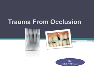
  • 1. Trauma From Occlusion DR.SWAPNA EDIGA By- DR.SWAPNA.E
  • 2. Contents  Introduction  Definition & Terms  Classification  Stages of tissue response to increased occlusal forces  Role of occlusion in pathogenesis of Periodontal disease  Human studies & Clinical trials  Animal experiments  TFO & Plaque Associated Periodontal Disease  Diagnosis  Treatment  Conclusion  References
  • 3. Introduction • To function occlusal harmony- masticatory apparatus- teeth & supporting tissues, TMJ & associated neuromuscular skeletal structures must operate in an integrated & dynamic manner • Loss of integrated function & homeostasis in response to functional demand may lead to exacerbation of existing periodontal condition. Angle defined occlusion as the normal relation of the occlusal inclined planes of the teeth when the jaws are closed
  • 4. Teeth transmit occlusal forces- mastication & swallowing Magnitude of occlusal forces controlled by muscles of mastication. Majority of occlusal forces are directed axially. The periodontium is designed to resist axial forces but some lateral forces can be tolerated
  • 5. Definitions- Trauma from Occlusion • A condition where injury results to the supporting structures of the teeth by the act of bringing the jaws into closed position- (Stillman 1917 ) • When the occlusal forces exceed the adaptive capacity of the periodontal tissues, tissue injury results. The resultant injury is termed as trauma from occlusion. (Orban and Glickman 1928,1933 )
  • 6. • Damage in the periodontium caused by stress on the teeth produced directly or indirectly by teeth of opposing jaw” (WHO 1978) • “An injury to the attachment apparatus as a result of excessive occlusal forces” (Glossary of Periodontic terms AAP 2001)
  • 7. Traumatogenic Occlusion • Any occlusion (e.g malocclusion) that produces forces that cause an injury to the attachment apparatus. • Other terms: ▫ Traumatic occlusion ▫ Occlusal disharmony ▫ Functional imbalance ▫ Occlusal dystrophy
  • 8. Occlusion Traumatism • The overall process by which a traumatogenic occlusion produces injury in the periodontal attachment apparatus.
  • 9. • Traumatizing Occlusion may act on individual tooth/groups of teeth • In premature contact relationship • In conjunction with parafunction- clenching, bruxism • In conjunction with loss of migration of premolars & molar teeth with gradually developing spread of anterior of teeth of maxilla
  • 10. Adaptive capacity of Periodontium to occlusal forces • Varies in different persons & in same person at different times • Influenced by Magnitude : when magnitude of occlusal forces is increased ,there is widening of PDL space, increase in No.& width of PDL fibers, increase in density of alveolar bone. Direction: changing direction of occlusal forces s re-orientation of stresses & strains within periodontium(Glickman 1970). Principal fibers accommodate occlusal forces along long axis of tooth. Lateral(horizontal) forces & torque(rotational) forces are more likely to injure periodontium. Duration : constant pressure on bone is more injurious than intermittent forces. Frequency: more frequent intermittent forces more injurious .
  • 11. Physiologically normal occlusal forces in chewing and swallowing : • Positive stimulus in maintaining the periodontium and the • Alveolar bone in a healthy and functional condition. Impact forces : mainly high & short duration periodontium can sustain high forces for short period • Exceeding the viscoelastic buffer capacities of PDL results  in loss of tooth and bone. Continuous forces :Very low forces (eg, orthodontic forces), • continuous in one direction  displacing tooth by remodeling alveolus. Jiggling forces : Intermittent forces in 2 directions (premature contacts eg, crowns, fillings) result in widening of the alveolus and increased mobility. TYPES OF OCCLUSAL FORCES
  • 12. Forces in one direction cause tipping of the tooth in the opposite direction or tooth displacement parallel to the force resulting in a ‘bodily movement . The clinical result is a (temporary) increased mobility. However, there are no changes in the supracrestal fibres, no loss of periodontal attachment, or an increased probing pocket depth. The increased tooth mobility is functional adaptation to the forces exerted on that tooth. In the compression zone, pressure stimulates osteoclasts in the adjacent bone and the alveolar wall is resorbed until a new connection is formed with the hyalinised bone (‘undermining resorption ’). In the tension zone, bone apposition and rupture of the collagen fibres occur. After removal of the force the periodontal ligament is reorganised and after some time develops a normal histological appearance. Forces too high (above the adaptation level), aseptic necrosis of the PDL (hyalinisation) root resorption occur resulting in shorter roots.`
  • 13. Jiggling forces different and opposite directions. Therotically same events occurs. However, they are not clearly seperated. Histologically : Aposition & resorption on either sides of PDL  widening of PDL space ( observed on radiographs) This phenomenon the increased mobility without pocket formation, migration and tipping. Hypermobility as long as the forces are exerted on the tooth not adaptation. not a sign of an ongoing process but result of a previous jiggling force.
  • 14. Reasons of deflective occlusal forces. Patterns of mastication, Loss of teeth, Loss of periodontal support, Dental caries, Faulty restorations Flawed orthodontics, Defective occlusal adjustment, Occlusal habits, Inadequate form and position of the teeth.
  • 15. EFFECTS OF OCCLUSAL DISHARMONY Discomfort in the region of the TMJ Food impaction Abnormal habits Pain in and around teeth with improper restorations Pulpal disturbances Facial pain of obscure origin Periodontal abscess
  • 16. Cheek biting Sensitivity of occlusal/incisal surfaces of teeth due to their abnormal wear Mobility of teeth Gingival enlargement, Gingival recession Bruxism; due to faulty occlusion, Unilateral mastication Restricted excursion of the mandible resulting in insufficient wear Extreme excursions of the mandible resulting in excessive wear. Migration of upper and lower teeth
  • 17. Terminologies • Occlusal trauma can be divided into 3 general categories: Occlusal Trauma: An injury to the attachment apparatus as a result of excessive occlusal force . Occlusal trauma is the tissue injury, and not the occlusal force. • Injury resulting from excessive occlusal forces applied to a tooth or teeth with normal support . Examples include high restorations, bruxism, drifting or extrusion into edentulous spaces, and orthodontic movement. Primary Occlusal Trauma: • Injury resulting from normal occlusal forces applied to a tooth or teeth with inadequate support . Secondary Occlusal Trauma: • Injury from an excessive occlusal force on a diseased periodontium . In this case, there is gingival inflammation, some pocket formation, and the excessive occlusal forces are generally from parafunctional movements. Combined Occlusal Trauma:
  • 18. • Traumatogenic Occlusion: Any occlusion that produces forces that cause an injury to the attachment apparatus. • Occlusal Traumatism: The overall process by which a traumatogenic occlusion produces injury in the periodontal attachment apparatus.
  • 20. ACUTE TRAUMA • Acute TFO develops from • An abrupt occlusal impact biting on a hard object. • Restorations, prosthetic appliances that interfere with or alter the direction of occlusal force • Clinical features • Tooth pain, sensitivity to percussion, and increased tooth mobility. • Outcome – • Injury heals and the symptoms subside if the force is dissipated 1) shift in the position of the tooth or 2) wearing away or correction of the restoration. • Injury may worsen and develops into necrosis accompanied by periodontal abscess formation, or • Persist as a symptom-free chronic condition • Result in cemental tears.
  • 21. CHRONIC TRAUMA • More common and is of greater clinical significance • Develops from- • Gradual changes in occlusion produced by • 1) Tooth wear, • 2) Drifting , • 3) Extrusion , • 4) With parafunctional habits such as bruxism and clenching, rather than as a sequel of acute TFO.
  • 22. PRIMARY TFO • When TFO is the result of alterations in occlusal forces • Insertion of a “high filling” • Insertion of a prosthetic replacement that creates excessive forces on abutment and antagonist teeth. • Orthodontic movement of teeth into functionally unacceptable positions. • Drifting movement or extension of teeth into spaces created by unreplaced missing teeth, OR • Changes produced • Do not alter the level of connective tissue attachment and do not initiate pocket formation. • Probably because the supracrestal gingival fibers are not affected and therefore prevent apical migration of the junctional epithelium.
  • 23. SECONDARY TFO •Occurs when the adaptive capacity of the tissues to withstand occlusal forces is impaired by bone loss resulting from marginal inflammation. • This reduces the periodontal attachment area and alters the leverage on the remaining tissues. •The periodontium becomes more vulnerable to injury and previously well-tolerated occlusal forces become traumatic.
  • 24. Stages of Tissue response to increased occlusal forces • 3 stages: • Stage I: injury • Stage II: repair • Stage III: adaptive remodeling of periodontium
  • 25. Stage I: Injury- increase in areas of resorption, decrease in bone formation Injury- excess occlusal forces Repair- forces are diminished. Furcation- most susceptible to injury Histological: temporary depression in mitotic activity & rate of proliferation & differentiation of fibroblasts in collagen formation & bone formation.
  • 26. Slightly excessive pressure: PDL widening, bone resorption. Slightly excessive tension: elongation of PDL fibers, Apposition of alveolar bone Severe pressure: pressure forces root to bone- undermining resorption: necrosis of PDL & bone. Severe tension:PDL widening, thrombosis,hemorrhage, PDL tearing,bone resorption.
  • 27. • Increased pressure areas: blood vessels numerous & reduced in size. • Areas of increased tension: enlarged blood vessels(Zaki 1963). • Greater pressure: ▫ compression of PDL fibers, areas of hyalinization(Rygh 1974). ▫ injury to fibroblasts & connective tissue –necrosis of ligament. ▫ Vascular changes:-within 30 mins-impairment & stasis of blood flow, ▫ 2-3 hours blood vessels packed with erythrocytes fragment , ▫ 1- 7 days disintegration of blood vessels & release contents into surrounding tissue. ▫ Increased resorption of alveolar bone & tooth surface occur. Histoligicaly
  • 28. Stage II : Repair-decreased resorption, increased bone formation • Damaged cells are removed & new CT cells & fibers , bone , Cementum are formed in an attempt to restore injured periodontium • Buttressing bone formation: • Central: occurs within jaw. Endosteal cells deposit new bone, restore the bony trabeculae, reduces the size of marrow spaces. • Peripheral: on bone surface.On facial & lingual bony plates.Lipping- Shelf like thickening of alveolar margin
  • 29. Stage III: Adaptive remodeling of periodontium: resorption & formation return to normal • If repair process does not keep pace with destruction caused by occlusion, periodontium is remodeled in an effort to create structural relationship • Thickened PDL- funnel shaped at crest, angular defects in bone with no pocket formation. Tooth is loose. Increased vascularization
  • 30. Role of occlusion in pathogenesis of Periodontal Disease • Over 100 years occlusal trauma is associated with PD • 1901 Karolyi: Association excessive occlusal forces & Periodontal destruction • 1917-1926:Stillman- excessive occlusal force was primary cause of PD • End of 1930: Excessive occlusal forces were a causative factor for PD & Occlusal discrepancies should be prophylactically treated to prevent PD • 1933- Orban & Weinman , 1941- Weinmann- effect of excessive occlusal forces on periodontium and Concluded -: no relationship & cause was gingival inflammation • 1950’s- 1960’s( Stahl 1962,Weinmann 1954,Ramjford 1971,) animal research- rats ,monkeys, dogs- effect of occlusal forces on periodontium: results : no relation
  • 31. Glickman & co workers 1960 • 1960 Glickman & co workers : animal models & human Autopsy material : dogs & monkeys high restorations : no association • Rhesus monkeys & Human autopsy models: phenomenon :- “Altered pathway of destruction” when occlusal forces were present. • Altered pathway of destruction : change in orientation of periodontal & gingival fibers which occurred in presence of excessive gingival inflammation to extend along PDL • This pathway caused vertical bony defects
  • 32. Glickman & co workers conclusion • Excessive forces in presence of plaque associated inflammation caused a change in the alignment of periodontal ligament ,allowing a altered pathway of inflammation/ destruction resulting in vertical bony defects • Process was termed “ Co destructive effect”
  • 33. • Zone of irritation: marginal & interdental gingiva. gingival inflammation cannot be induced by TFO but is result of irritation from microbial plaque. First involvement of alveolar bone & later PDL. Progression : an even (horizontal)bone destruction • Zone of co-destruction: • PDL, Cementum, Alveolar bone. Coronally demarcated by trans-septal (interdental) & dento-alveolar collagen fiber bundles.the tissue in this zone becomes a lesion of TFO • Fiber bundles which separate zone of co destruction affect from 2 directions: 1. Inflammatory lesion maintained by plaque in zone of irritation 2. Trauma induced changes in zone of co- destruction Glickmans Concept (1965,1967) Periodontal structures divided into 2 zones
  • 34. Waerhaug(1979) concept • “Plaque front”- was always in close approximation to epithelial attachment level & always followed morphology of bony defect. • Also found excesssive occlusal forces bore no relationship to underlying bony defect & that vertical bony defects were found equally around traumatized & non traumatized teeth • Conclusion: bone loss was always associated with down growth of plaque & there was no relationship between excessive occlusal forces & vertical bone loss.
  • 35. 2 extensive studies- Polson & Lindhe( 1970) Polson (1982)& co workers Lindhe (1982)& co workers • squirrel monkeys- • mesiodistal compression forces comparable to orthodontic forces • beagle dogs, • buccolingual forces using a high occlusal contact and a finger spring Effect of plaque & excessive occlusal forces in animal models Results: similar results Conclusion: excessive occlusal forces in absence of plaque cause loss of bone density & mobility of affected tooth but no evidence was found that occlusal force alone could cause attachment loss Conclude: excessive force alone does not cause loss of attachment, with plaque it causes.
  • 36. Human studies & clinical trials • World workshop in Periodontics “prospective studies on effect of occlusal forces on progression of Periodontics are not ethically acceptable in humans” Gher 1996 • Human studies are limited to retrospective & observational research. Rosling et al 1976 Advanced PD, multiple vertical defects, mobile teeth subjected to antimicrobial therapy Infrabony pocket at Hyper mobile teeth exhibited same degree of healing as firm teeth Fleszar et al 1980 Tooth mobility on healing following periodontal therapy Pockets of clinically mobile teeth do not respond well to those of firm teeth exhibiting same degree of disease severity. Philstrom et al 1986 TFO & periodontitis Teeth with increase mobility had deeper pockets, attachment loss, bone loss compared to teeth without.
  • 37. Clinical trials • Wang & Ramjford 1994-molars with furcation involvement & mobility have greater probing depths compared to molars that are clinically immobile. • Ramjford & Burgett 1992: patients who received occlusal adjustment as part of their periodontal treatment had greater attachment gain than patients who did not. • These studies suggest that occlusal adjustment should be performed where indicated as part of periodontal treatment
  • 38. • Mc Guire & Nunn 1996: mobility & para functional habits that are not treated with biteguard are associated with increased attachment loss, worsening prognosis & tooth loss. • Cortelleni & Tonetti 2001- mobile teeth treated with regenerative surgery did not respond compared to non mobile teeth. • Harrel & Nunn 2001: occlusal discrepancies appeared to be a significant risk factor that contribute to more rapid periodontal destruction & that treatment of occlusal discrepancies seemed to slow periodontal destruction.
  • 39. Animal Experiments Orthodontic type trauma Muhlemann 1961, Waerhaug 1966, karring 1982 Teeth exposed to unilateral forces of magnitude, frequency/duration that they are unable to withstand Certain well defined reactions develop in PDL leading to bone resorption.
  • 40. Jiggling type trauma • Healthy periodontium with normal height Supralaveolar CT was not influenced by occlusal forces: reason: tissue compartment is bordered by hard tissue only on one side- gingiva was non inflamed at start of experiment remained non inflamed & not aggravated by jiggling forces.
  • 41. Healthy periodontium Reduced height Within certain limits a healthy periodontium with reduced height has capacity similar to periodontium of normal height to adapt to altered functional demands
  • 42. Plaque associated Periodontal disease Occlusal forces which allow adaptive alterations to develop in the pressure/tension zone of PDL will not aggravate a plaque associated PD
  • 43. Lindhe & Svanberg 1974 If magnitude & direction of occlusal forces are not adapted by tissues-injury in Co-destruction is permanent. Angular bone loss continues Progressive mobility Plaque associated lesion in area of irritation & inflammatory lesion in area of co- destruction merged, the epithelium proliferated in an apical direction & PD aggravated.
  • 44. Effects of insufficient occlusal forces • May also be injurious • Insufficient stimulation causes thinning of PDL • Atrophy of fibers • Osteoporosis of alveolar bone • Reduction in bone height • Hypo function causes: ▫ Open bite relationship ▫ Absence of functional antagonists ▫ Unilateral chewing habits
  • 45. Reversibility of traumatic lesions TFO is reversible The injurious force must be relived for repair to occur(Glickman 1961, Polson 1976). Presence of inflammation due to plaque accumulation in periodontium may impair reversibility of traumatic lesions
  • 46. Effects of excessive occlusal forces on pulp Disappearance of pulpal symptoms after correction of excessive occlusal forces Animal subjects(Cooper 1971, Landay 1970): pulpal reactions to increased occlusal forces. No response to minimum or short periods.
  • 47. Diagnosis Clinical symptoms Clinical signs Examination Increased tooth mobility is not always indicative of trauma from occlusion. It is important, however, that hypermobility which does occur as a result of trauma from occlusion is detected in patients with reduced periodontal attachment. The reason for this is that trauma from occlusion may accelerate further reduction in attachment in a patient with active periodontitis. A clinical diagnosis of occlusal trauma can only be confirmed where progressive mobility can be identified through a series of repeated measurements over an extended period. This means that simple but reliable monitoring needs to be undertaken. A simple monitoring protocol is needed
  • 48. • SIGNS MOBLITY + FREMITUS CHANGE IN PERCUSSION MIGRATION WEAR PATTERNS PDLABCESS GINGIVAL CHANGES HYPERTONICITY OF MUSCLES
  • 49. Radiographic features 1. Widening PDL space, thickening of lamina dura in lateral aspect of root, in apical regions & bifurcation areas. 2. Vertical rather than horizontal destruction of interdental septum 3. Radiolucency & condensation of alveolar bone. 4. Root resorption
  • 50. Detection of occlusal trauma clinically • Analysis of patients occlusal relationship- Angle’s classification. • The relationship between cusps is the most important factor in transmittal of occlusal forces to the periodontal structures. • Initial contact between teeth is detected by gentle manipulating patient's mandible into retruded position • Retruded position: both right & left condyles are firmly placed in fossa of TMJ • Patient is asked to close until patient feels first contact between teeth. • Centric relation: initial contact in retruded position. • Patient is asked to continue to close the jaws together until maximum contact between the teeth is achieved. • Centric occlusion: jaw position of maximum tooth contact
  • 51. Treatment • Occlusal adjustment • Management of parafunctional habits • Temporary, provisional or long term stabilization of mobile teeth with appliances • Orthodontic tooth movement • Occlusal reconstruction • Extraction of selected teeth
  • 52. Occlusal adjustment (Selective Grinding) • Non reversible. Reshaping of occlusal surfaces to create harmonious contact relationships between the upper and lower teeth. • Thorough occlusal analysis must be embarked: • Clinical assessment of occlusion:- comprehensive assessment of teeth • Interarch relationships, jaws & associated muscles TMJ & their movements, • examination of articulate study casts: semi adjustable articulator with models mounted in retruded position
  • 53. Occlusal adjustment Indications Contraindications • To reduce traumatic forces to teeth • To reduce damage from parafunctional habits • To reshape teeth: reduce soft tissue injury • To reduce food impaction • Occlusal adjustment without ▫ pre-treatment study & patient education ▫ Signs & symptoms of occlusal trauma ▫ Microbial induced inflammatory PD treatment. • Bruxism Rx based on patient history without evidence of damage, pathosis/pain • When patients emotion precludes satisfactory result. • Treatment for severe extrusion, mobility/ malposition teeth that would not respond to occlusal adjustment alone.
  • 54. Equilibrating mobile teeth • In a patient with mobile teeth, it may be necessary to temporarily stabilise those teeth before equilibration is possible. • If a tooth is mobile, it is very difficult if not impossible to effectively modify its shape with the aim of reducing the occlusal forces acting upon it (equilibration).
  • 55. • Whether the inflammatory periodontitis has been treated successfully. • If there is an inflammatory periodontal process this should be treated initially. • Subsequently when the periodontal condition is stable, occlusal therapy may be necessary for some patients and could involve either occlusal equilibration or splinting. • The radiographic appearance of the periodontal support. occlusal equilibration is indicated will depend upon:
  • 56. • Occlusal equilibration is considered an effective form of therapy for teeth with increased mobility which has developed together with an increase in the width of the periodontal ligament (PDL). • Reducing the occlusal interference on a tooth with normal bone support will normalize the width and height of the PDL. • Eliminating any occlusal interferences for a tooth which has a reduced bone height as a result of periodontal disease will result in bone formation and remodelling of the alveolus only to the pre-trauma level
  • 57. • It is generally accepted that occlusal adjustment directed solely at establishing an ideal conceptualized pattern is contraindicated. • Rather, it should only be performed when the objective is to facilitate treatment or intercept actively destructive forces. • When occlusal therapy is planned as part of periodontal treatment, it is usually deferred until initial therapy aimed at minimizing inflammation throughout the periodontium has been completed. • This is based upon the fact that inflammation alone can contribute significantly to a tooth’s mobility.
  • 58. Splinting 1) Stabilize teeth with increasing mobility that have not responded to occlusal adjustment and periodontal treatment. 2) Stabilize teeth with advanced mobility that have not responded to occlusal adjustment and treatment when there is interference with normal function and patient comfort. 3) Facilitate treatment of extremely mobile teeth by splinting them prior to periodontal instrumentation and occlusal adjustment procedures. 4) Prevent tipping or drifting of teeth and extrusion of unopposed teeth. 5) Stabilize teeth, when indicated, following orthodontic movement. 6) Create adequate occlusal stability when replacing missing teeth. 7) Splint teeth so that a root can be removed and the crown retained in its place. 8) Stabilize teeth following acute trauma.
  • 59. 1) When the treatment of inflammatory periodontal disease has not been addressed. 2) When occlusal adjustment to reduce trauma and /or interferences has not been previously addressed. 3) When the sole objective of splinting is to reduce tooth mobility following the removal of the splint. Contra-indications This means that if periodontal treatment results in a stable periodontal condition which is comfortable, splinting is not needed.
  • 60. OCCLUSAL PARAFUNCTION: BRUXISM • Bruxism : clenching or grinding of the teeth when the individual is not chewing or swallowing – Ramfjord and Ash - 1966 Bruxism can occur as brief, rhythmic strong contractions of the jaw muscles during eccentric lateral jaw movements or in maximum intercuspation, which is called clenching. Bruxism may also take the form of tapping. Bruxism often occurs without any neurologic disorders or defects and can be viewed as a phenomenon present in healthy individuals. Bruxism may lead to tooth wear, fractures of the teeth or dental restorations, or uncosmetic muscle hypertrophy.
  • 61.  Presence of tooth wear in patients is not necessarily the cause for signs and symptoms of TM disorders.  Data from Rugh indicate that 83% of a group of bruxers, performed bilateral muscle contraction, whereas 17% performed unilateral contractions. Nocturnal bruxism & Daytime (diurnal) bruxism.  Most people are not aware of a bruxism habit until it is brought to their attention.  When active tooth gnashing occurs, the enamel rods are fractured and become highly reflective to light. Thus shiny, bright facets
  • 62. TREATMENT OF BRUXISM 1. Behavioural modality – explanation and arousal of the patients awareness of the habit. • If pain and stiffness are associated, physical therapy. • Anti-anxiety drugs.. • Ware – 1982 – advocated use of antidepressants as a means of inhibiting the REM sleep. 2. Night guard appliance (max stabilization splint). • Aim is to protect the tooth surface and to dissipate forces. • More practical for treating nocturnal bruxism..
  • 63. • Splints are more significant in the management of the destructive effects of bruxism. • Patient instructed to wear the splint during sleep. • Should be adjusted again in 2-3 weeks.. • Should be observed for bruxofacets. • If present should be burnished away. • Balancing should be completed before patient dismissal 3. Coronoplasty – recently placed dental restorations or other occlusal treatments. • Occlusal adjustment, reconstruction, or orthodontic treatment are contraindicated as a means of controlling bruxism.
  • 65. TERMINOLOGY Intercuspal position (ICP): Synonyms: centric occlusion (CO), habitual occlusion, acquired centric, habitual centric, maximum intercuspation. Median occlusal position (MOP): a dynamic contact position of the teeth that may be obtained on command by a snap jaw closure foll moderate jaw opening Retruded position (RP): Any position of the mandible on the terminal hinge path. Synonyms: centric relation (CR), Terminal hinge position. Retruded contact position (RCP): The end point of terminal hinge closure Synonym: centric relation contact (CRC).
  • 66. Laterotrusion : Synonym: working movement. Mediotrusion: Synonyms: non-working side movement, balancing movement. Laterotrusive side: Synonyms: working side, functional side. Mediotrusive side: Synonyms: nonworking side, balancing side, nonfunctional side, idling side.  According to Ramfjord, “centric occlusion is a tooth to tooth and jaw to jaw relationship, in which the teeth are in ideal intercuspation and all components of masticatory system – the TMJ, the neuromuscular elements and occlusal surfaces are in harmonious relationship.”
  • 67. Centric Relation • The most orthopedically stable joint position is when the condyles are in their most superoanterior position in the articular fossae, resting against the posterior slopes of the articular with the discs propoerly interposed . • All elevator muscles activated … no occlusal influences • Musculoskeletal stable position
  • 68. PATIENT COUNSELING • PROPER DIAGNOSIS • Point out loose teeth • Relate wear problems • Study the occlusal relationship • Demonstrate on the mounted casts • Tell the patient to expect further adjustments. Never start coronoplasty unless both the dentist and the patient are committed to complete it.
  • 69. TIMING IN RELATION TO SURGERY Preliminary grinding Definitive grinding Check grinding
  • 70. PRELIMINARY GRINDING • First step in treatment • Done when the patient complains of pain or discomfort or when normal function is prevented by excessive mobility. • Spot grinding
  • 71. DEFINITIVE GRINDING • Progressive tooth mobility. • Radiographic signs of trauma. • Performed after eliminating the inflammation in periodontal tissues.
  • 72. CHECK GRINDING • Performed as a final measure. • Usually a month after surgery, the mouth is checked for trauma that may have resulted from a slight shifting of the teeth.
  • 73. FOR LOCATING CR: Begin with locating the musculoskeletally stable position – CR if for any reason the condylar position is in question selective grinding shouldn’t be carried out until a stable reproducible position has been achieved Kinematic face bow Semi or fully adjustable articulator Gothic arch & extraoral tracer
  • 74. STEPS IN OCCLUSAL ADJUSTMENT I. Initial grinding II. Harmonization in terminal hinge occlusion, III. Harmonization in protrusive position and movement, IV. Harmonization in lateral occlusal position and lateral excursion, V. Reestablishment of physiologic occlusal anatomy and careful polishing of all ground surfaces.
  • 75. INITIAL GRINDING 1. Narrowing of bucco lingual diameters 2. Shortening of extruded teeth 3. Improvement of esthetics 4. Correction of marginal ridge relationships 5.Reduction of plunger cusps 6. Correction of rotated, malposed, or tilted teeth 7. Correction of facets and abraded teeth
  • 76. ABNORMAL WEAR • The occlusal table is made smaller • Occlusal forces will become centered over the tooth and will tend to be transmitted along the long axis of the tooth • This step is indicated only when such narrowing would neither disturb vertical dimension (by removing cusp tips that contact in centric) nor induce cheek biting.
  • 77. EXTRUSION • Unesthetic appearance • May be the premature tooth in many movements. • The plane of occlusion is disturbed
  • 78. ESTHETICS • When individual anterior teeth are disproportionately longer than the same teeth on the other side of the mouth, they may be ground to a more symmetric & regular form.
  • 79. UNEVEN MARGINAL RIDGES • Marginal ridges maybe ▫ unequal in height; ▫ may not meet at the contact area • Correct them by grinding or restorative dentistry
  • 80. PLUNGER CUSP • The elongated distobuccal cusp of the maxillary molar wedges the lower molars and forces them apart. • Food impaction occurs during mastication • Shortening & rounding without taking the tooth out of CO
  • 81. ROTATED,MALPOSED OR TILTED TEETH • Esthetics a problem • Careful grinding to improve the crown form
  • 82. WEAR FACETS AND ABRADED TEETH • Teeth subject to masticatory and parafunctional activity tend to wear. • Abraded teeth need more force in mastication so it need to be reduced
  • 83. SHARP EDGES • Can cause irritation to the tongue and cheek . • Sharp edges of restorations protruding beyond the enamel surface. • Undermining or chipping of enamel exposing sharp edges .
  • 84. ANTERIOR OPEN BITE Never grind posterior teeth to bring teeth in contact.
  • 85. Developing an acceptable centric relation contact position The goal of this step is to create desirable tooth contacts when the condyles are in their musculoskeletally stable position. In many patients an unstable occlusal condition exists in CR and creates a slide to the more stable ICP. A major goal of SG is to develop a stable intercuspal contact position when the condyles are in the CR position A slide of the mandible is created by the instability of the contacts between the opposite tooth inclines. When the cusp tip contacts a flat surface in and force is applied by the elevator muscles, no shift occurs. Thus the goal in achieving acceptable contacts in ICP is to alter or reshape all inclines into either cusp tips or flat surfaces. Cusp tip-to-flat surface contacts are desirable as they effectively direct occlusal forces through the long axes of the teeth.
  • 86. Achieving the centric contact relation The patient reclines in the dental chair, and the CR is bimanually located. The teeth are lightly brought together and the patient identifies the tooth that is felt to contact first. The mouth is then opened, and the teeth are are thoroughy dried with an air syringe or a cotton roll Thin articulating paper is then held with forceps on the side identified as the first contact. The mandible is again guided to CR and the teeth contact, lightly tapping on the paper. The contact areas are located for the maxillary and mandibular teeth. With light pressure, encourage small hinge movements. Exert more pressure as the jaw falls downwards and backwards to the retruded position, try to seek ligamentous resistance of the TMJs. If not try again. Talk in low tones, using repetitive phrases – “just let it go”
  • 87. A. In CR, a mesial incline of the maxillary tooth contacts a distal incline (arrow) of the mandibular tooth B. the contact closest to the cusp tip is located on the mandibular tooth. This incline is elimainated allowing only the cusp tip to contact C. during the next closure this mandibular cusp tip contacts mesial inclines(arrow) of the maxillary cusp. D. This incline is reshaped into a flat surface (i.e. hollow grinding) E. On the next closure the mandibular cusp tip can be seen to contact the maxillary flat surface and the treatment goals for this pair of contact is achieved Mesial view
  • 88. • A. the mandibular buccal cusp prematurely contacts, preventing the contact of the maxillary lingual cusp. • B. no contact during the laterotrusive movements (large arrows) • C. no contact during mediotrusive movement (large arrows) • D. the fossa area opposing the mandibular buccal cusp is reduced • E. this reduction allows contact of the maxillary lingual cusp tip. In case there is a contact during the laterotrusive and mediotrusive contacts  the mandibular buccal cusp is shortened.
  • 89. Static occlusion Static occlusion is described as occlusion which occurs in intercuspal position. It is an occlusion in centric relation with maximum intercuspation.
  • 90. Dynamic Occlusion Dynamic occlusion refers to the occlusal contacts that are made when the mandible is moving relative to maxilla. The mandible is moved due to muscles of mastication and the pathway along which it moves is determined not only by muscles of mastication but by two other guiding systems.
  • 91. Posterior guidance system is provided by the temperomandibular joint. Anterior guidance system by teeth. Canine guidance Group function
  • 92. Ideal Occlusion In ideal static occlusion, occlusion occurs in centric relation and there is provision for free centric movements.
  • 93. In protrusive movement there is contact in canines and disclusion in the remaining posterior teeth(Canine Guidance)
  • 94. In lateral movement there are multiple contacts on working side i.e. premolars and molars, and disclusion on the non working side (group function).
  • 95. Developing an acceptable lateral and protrusive guidance • Technique: • After the CR contacts are established they should never be altered. All adjustments for the eccentric contacts occur around the CR contacts without altering them. • The patient closes in CR, and the relationship of the anterior teeth is visualized. It is then determined whether immediate canine guidance is possible or a group function guidance is necessary
  • 96. • When a group function is indicated, the teeth that can assist in the guidance must be selected. The patient moves the mandible through the various lateral and protrusive excursions to reveal the most desirable contacts. • In some instances gross mediotrusive contacts will actually disocclude to visualize the best guidance e.g during a right laterotrusive movement there is significant mediotrusive contact on the left third molars. • This disoccludes the right side and must be eliminated before the type of laterotrusive guidance on the right side can be evaluated.
  • 97. When the desirable guidance contacts been determined, they are refined and the remaining eccentric contacts eliminated. To ensure that the already established CR contacts are not altered, two different marking paper are used. The teeth are dried, and blue marking paper is placed between them. The patient closes and taps on the posterior teeth. Then from CR position a right excursion is made with return to centric. Finally, a straight protrusive movement is made with return to centric.
  • 98. The mouth is then opened, the blue paper is removed and replaced with red paper and patient closes and taps on the CR contacts. The red paper is removed, and the contacts are inspected. All eccentric contacts are now marked in blue, and the CR contacts are marked in red. The blue eccentric points are adjusted to meet the determined guidance condition without altering any red CR contacts. L T C R MT
  • 99. • A red dot with a blue streak extending from it is typically seen. • This type of marking reveals that the red centric cusp tip contacts an opposing tooth incline during a particular eccentric movement. Procedure for canine guidance: when the anterior tooth relationship provides for canine guidance, all blue marks on the posterior teeth are eliminated without alteration of the established CR contacts (red). Once this is accomplished, the teeth are redried and the blue eccentric and red centric marking procedure is repeated. Often several adjustments are necessary to achieve the desired results. At the completion of this procedure the posterior teeth reveal only red CR contacts on the cusp tips and flat surfaces. The canine reveal the blue laterotrusive contacts and the incisors (with possibly the canines) reveal the blue protrusive contacts
  • 100. Procedure for group function guidance When the anterior tooth relationship is such that a group function is necessary for the guidance, all the blue contacts on the posterior teeth are not eliminated. Because selected posterior teeth are necessary to assist in the guidance, care must be taken not to eliminate these contacts. The desirable contacts are the laterotrusive on the buccal cusps of the premolars and the mesiobuccal cusp of the first molar. When the selective grinding procedure is completed. The occlusal condtition reveals only the red CR contacts on the posterior teeth (except for the blue laterotrusive contacts on the buccal cusps that are necessary to assist in guidance). The canines reveal the blue laterotrusive contacts as the movement becomes great enough to disocclude these teeth.the incisors reveal the blue protrusive contacts.
  • 101. Evaluation in the upright head position (Alert feeding position) • The selective grinding procedure is not complete until the upright head position has been evaluated. • Because most such procedures are performed in a reclined position. • Evaluation of postural changes of the mandible must be accomplished before the patient is dismissed. • In the upright position with the head tilted forward app 30 degrees, the patient closes on the posterior teeth. • Determining whether a postural change in the mandibular position has occurred that will cause anterior tooth contacts to be heavier than posterior tooth contacts is important.
  • 102. If this has occurred., the anterior tooth contacts are reduced slightly until the posterior teeth contact more heavily. Care must be taken in questioning the patient that the information received is valid. When the question is asked merely whether the anterior teeth contact more heavily, the patient may protrude slightly onto the guidance and check for contact; in this position the anterior tooth contacts will feel heavier and the patient will therefore answer affirmately, with the result that a porion of the established guidance will be unnecessarily removed. The most successful way to question a patient in the alert feeding position is to ask him or her to close the mouth and then tap the posterior together. While this is being done, the patient is asked whether the posterior teeth contact predominantiy, or both anterior and posterior teeth contact equally.
  • 103. As soon as the posterior teeth are felt more predominantly the selective grinding procedure is complete. Normally one or two adjustments will accommodate for this postural change of the mandible. Any red CR contacts on the anterior teeth are slightly reduced until the patient reports feeling predominantly the posterior teeth contacting. The patient again taps on the posterior teeth. In this upright position the anterior teeth are dried and red marking paper is placed between them. If however, the anterior teeth are contacting heavily or both anterior and posterior teeth are contacting evenly final adjustment in the alert feeding position is necessary. If the posterior teeth are contacting predominantly , minimal postural change has occurred and the selective grinding procedure is complete.
  • 104. REESTABLISHMENT & POLISHING • Occlusal tooth anatomy should be re-established. • Sluiceways and embrasures are to be formed • Sharp edges should be rounded slightly. • All the tooth surfaces that were ground should be polished and made comfortable to tongue and cheek
  • 105. Patient instructions After the selective grinding procedure, the patient’s muscles may feel tried. This is a normal finding especially when the procedure has been accomplished during a long appointment. The patient can be informed that some teeth may feel gritty when rubbed together but these will become smooth and polished within a few days. Patients need not concentrate on any mandibular positions or tooth contacts to assist in the effectiveness of this procedure. Those who make a conscious effort to explore the occlusal conditions may likely find contacts not identified during the procedure and become concerned. The overall effect of such activity is generally muscle hyperactivity. Asking the patient to relax the muscles and keep the teeth from contacting is often the best advice.
  • 106. Conclusion • The treatment of occlusal discrepancies/occlusal trauma should be viewed in context of control of one of risk factors contributing to periodontal disease. • The successful treatment of periodontal disease requires control of all controllable risk factors. • Occlusal treatment should be performed, where indicated as a routine part of periodontal treatment.
  • 107. References • Textbook of Clinical Periodontology. Newmann, Klokkevold, Takaei, Carranza- 10th Edi • Clinical Periodontology & Implant Dentistry.Jan Lindhe- 5th Edition • Stephen K Harrell. Occlusal Forces as Risk factor for Periodontal Disease. Periodontology 2000; vol 32;2003:111-117 • William Hallm0n, Stephen K Harrell. Occlusal Analysis, diagnosis & management in the practice of Periodontics. • Dr. K. Malathi1 coronoplasty-IOSR Journal of Dental and Medical Sciences (IOSR-JDMS ) • Okeson-textbook of occlusion and temporomadibular joint disorders.

Editor's Notes

  1. The periodontium can sustain high forces during a short period; however, forces exceeding the viscoelastic buffer
  2. Jiggling forces, coming from different and opposite directions, cause more complex histological changes in the ligament. Theoretically the same events (hyalinization, resorption) occur, however, they are not clearly separated. There are no distinct zones of pressure and tension. Histologically, there is apposition and resorption on either side of the periodontal ligament, resulting in a widening of the periodontal space .This may be observed on radiographs. This phenomenon explains the increased mobility without pocket formation, migration and tipping. The clinical phenomena are not only dependent on the magnitude of the forces, but also on the crown –root relationship, the position in the arch, the direction of the long axis, and the pressure of tongue and cheek musculature. The interarch relationship (for example,deep bite)influences the extent of the trauma caused by jiggling forces. The hypermobility is found as long as the forces are exerted on the tooth: there is no adaptation. Hypermobility is therefore not a sign of an ongoing process, but may be the result of a previous jiggling force. Jiggling forces exerted on the teeth in this condition result in a pronounced increase in tooth mobility because the point of rotation (fulcrum) is closer to the apex than normal  
  3. and occlusion is considered the primary etiologic factor in periodontal destruction
  4. and occlusion is considered the primary etiologic factor in periodontal destruction
  5. Benzodiazepines- diazepam (Valium) : 2.5-5 mg not more than 7 days (since dependency) If only this drug is given then can be prescribed for 2 weeks Other drugs – alprazolam(xanax) , clonazepam (klonopin) Antidepressants – low dose of aminotrypline 10mg  decrease frequent awakening, increase stage IV (delta) sleep and markedly decrease the time spent in REM sleep
  6. must be integrated with the course of periodontal treatment. At three times in the course of periodontal treatment, occlusal interventions may be undertaken:
  7. Manual method may be used to locate CR
  8. These steps presume that the patient has a relatively normal occlusion or an Angle class I or class II, division 1, occlusion.
  9. Initial grinding should precede adjustment in hinge position. Many interferences are eliminated during this step, In initial grinding, the teeth are carved to obtain as ideal an arch and occlusal plane as possible. Need for further adjustment may be reduced.
  10. Narrowing of occlusal table:so that the rounded extreme edges of the occlusal surface will be unable to participate in transmitting torquing forces.
  11. Asymmetric anterior teeth,ragged ,abraded incisal surfaces,rotated or slightly overlapped
  12. 2) (because of rotation or malposition); 3) (because of poor restoration or wear). may have faulty marginal ridge and sluiceway form
  13. This is especially true whenthe antagonist is firm & the opposing teeth are mobile.
  14. Buccal surface of upper & lingual surface of lower
  15. Caused by tongue thrust swallowing. Never grind posterior teeth to bring teeth in contact.