1. Treatment planning and patient positioning for MR-guided high intensity focused ultrasound treatment: a systematic approach
David Kinnaird1,2, Doug Wackerle2,3, Daniel Yang2,4, Avinash Eranki2, Matthew Oetgen2, Aerang Kim2, Karun Sharma2, Harry Kim5, Peter Kim2, Pavel S. Yarmolenko2, Haydar Celik2
1University of Pennsylvania, Philadelphia, PA, USA. 2Sheikh Zayed Institute for Pediatric Surgical Innovation, Children’s National Medical Center, Washington, DC, USA.
3School of Medicine and Health Sciences, George Washington University, Washington, DC, USA. 4Princeton University, Princeton, NJ, USA. 5Texas Scottish Rite Hospital for Children, Dallas, Texas, USA
Acknowledgements
Summary and Conclusions
I would like to thank the Sheikh Zayed Institute at Children’s National Medical Center for this
opportunity to work on this team. I would also like to thank Pavel Yarmolenko and Haydar Celik
for being my mentors, as well as the rest of the team for aiding my project. I would also like to
acknowledge the use of Google Sketchup, for creation of certain 3D images, and Avizo
Standard 8.1.1 from the FEI Visualization Sciences Group, for modeling the patient’s
extremities.
Methods
Algorithm:
Using the MR-HIFU system characteristics, determine:
• Gel geometry
• Grid coordinates that define table location of the fiducials (joints and/or markers)
Physician’s Guide:
Obtain multiple maximum intensity projections (MIP) that show maximal
separation of target tissue from tissue that must be spared
Provide explicit instructions for limb positioning, assuming HIFU is aimed using
orientation from optimized MIPs
Cutting Apparatus:
•Design:
Simple, easy-to-use design that allows for oblique gel cutting
•Use:
Cutting the gel at specific angle for maximal separation
Cutting the groove that holds the patient’s extremity
Introduction
Approach – Increase precision of initial position:
1. A concise patient positioning guide based on patient-specific 3D imaging data
2. A grid, spatially referenced with the guide, overlaid on the table
3. A custom shaped gel, precisely cut to accommodate patient and tumor geometry
Objective:
Systematically design a flexible patient positioning algorithm for MR-HIFU treatment of
extremity lesions.
Motivation – Optimize patient positioning:
1. Treatment time is limited (<4 hours) due to risks associated with general anesthesia and cost
2. Incorrect patient position may make parts of tumor inaccessible
3. Repositioning takes time, reduces range of tumor sizes that can be treated
4. Complex cases require advanced planning
1. Patient-specific measurements used to set height and angle of the target limb relative to
the HIFU membrane.
Used to prepare gel pad that optimally positions patient’s limb
2. Algorithm-generated grid coordinates ensure tumor positioning over center of the HIFU
window to minimize the need for repositioning.
3. This systematic approach to reducing total treatment time may allow for treatment of a
wider variety of lesion sizes and locations.
4. Analysis of especially challenging solid tumors of the extremity from our institution
demonstrates the need for a rational treatment planning approach.
Physician’s Guide
Positioning Solution
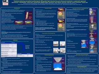
Customization of Gel Standoff Pad
Materials:
Nickel-plated steel wire (0.2mm, 3 ohms total resistance) held at variable height by
clamps on two metal rods
Resistance heating with a variable power supply
Methods:
1. Adjust wire to pre-calculated angle (Θg) to cut the gel
2. Apply current onto wire
3. Slide gel across heated wire
Treatment Planning Challenges:
Tumor behind knee:
•Bones limit access to the tumor
•Leg thickness varies around the tumor
•Tumor major axis must be parallel to tabletop to enable complete
treatment without repositioning
Excessive tumor volume:
•Large tumor size requires as much time for ablation as possible
•Tumor is on a slight slant within the leg, must be parallel to the
tabletop to enable complete treatment without repositioning
Tumor adjacent to shoulder joint:
•Shoulder and tumor geometry prevent usage of flat or U-shaped
coupling gel
•Shoulder must be raised so that tumor center is far enough away
from HIFU membrane
Pre-treatment planning and physical aid manufacturing are
required
Methods:
1. 3D segmentation
2. Patient positioning optimization
3. Reduction of variables
4. Custom positioning aids
Results:
Patient-specific coupling gel, reference grid,
and pre-determined padding
Systematic Planning in Challenging Cases
Treatment Planning Challenge:
• Relative location of tumor to bones defines
optimal target limb positions
• To minimize need for possible
repositioning, tumor must be contained
within the volume accessible to the
HIFU focus
•Pictured is the design of the cutter
•Both ends of the wire are at adjustable heights
•Gel is cut sideways for the angle cut, straight on for
cutting the U-shape hold for the extremity
Included in Guide:
Generic illustrations used to clearly describe positions of any
target extremity
Patient specific 3D segmented image data used to generate
cross-sectional views that show optimal rotations of the limb
around the long bone axis
Instructions provided for where to place the distal and
proximal fiducials and how to properly position limb.
Guide will:
Accompany physician into room
Suggest optimal lying position
Suggest viable rotations, ranking them in order of best
treatment and easiest to position
Provide troubleshooting solutions for challenging cases
Physician is responsible for spatial location of patient,
specifically rotation of the limb and lying position of patient;
guide will provide suggestions.
Requisite Position:
•Extremity must be aligned so that the focal point of the ultrasound
beam is at center of tumor
•Ensures maximum maneuverability of the transducer around the
tumor
Coordinates:
Ensures placement of the tumor over center of
the HIFU transducer window
o Maximizes the treatable volume
Coupling Gel:
Ensures extremity is at optimal angle and height
Guide:
Provides several optimized options for target
limb positioning
Helps lay the patient on the table correctly to
minimize discomfort
Helps pick the best rotation of the extremity
Addresses potential uncertainty with minor
factors in patient positioning
Treatment Planning Algorithm and Development of Physical Aids:
Input Variables Abbreviation
Available Treatment Volume Va
Transducer Extent of Motion Lx, Wx
Transducer Focal Deflection Radius Df
Dimensions of the Extremity Le, We, He
Dimensions of the Tumor Lt, Wt, Ht
Location of the Tumor
(Relative to Distal Joint)
Dt
Angle of Extremity Section
(Relative to tabletop)
Θg
Output Variables Abbreviation
Gel Side Heights H1, H2
Fiducial Grid Coordinates “A1”
Treatment
Planning
Algorithm
Background – Pediatric MR-HIFU Ablation:
• MR-guided high intensity focused ultrasound (MR-HIFU) allows for non-
invasive focal heating and mechanical destruction using an external
transducer
• Treatment used in adults for uterine fibroids, osteoid osteomas,
prostate tumors, brain tumors
• Largely unexplored in children
• Need for precise treatment planning
Several examples of challenging cases were examined: