1. Does Bi-articular muscle fatigue result in a
decrease in ankle joint extension kinetics in the
squat jump?
Daniel Yazbek
ABSTRACT
Purpose: Compare the foot segment kinetics in the squat jump before and after Gastrocnemius
fatigue. Methods: 5 healthy trained male basketball players (age 20-22) performed maximal squat
jumps before and after standing calf raises. Each subject performed 8 sets of 12 calf raises with 45
seconds rest between sets with a load of 75% Of 1RM. Jumping motion was captured using one high
speed pentax fastcam PCI R2 Photron at 125 Hz. Kinematic and kinetic data was expressed as thigh,
leg, and foot to determine angular velocity, moment and power. Results: There was no significant
difference between foot segment power after gastrocnemius fatigue (0.182 > .05) as indicated through
a paired samples t-test using SPSS data analysis. Conclusion: Bi-articular muscle fatigue does not
reduce segmental kinetics as there role is to transfer energy in a proximal-distal sequence pattern,
rather than generate work.
Keywords: Bi-articular, energy-transfer, fatigue, work
Introduction: The squat jump requires
maximal extension power of the hip, knee
and ankle. The gastrocnemius is regarded
as a powerful plantar flexor of the foot and
it would be expected that fatigue of this
muscle would compromise the explosive
power of a squat jump. Athletes must be
able to achieve maximal height with each
jump despite muscular fatigue
Our study aims to identify the differences
in the magnitude of foot angular velocity,
moment and power before and after
fatigue of the gastrocnemius, when
performing a squat jump.
There has not been a study examining the
direct effects on bi-articular muscle fatigue
on performance. However, there have
been studies in which mono and bi-
articular muscles have been examined, in
order to understand there functions in
movement and there have been studies
regarding the effects of muscle fatigue on
performance.
However, the effects of muscle fatigue
were examined in muscles which mostly
involved mono-articular muscles.
Our study aims to bring both variables
within the context. Therefore our study
aims to address the following question. If
Bi-articular muscles do not undergo a
significant change in muscle length and if
work is defined as force acting over a
distance, then bi-articular muscles that are
fatigued should not contribute to
decreased segment work or power?
Overall, we hypothesize that ankle joint
plantar flexion velocity, moment and
power will not be significantly different
after the fatiguing protocol.
Literature review: The squat jump is an
essential athletic component that is
performed by many athletes in a variety of
sports such as basketball and high jump
(Stone et al, 2003). The summation of
work and power of the muscles crossing
joints and the amount of energy
2. transferred from proximal to distal
segments is critical to translating angular
momentum into upward linear momentum
of the bodies’ centre of mass (Scehnau &
Bobbert, 1987).
It has been shown that muscles that span
across one joint, generate work and cause
rotation about its joint axis and in doing so,
transfer energy to the more distal
segments through the two joint muscles
(Schenau, Jacobs & Bobbert, 1996).
In a study by Barret & Neal (2001), the
major source of work generated during the
vertical jump was 92-94%. The mono-
articular hip and knee extensors, gluteus
Maximus and vastus intermedius
respectively, were shown to play a large
role in terms of work generation and
contributed to 80% of the total work done
by the muscle tendon units. Conversely,
Bi-articular muscles rectus femoris
hamstrings and gastrocnemius generated
relatively little work themselves but played
a role in transferring mechanical energy
between joints. The amount of energy
transferred between joints via biarticular
muscles was between 11-29%.
Additionally, an analysis of the lower
extremity during a full squat showed that
the gastrocnemius was heavily recruited
but performed no positive work because it
was contracting eccentrically and could
have been involved with transferring
energy distally due to its bi-articular nature
(Robertson, Wilson & Pierre, 2008).
However, the conclusions for this study
were not specific to a countermovement
jump nor did it employ a fatiguing protocol
to the gastrocnemius.
It is clear that muscle fatigue causes a
decrement in jumping performance which
was shown by Toumi et al (2006), in which
maximal concentric power and muscle-
tendon stiffness was reduced in the squat
jump following isometric leg press fatigue
and drop jump stretch shortening
repetitions till fatigue. Ground contact time
was increased equally following both
fatiguing protocols. However, in this study,
the fatiguing protocols did not isolate the
bi-articular muscles from the mono-
articular muscles, which if it did might
have resulted in a different conclusion due
to their differing functions.
JI et al, (2000) studied the effect of cycling
induced fatigue on joint power in the squat
jump. The fatigue protocol consisted of
one interval of 30 second pedalling on a
monark cycle at a load of 0.075kg per one
kilo of bodyweight. There was a significant
reduction in knee joint power after fatigue.
However, there was a smaller reduction of
Hip and ankle joint power compared to
knee joint power after fatigue. This study
did not explain why a reduction in fatigue
affected knee joint power rather than the
hip or ankle, nor did it fatigue just the two
joint muscles.
In another study, Tomioka, Owings &
Grabiner (2001), showed that lower
extremity coordination may be more of a
factor in contributing to maximal jump
height than lower extremity strength and
that maximal vertical jump can be
diminished by altered hip-knee
coordination. It is not known however if
fatigue of the gastrocnemius might alter
the hip-knee angle coordination and
consequently affect the jump performance.
3. Methods:Participants of 20-21 years (5
male), volunteered to be involved in the
study.
Protocol - Markers were placed on the
the lateral malleolus and the 5th
metatarsal
of the foot. This placement of markers
defined the foot segment. The participant
performed a counter movement jump with
arms crossed across the chest. The
angular velocity, moment and power were
calculated.
Following the jump, subjects performed 8
sets of 12 repetition calf raises using a
load of 75% of their 1RM on a 35cm step.
The subject would rest 45 seconds before
continuing the next set, for a total of 8
sets. To ensure the gastrocnemius was
under large fatigue, a standing calf raise
jump was performed before and at the
start of fatigue protocol. Failure of the toes
to clear the ground compared to
successful clearance of the toes at the
start of the protocol indicated muscle
fatigue. Following this, a second
countermovement jump was immediately
performed.
Segment angle was defined as the
horizontal line from the distal joint axis and
the line intersecting the segment parallel,
above that joint.
Power was calculated from foot moment
and foot angular velocity
Schematic model:
Results:
-20
-15
-10
-5
0
5
10
15
1
7
13
19
25
31
37
43
49
55
61
67
73
79
85
91
97
pre
post
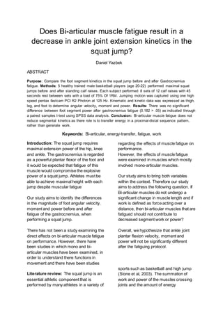
Figure 2: An exampleofone participant ofthe anklejoint plantar flexionpowerversustime, during theascending phase of
the vertical squat jump. The Blueand red linesrepresentthesquat jump before and after fatiguerespectively. This datawas
resampled to comparemean segment power. The preand post foot segment power ofthejump was12.5 J.S-1. Net Plantar
flexion power wasdefined aspositiveand in theclockwisedirection. All preand post valuesfor participantswerenot
significantly different (0.182>0.05)
Foot segmentpower
Power (J.S-1
Time (frames / seconds) (125/s)
𝑃 = 𝑀 ∙ 𝜔
Figure 1: Modelofthe Hip,knee& ankle withrespectivethigh,leg & foot segments. The Gastrocnemiusisrepresented asthe
line joining thedistal posterior thigh to theposteriorplantar foot. Diagram on the left is
the descent phase and rightside istheascent phase ofthe
squat jump.
4. Although peak plantar flexion power was
not significantly different (0.182>0.05) in
the 5 subjects, this participant showed no
difference in peak plantar flexion power of
the squat jump. Peak foot segment power
was 12.5 J.S-1 before and after fatigue.
Discussion: Our results imply that the
gastrocnemius must have acted passively
to confine segment direction and
magnitude.
Maximal muscular fatigue of the bi-
articular gastrocnemius did not affect the
foot segment power during the squat
jump. It appears that our results do
support the mechanical function of bi-
articular muscles.
The gastrocnemius must have behaved
primarily as a segmental link between the
thigh and foot, rather than generate work,
in which it’s coupling action to plantar
flexion power, was dependent on the
muscles proximally, such as the one-joint
vasti and gluteus Maximus.
One flaw to this study is the failure to
induce a fatiguing protocol that is specific
to the speed of plantar flexion during the
squat jump. Had we incorporated
plyometric calf raises to the protocol,
perhaps the foot segment kinetics would
have changed. Further studies need to
address these effects.
Conclusion: Maximal muscle induced
fatigue to the gastrocnemius does not
decrease foot segment power during the
squat jump.
References:
Barret,R., & Neal,R. (2000). Energeticsof lowerextremitymovementspredictedusingan
EMG-Drivenmuscle model.InternationalSymposiumon Biomechanics.18:1-4.
JI, Q., JI, Z. & Liu,R. (2000).Joint power and its relationship to the fatigue of human body during
Vertical jumps.InternationalSymposiumon Biomechanics.18: 1-4.
Robertson,D.E.,Wilson,J.J.,&ST Pierre,T.A.(2008). Lowerextremityfunctions duringfull
squats.Journalof Applied Biomechanics.24: 333-339.
Stone, M.H., O’Bryant, H.S., McCoy, L., Coglianese,R., Lehmkuhl,M. & (2003). Power and maximum
strength relationships during performance ofdynamic and static weighted jumps. Journal ofstrength and
conditioning Research. 17(1):140-14.
Tomioka,M., Owings,T.M.,& Grabiner,M.D. (2001). Lower extremitystrengthand
coordinationare independentcontributorstomaximal vertical jumpheight. Journalof Applied
Biomechanics.17: 181-187.
Toumi,H, Pourmarat,G., Best,T.M., Martin,A.,Fairclough,J.,& Benjamin,M.(2006).
Fatigue andmuscle-tendonstiffnessafterstretch-shorteningcycle andisometricexercise.Applied
Physiology,Nutrition &Metabolism.31(5):565-572.
5. VanIngenSchenau,G.J.,Bobbert,M.F.,& Jacobs,R. (1996). Mechanical outputfrom
individualmusclesduringexplosive legextensions:The role of Bi-articularmuscles. Journalof
Biomechanics.29(4): 513-523.
VanIngenSchenau,G.J.,Bobbert,M.F.& Rozendal,R.H.(1987). The unique actionof Bi-
articularmusclesincomplex movements. Journalof anatomy.155: 1-5.