Complex Odontogenic Infection
•Download as PPTX, PDF•
12 likes•1,500 views

Serious Life-threatening conditions Copyright (c) Department of Oral and Maxillofacial Surgery University of Dental Medicine, Yangon

Recommended
Recommended
More Related Content
What's hot
What's hot (20)
Similar to Complex Odontogenic Infection
Similar to Complex Odontogenic Infection (20)
More from Cing Sian Dal
More from Cing Sian Dal (20)
Recently uploaded
Recently uploaded (20)
Complex Odontogenic Infection
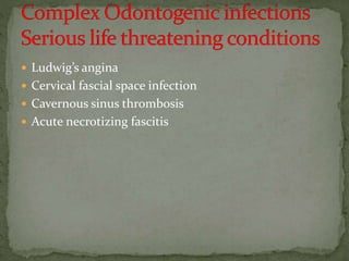
- 1. Ludwig’s angina Cervical fascial space infection Cavernous sinus thrombosis Acute necrotizing fascitis
- 2. Bilateral submandibular , sublingual and submental spaces = 6 spaces Impending Ludwig ( close to ) Rapidly and aggressively spreading cellulitis involving bilateral submandibular, sublingual and submental spaces
- 4. extension of infection from the the mandibular teeth usually second and third molars infection erodes through the medial aspect of the mandible inferior to the mylohyoid line as a result of haemolytic strepto cocci , aerobic and anaerobic organism virulence property - hyaluronidase , collagenase , fibrinolysin , enzyme that cause tissue destruction or promote bacterial spread
- 5. Submental space lies between the anterior bellies of the digastric and between the mylohyoid muscle and overlying skin , by mandibular incisors
- 6. Sublingual space lies between the oral mucosa of the floor of the mouth and the mylohyoid muscle , most commonly seen with premolars and first molar , its posterior border is open and communicate with submandibular space ,
- 7. Submandibular space liesbetween the mylohyoid muscle and overlying skin and superficial fascia , posterior boundary communicates with the secondary spaces
- 8. chill , fever , increased pulse rate and respiratory rate - toxic appearance - fatigue , feverish , malaise - painful brawny swelling of the upper part of the neck and the floor of the mouth on both sides - induration , board like and do not pit on pressure , no fluctuation , tissue may become gangrenous , when cut - lifeless , sharp demarcation from the surrounding normal tissue - typical open mouthed appearance , tongue is protruded and elevated - sublingual space involvement , limited tongue movement - drooling of saliva , increased salivation
- 9. may has severe trismus ( < than 10mm ) - difficulty in swallowing ( dysphagia ) -respiratory obstruction due to odema of the glottis ,noisy breathing ( stridor ) , restlessness, respiration using acessory muscles , cyanosis , asphyxiation - die from asphyxia , toxaemia , septicaemia , infection to the mediastinum
- 10. Surgical intervention - surgical incision should be under taken early before respiratory obstruction develops , Aim to release of tissue tension , adequate exposure of deep compartment , to provide drainage primary as well as secondary spaces , more than one drain ( antibiotic alone cannot eleminate the pus ) , pus for C & S , G.A. is hazardeous , Local anaesthesia is more safer
- 11. Incision in the submental area should be extended through the mylohyoid muscle to the mucous membrane Emergency tracheostomy if respiration becomes embrassed , Gross swelling may distort the normal anatomy of the face and neck Parallel incision medial to the lower border of the mandible which extended upward to the base of the tongue in the submandibular area
- 12. Pretracheal space Lareral pharyngeal space Retropharyngeal space Danger space Prevertebral space
- 13. Ant; Sup. and mid. pharyngeal constrictor m. Post; Carotid sheath and scalene fascia Sup; Skull base Inf; Hyoid bone Likely cause; Lower third molars, Tonsillar infection in neighboring spaces Contents; Carotid a., Internal jugular v., Vagus n., Cervical sympathetic chain Neighbouring space; Pterygomandibular, Submandibular, Sublingual, Peritonsillar, Retropharyngeal
- 14. Severe trismus – involvement of the lateral pterygoid muscle Difficulty in swallowing Lateral swelling of the neck Direct effect of the infection on the contents of the space; grave problems ; Thrombosis of the IJV Erosion of the carotid artery or its branches Interference with IX, X & XII CN Infection progresses to retropharyngeal space
- 15. Ant; Sup. And mid. Pharyngeal constrictor m. Post; Alar fascia Sup; Skull base Inf; Mediastinum (Fusion of alar and prevertebral fasciae at variable level between C6-T4)
- 16. Xray - Retropharyngeal soft tissue shadow is narrow (3- 4mm ) and located at C2 and at C6 when retropharyngeal space is involved , soft tissue becomes substantially thicker , space enlarge and compromising the airway No important contents Posterosuperior mediastinum may also become involved secondarily Mediastinum ; the space in the thorac between two pleural sac , contains heart , aorta, trachea , oesophagus and thymus Progressive involvement of the prevertibral spaces
- 17. Ant; alar fascia Post; prevertibral fascia Sup; cranial base Inf; diaphragm Mostly risk to the involvement of entire mediastinum
- 18. Three greatest potential complications Serious possibility of upper airway obstruction as a result of anterior displacement of the posterior pharyngeal wall into the oro pharynx Rupture of the retropharyngeal space abscess with aspiration of pus into the lungs and subsequent asphyxiation Infection spread into the mediastinum which results in severe infection in the thorax
- 19. Ant; Sternothyroid- thyrohyoid fascia Post; Trachea Sup; Thyoid cartilage Inf; Superior mediastinum
- 23. An extension of the infection into the area not detected at first treatment may have to be I&D
- 25. Cavernous sinus contents O TOM CAT: O TOM are lateral wall components, in order from superior to inferior. CA are the components within the sinus, from medial to lateral. CA ends at the level of T from O TOM. See diagram. Occulomotor nerve (III) Trochlear nerve (IV) Ophthalmic nerve (V1) Maxillary nerve (V2) Carotid artery Abducent nerve (VI) T: When written, connects to the T of OTOM.
- 26. Cavernous sinus is so called because it is divided into caverns by fibrous septa , sponge like appearance. It lies along side the body of the sphenoid bone in the middle cranial fossa and it is formed in between the outer layer of the dura covering the body of the sphenoid bone and inner layer of dura , two cavernous sinuses are connected by anterior and posterior intercavernous sinus , may readily spread from one sinus to other
- 28. high mortality even today - superior spread of infection via a haematogenous route , septic thrombosis of the cavernous sinus - veins of the face and orbit lack valves which permits blood flow in either direction -
- 29. posteriorly via - pterygoid plexus and emissary veins ( are communications between intracranial venous sinuses and extracranial vein , foramen ovale and or sphenoidal foramen / Vesalires) pterygoid plexus also anastomoses with the inferior opthalmic vein by a vein treansversing the inferior orbital fissure ,
- 30. anteriorly via angular vein and inferior or superior opthalmic veins ( supratrochlear and supraorbital unite at the medial corner of the eyelid ) angular vein which then continue as across the face as a facial vein , communication of the angular vein with the superior opthalmic vein often called nasofrontal vein , superior opthalmic vein is tha main tributary of the cavernous sinus , large communication from the facial vein via the deep facial vein to the pterygoid plexus
- 32. ; six features (1) known site of infection- boils , furunculosis and infected hair follicles (staphylococci infection) of the face that is drained by the facial vein ( danger trigone ) hardening along the course of vein , odontogenic infection , infections from eyes ( via the superior , inferior opthalmic vein - direct and
- 33. indirect through pterygoid plexus ), ears ( through petrosal sinus ), paranasal sinuses , pharynx ( pharyngeal plexus communicate with cavernous sinus by emissary veins ) , tonsillar and paratonsillar abscesses (2) evidence of blood stream infection - signs of systemic involvement - fever, increased pulse rate & respiratory rate , toxic appearance , blood culture positive of Staphylococcal aureus (3) early sign of venous obstruction in the retina conjuntiva or eyelid- papillodema (Ophthalmoscope) chemosis ( odema of the occular conjuntiva ) , orbital cellulitis and abscess , 50 % motality loss vision , one or both eyes , impairment of the vision is due to odema of the optic nerve with congestion of the central vein of the retina
- 34. Superior Orbital syndrome - is characterized by opthalmoplegia , ptosis , proptosis of the eyes , dilated and fixity of pupil , sometime blood stained tear trickled down the cheek , anaesthesia of the eyelid and forehead Orbital apex syndrome - involvement of the optic nerve , blindness (4) paresis of the 3,4 & 6 CN resulting from inflammatory edema - voluntary movement of all extrinsic occcular muscles are abolished ( opthalmoplegia ) , early one eye involvement , later other one
- 35. 6 CN being the more exposed position within the sinus and often the first to be involved loss of abduction ( away from the midline - lateral rectus ) - 4 C N - supplies superior oblique - impairment leads to loss of downward movement of the eyeball if it is adducted(move toward the midline) - 3 C N - supplies all muscles except lateral rectus and superior oblique , 3 C n involvement leads to loss of adduction ( toward midline - medial rectus ) , elevation ( superior rectus , depression (inferior rectus ) , elevation abduction ( inferior oblique ) ,elevation of upper eyelid ( levator palpebra superioris)
- 36. (5) abscess formation in the neighbouring soft tissue (6) evidence of meningeal irritation - head ache , vomitting , photophobia , irritable , evokes reflex spasm in the paravertebral muscles resulting in neck stiffness in the cervical area and positive Kernig’s sign in lumbar area muscle spasm
- 37. Signs of meningitis – Neuchal rigidity (unable to do flexion of the neck) due to spasm of paravertebral muscles Kernig’s sign (in supine position when the thighs are held at 90 degree, the legs are unable to extend at the knees) due to stiffness of hamstring muscles at the legs Brudzinski’s sign( at supine position, when flexion of neck, the hips and knees also flex involuntarily)
- 38. patient in supine , flex the neck until the chin touches the chest Brudzinski’s sign - flexion of hips and knees in response to passive neck flexion
- 39. Kernig’s sign - patient in supine , both legs extended , contraction of hamstrings in response to knee extension while hip is flexed
- 40. die from , septicaemia , meningitis , enchephalitis - Treatment - antibiotic therapy ,corticosteroid are recommended to prevent circulatory collapse secondary to pituitary dysfunction , controversy - use of anticoagulant because of spread of infection , surgical access through eye enucleation , neuro surgical management
- 41. is a rapidly spreading soft-tissue infection that involves the subcutaneous tissues produces morbidity and in some instances mortality Most cases occur in the extremities, abdomen and perineum a rare complication from dental infection Sometimes as a result of minimal skin trauma or a simple tooth extraction
- 42. polymicrobial bacteria involved are the same species as those that cause chronic dental infections in the gingival crevice or periapical infections of the jaw immunocompromised, but also can occur in healthy people Obesity????
- 43. typically is febrile elevated WBC counts also might be hypotensive and tachycardia pain is severe and out of proportion to the clinical findings can be hypo aesthetic or anesthetic
- 44. within 24 - 48 hours , the area become red , edematous and painful , but soon becomes anaesthetic , well or ill demarcated , becomes dusky , purplish and black 4- 5 days necrosis of the skin appears , release of brownish exudate with gas bubbles the necrotic tissue starts to separate within 8 - 10 days
- 45. Rapid surgical debridement is warranted to stop the necrosis from spreading radical surgical debridement of necrotic tissue definitively by inspecting the tissue and performing a biopsy incision in advance of the line of necrosis to prevent subcutaneous spreading along fascial planes . The practitioner should make incision into the affected tissue produces virtually no bleeding Drainage appears dishwater like Blood vessels are thrombosed Fetor odor indicating necrotic tissue is characteristic
- 46. tracheostomy or endotracheal intubationto protect the patient’s airway owing to severe neck swelling Ventilator support is required in patients with severe cases of CNF, owing to acute respiratory failure Skin graft may be necessary later in case of large skin defect
- 47. A delay in seeking treatment for odontogenic infection is a common finding early stages may resemble odontogenic cellulitis or as abscess
- 50. transcervical neck incision to create a wide exposure
- 51. can lead to involvement of the neck, mediastinum and chest wall
- 54. in the submandibular region to open the fascial planes from the mandible to the clavicle
- 55. Cardiovascular intensive measures such as intravenous (IV) fluids and medication to support the patient’s blood pressure and heart rate. Hyperbaric oxygen therapy(HBO) is an adjunctive treatment for CNF. It has shown a beneficial effect.
- 56. Overwhelming sepsis, mediastinitis and multiple organ failures If mediastinum involvement occurs, the mortality rate is approximately 50 percents.
- 57. Must be hospitalised Surgical and medical management require more extensive and aggressive treatment
- 58. Medical support of the patient with special attention to correcting host defense compromises where they exist Administration of the proper antibiotics in appropriate doses Surgical removal of the source of infection as early as possible Surgical drainage of the infection with placement of proper drains Constant reevaluation of the resolution of the infection
- 59. Surgeon must not wait for unequivocal evidence of pus formation I&D must be extensive , various sites At OTh Aggressive exploration of the involved fascial space One or more drain require to provide adequate drainage and decompression of the infected area Removal of the source of infection as early as possible , removal of drain should not be done prior to the extraction of the causative tooth
- 60. Support host defense mechanism including analgesics , fluid requirements and nutrition High dose bacteriacidal antibiotics Almost always administered intravenously Mouth rinses - 0.02 % Chlorhexidine gluconate, bland M/W Mouth opening exercise - active and passive
- 61. Airway continually monitored If respiration becomes embrassed surgical airway established if warranted Emergency tracheostomy , gross swelling may distort the normal anatomy of the face and neck