Reversal of neurological defects in a mouse model of rett syndrome
USPD 2014
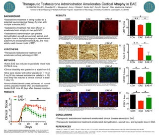
1. 0 10 20 30 40
0
1
2
3
4
Days
ClinicalScore
EAE+T
EAE
*
Therapeutic Testosterone Administration Ameliorates Cortical Atrophy in EAE
SHANNON WAILES1, Chandler R. L. Mongerson1, Amy J. Wisdom2, Noriko Itoh2, Rory D. Spence1, Allan MacKenzie-Graham1
1Division of Brain Mapping or 2Multiple Sclerosis Program, Department of Neurology, University of California, Los Angeles, CA 90095
BACKGROUND
•Testosterone treatment is being studied as a
potential neuroprotective therapy for men with
multiple sclerosis (MS)1.
•Testosterone treatment has been shown to
ameliorate brain atrophy in men with MS1.
•Testosterone administration can prevent
demyelination as well as neuronal, axonal, and
synaptic loss in the hippocampus in experimental
autoimmune encephalomyelitis (EAE), the most
widely used mouse model of MS2.
HYPOTHESIS
•Therapeutic testosterone treatment will
ameliorate cortical pathology in EAE.
METHODS
•Active EAE was induced in gonadally intact male
C57BL/6 mice.
•Clinical disability was graded on a scale from 0-5.
•Mice were treated with either placebo (n = 10) or
5 mg 90 day release testosterone pellets (n = 10)
once they had reached a clinical score of at least
1 (day 12).
•Immunohistochemistry was performed on normal
control, placebo-treated EAE, and testosterone-
treated EAE mice 40 days after disease induction.
RESULTS
CONCLUSIONS
•Therapeutic testosterone treatment ameliorated clinical disease severity in EAE.
•Therapeutic testosterone treatment ameliorated demyelination, axonal loss, and synaptic loss in EAE.
REFERENCES
1) Sicotte, N. L., Giesser, B. S., Tandon, V., Klutch, R., Steiner, B., Drain, A. E., et al. (2007). Testosterone treatment in multiple sclerosis: a pilot study. Archives of Neurology, 64(5), 683–688.
2) Ziehn, M. O., Avedisian, A. A., Dervin, S. M., Umeda, E. A., O'Dell, T. J., & Voskuhl, R. R. (2012). Therapeutic Testosterone Administration Preserves Excitatory Synaptic Transmission in the Hippocampus during
Autoimmune Demyelinating Disease. Journal of Neuroscience, 32(36), 12312–12324.
MBP
A
NF200/DAPI
A
CON EAE EAE+T
0
25
50
MBP%Area
** *
*
CON EAE EAE+T
0
5
10
TypeIILesions
** *
CON EAE EAE+T
0
20
40
NF200%Area
* *
CON EAE EAE+T
0
20
40
60
PSD%Area
* *
Figure 2. Therapeutic testosterone treatment ameliorated demyelination in the cortex. Myelin basic protein (MBP) expression in normal control
(A), placebo-treated EAE (B), and testosterone-treated EAE mice (C). MBP expression was significantly greater in testosterone-treated EAE mice
compared to placebo-treated EAE mice, however, testosterone-treated EAE mice also demonstrated significantly less MBP than normal control mice
(D). n = 8 for each group. *p < 0.05. **p < 0.01. Welch’s t-test. Scale bar = 44µm.
PSD/DAPIPLP
Figure 1. Therapeutic testosterone treatment ameliorated clinical disease. Mice with EAE were treated with
either placebo (red) or testosterone (green) 12 days (arrow) after disease induction. Testosterone treatment
significantly decreased clinical disease from day 21-40. n = 10 per group. *p < 0.05. Repeated Measure ANOVA.
A
CON
B C D
EAE EAE+T
CON EAE EAE+T
Figure 3. Therapeutic testosterone treatment ameliorated type II lesions in the cortex. Myelin proteolipid protein (PLP) expression was used to
quantify type II lesions (black arrow) in normal control (A), placebo-treated EAE (B), and testosterone-treated EAE mice (C). Type II lesions were
significantly less in testosterone-treated EAE mice compared to placebo-treated EAE mice, however, testosterone-treated EAE mice also
demonstrated significantly more lesions than normal control mice (D). n = 8 for each group. *p < 0.05. **p < 0.01. Welch’s t-test. Scale bar = 19µm.
RESULTS
A B C D
CON EAE EAE+T
A B C D
CON EAE EAE+T
A B C D
Figure 4. Therapeutic testosterone treatment ameliorated axonal loss in the cortex. Neurofilament 200 (NF200) expression (green) was used to
quantify axonal loss in normal control (A), placebo-treated EAE (B), and testosterone-treated EAE mice (C). Axonal loss was significantly less in
normal control and testosterone-treated EAE mice compared to placebo-treated EAE mice (D). Dapi (blue) shows cell nuclei. n = 8 for each group.
*p < 0.05. Welch’s t-test. Scale bar = 27µm.
Figure 5. Therapeutic testosterone treatment ameliorated synaptic loss in the cortex. Postsynaptic density protein (PSD95) expression (red)
was used to quantify synaptic loss in normal control (A), placebo-treated EAE (B), and testosterone-treated EAE mice (C). Synaptic loss was
significantly less in normal control and testosterone-treated EAE mice compared to placebo-treated EAE mice (D). Dapi (blue) shows cell nuclei. n =
8 for each group. *p < 0.05. Welch’s t-test. Scale bar = 19µm.