1. Spinal cord injury (SCI):.
• Female Sprague-Dawley rats were anesthetized and a laminectomy was performed at vertebral level T8 followed by
lateral compression of the spinal cord using modified forceps.
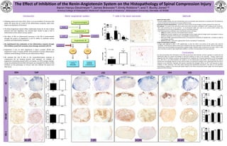
Eriochrome cyanine histochemistry: myelin stain used to assess the percentage of myelin sparing at the injury site.
IHC analyses: At various times post-injury, spinal cords were harvested, embedded in OCT compound and stored at -
80C until sectioning into 10 m sections. 0.1 M phosphate buffer was used as our primary buffer solution, and
biotinylated horse anti-mouse (BHAM) was used as a secondary antibody.
• The following primary antibodies were used identify various cell types:
• GFAP (anti-GFAP): astrocyte intermediate filament protein
• OX-42 (anti-CD11b): recognizes most macrophages via the integrin alpha M antigen which participates in various
adhesive interactions of monocytes, macrophages, and granulocytes.
• ED-1 (anti-CD68): expressed predominantly on the lysosomal membranes of myeloid cells – provides an index of
macrophage activation
• OX-19 (anti-CD5): reacts with a glycoprotein found on peripheral T-lymphocytes
Image and quantitative Analyses:
• Images were digitized at 40X or 50X magnification so that the entire cross section of the spinal cord could be
visualized. Three sections (200 m apart) were analyzed at the injury epicenter for each animal. The area of positively-
stained tissue was thresholded and quantified using NIH Image J software. Data is reported as the mean SEM. T-cells
were manually counted at high magnification (200X) in 3 sections corresponding to the injury epicenter.
The Effect of Inhibition of the Renin-Angiotensin System on the Histopathology of Spinal Compression Injury
Aaron Hanyu-Deutmeyer , James Brewster , Emily Robbins , and T. Bucky Jones+,
Arizona College of Osteopathic Medicine, Department of Anatomy+, Midwestern University, Glendale, AZ 85308
Introduction Methods
• Following spinal cord injury (SCI), there is an accumulation of immune cells
within the injury site as a result of the inflammatory response, which then
leads to an expansion of the lesion.
• The Renin-Angiotensin System (RAS), traditionally known for its role in blood
pressure and fluid regulation, has recently been shown to play a role in
inflammation of the central nervous system.
• The effect of RAS on inflammatory processes in the CNS is predominately
through the actions of Angiotensin II and its ability to modulate various
cytokines and inflammatory mediators.
• We hypothesized that modulation of the inflammatory response through
RAS inhibition would limit secondary tissue damage associated with SCI.
• Angiotensin II acts on both Angiotensin II type-1 receptor (AT1R) and
Angiotensin II type-2 receptor (AT2R); AT1R activation increases inflammatory
mediators while binding of AT2R has anti-inflammatory effects.
• We assessed the role of RAS on the neuroinflammatory response to
compression SCI by treating animals with captopril, an inhibitor of
angiotensin-converting enzyme (ACE), or losartan, an AT1R receptor blocker.
Using immunohistochemical (IHC) techniques we evaluated the effects of RAS
inhibition on the phenotype of immune cells that infiltrate the spinal cord
after injury.
Conclusions
Renin-angiotensin system
Our data demonstrate that modulation of the RAS by post-injury administration of either captopril or losartan did not
significantly affect the overall macrophage or astrocyte response to injury at the epicenter. Previous reports have
suggested that RAS inhibition produces neuroprotection by modifying the functional phenotype of CNS macrophages
(e.g., shift from a pro-inflammato44ry M1 profile to an anti-inflammatory M2 profile). The antibodies we used to
evaluate the macrophage response to injury did not allow us to determine whether such a phenotype shift occurred in
response to our treatment. Captopril and losartan treatment had divergent effects on the T-cell response to injury.
Inhibition of ACE enhanced T-cell influx while blocking AT1R significantly decreased T-cell influx at the epicenter.
Expanding our analyses to the rostral and caudal margins of the lesion may provide further insight into the therapeutic
potential of RAS inhibition.
ED-1OX-42GFAPEC
VEH CAP VEH LOS VEH LOS VEH LOS
7 DPI 14 DPI 28 DPI28 DPI
T-cells in the lesion epicenter
7 dpi
14 dpi
*
*
*
* p < 0.05