![Cecilia Young et al J Res Med Dent Sci, 2018, 6 (4):61-64
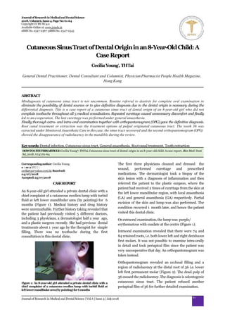
Figure 2: Orthopantomogram (OPG) showed retained roots of both
deciduous lower left first molar 74, and deciduous lower right first
molar 84 and apical radiolucencies at permanent lower left first
molar36
Re-appointment was scheduled on the next day for
follow-up.
One of the treatment options for this patient would be
removing the pulp through root canal treatment (RCT)
in multiple visits and observe the subsiding and recovery
of the surrounding bone and the healing of the fistula.
Since the infection has been there for over 6 months and
draining from the face, the use of a diode laser for
disinfection before obturation for root canal treatment
was also recommended. Another treatment option
would be an extraction of the tooth. Both treatment
options needed follow-up to monitor the recovery by
orthopantomogram (OPG).
Considering the multiple visits required by root canal
treatment and also cost and time involvement by the
patients, the patient finally had the tooth 36 extracted
under Monitored Anaesthetic Care. There were no
adverse and unanticipated events in the treatment.
During the review, the dentist explained the conditions
and OPG findings (Figure 3) to the father of the patient.
The bone was healed, and the lower left second molar
was likely to occupy the original space of the extracted
lower left first molar. The patient was symptom-free but
with a scar (Figure 4).
Figure 3: The radiolucency at distal root region of lower left first
permanent molar 36 disappeared. The developing lower left
second permanent molar 37 is migrating to the 36 space. It might
notfullyoccupythe space
Figure4:Scar
62
Journalof Research in Medical and DentalScience |Vol.6 |Issue 4 |July2018
DISCUSSION
Previous literature has shown misdiagnoses by
physicians and/or dermatologists as dermatological
problem are notuncommon[1-4], e.g.a simple cutaneous
abscess [1]. Often patients of cutaneous sinus tract are
misdiagnosed, and they seek help from dentist only after
the initial therapy failed [1-3]. The time taken between
the first presentation of symptoms and finally arriving a
correct diagnosis could be lengthy, possibly even over
15 years[4].
Odontogenicinfections canspread through fascial planes
[5] and intracranially [6]. Except for intraoral abscess, it
may also cause orbital cellulitis [7,8], may eventually
leading to blindness [7], cavernous sinus thrombosis [9],
brain abscess [6,10], and Ludwig’s angina [11,12].
Misdirected therapies without treating the source of
infection can be multiple to the patient, such as
antibiotics [1,3,13,14], steroids [3] and surgical excisions
[14]. An antibiotic course might result in a temporary
amelioration of symptoms [3,14], the condition will
recur unless the dental origin is cleared [3,5,14].
Treatments without removal of the source often delay
the appropriate treatment and recovery [1-3,5,13,14].
Differential diagnosis of cutaneous sinus tract include
suppurative apical periodontitis [3,15], osteomyelitis
[3,14,15], congenital fistula [3,15], salivary gland fistula
[3,14,15], an infected cyst [3,15] and deep mycotic
infection [3,7]. The most common cause of cutaneous
sinus tract of dental origin is periapical infection around
the root apices of nearby carious or traumatised teeth
[13].
Patients are usually not aware of intraoral symptoms
and do not relate the skin lesion to the dental origin.
Thus patients are likely to first seek help from physicians
instead of dentists [2,16]. The consultation and opinion
of a dentist are vital, as the periodontal disease [10] and
a dead pulp [16] are the main sources to head and neck
infection.
Referral to dentists, to perform complete extraoral and
intraoral examination dental radiography [1,2,5,17] and
pulp test [5,17] are procedures during the consultation;
to eliminate the possibility of dental sources or to give
definitive diagnosis due to dental origin is necessary
during the differential diagnosis [1,5,14].Proper referral](data:image/gif;base64,R0lGODlhAQABAIAAAAAAAP///yH5BAEAAAAALAAAAAABAAEAAAIBRAA7)
Recommended
Recommended
More Related Content
What's hot
What's hot (20)
Similar to Cutaneous sinus-tract-of-dental-origin-in-an-8yearold-child-a-case-report
Similar to Cutaneous sinus-tract-of-dental-origin-in-an-8yearold-child-a-case-report (20)
More from Cecilia Young 楊幽幽
More from Cecilia Young 楊幽幽 (20)
Recently uploaded
Recently uploaded (20)
Cutaneous sinus-tract-of-dental-origin-in-an-8yearold-child-a-case-report
- 1. JournalofResearchinMedicalandDentalScience 2018,Volume6,Issue4,PageNo:61-64 Copyright CCBY-NC4.0 AvailableOnline at:www.jrmds.in eISSNNo.2347-2367: pISSNNo. 2347-2545 Correspondingauthor:Cecilia Young e - m a i l : ceciliatyp@yahoo.com.hk Received: 04/07/2018 Accepted:25/07/2018 CASEREPORT An 8-year-old girl attended a private dental clinic with a chief complaint of a cutaneous swollen lump with turbid fluid at left lower mandibular area (by pointing) for 6 months (Figure 1). Medical history and drug history were unremarkable. Further history taking revealed that the patient had previously visited 5 different doctors, including 3 physicians, a dermatologist half a year ago, and a plastic surgeon recently. She had previous dental treatments about 1 year ago by the therapist for simple filling. There was no toothache during the first consultation in this dental clinic. Figure 1: An 8-year-old girl attended a private dental clinic with a chief complaint of a cutaneous swollen lump with turbid fluid at leftlowermandibulararea(bypointing)for6months The first three physicians cleaned and dressed the wound, performed curettage and prescribed medications. The dermatologist took a biopsy of the skin lesion with a diagnosis of inflammation and then referred the patient to the plastic surgeon, where the patient had received 2 times of curettage from the skin at the left lower mandibular region, with local anaesthesia (LA) and general anaesthesia (GA) respectively. Partial excision of the skin and lump was also performed. The condition recurred 1 month later, and hence the patient visited this dental clinic. Onextraoral examination,the lump was purple/ erythematous with exudate at the centre (Figure 1). Intraoral examination revealed that there were 74 and 84 retained roots, i.e. both lower left and right deciduous first molars. It was not possible to examine intra-orally in detail and took periapical film since the patient was very uncooperative that day. An orthopantomogram was taken instead. Orthopantomogram revealed an occlusal filling and a region of radiolucency at the distal root of 36 i.e. lower left first permanent molar (Figure 2). The dead pulp of 36 caused the radiolucency. The diagnosis is odontogenic cutaneous sinus tract. The patient refused another periapical film of 36 for further detailed examination. CutaneousSinusTractofDentalOrigininan8-Year-OldChild:A CaseReport CeciliaYoung* , THTai General Dental Practitioner, Dental Consultant and Columnist, Physician Pharmacist People Health Magazine, Hong Kong ABSTRACT Misdiagnosis of cutaneous sinus tract is not uncommon. Routine referral to dentists for complete oral examination to eliminate the possibility of dental sources or to give definitive diagnosis due to the dental origin is necessary during the differential diagnosis. This is a case report of a cutaneous sinus tract of dental origin of an 8-year-old girl who did not complain toothache throughout all 5 medical consultations. Repeated curettage caused unnecessary discomfort and finally led to un-cooperation. The last curettage was performed under general anaesthesia. Finally thorough extra- and intra-oral examination together with orthopantomogram (OPG) gave the definitive diagnosis. Root canal treatment or extraction was the treatment options of pulpal originated cutaneous tract. The tooth 36 was extracted under Monitored Anaesthetic Care in this case;the sinus tract recovered and the second orthopantomogram (OPG) showed the disappearance of radiolucency in the mandible during the review. Keywords:Dental infection, Cutaneous sinus tract, General anaesthesia, Rootcanal treatment, Tooth extraction HOWTOCITETHISARTICLE:Cecilia Young*, THTai, Cutaneous sinus tract of dental origin in an 8-year-old child: A case report, JRes Med Dent Sci, 2018, 6 (4):61-64 61 Journalof Research in Medical and DentalScience |Vol.6 |Issue 4 |July2018
- 2. Cecilia Young et al J Res Med Dent Sci, 2018, 6 (4):61-64 Figure 2: Orthopantomogram (OPG) showed retained roots of both deciduous lower left first molar 74, and deciduous lower right first molar 84 and apical radiolucencies at permanent lower left first molar36 Re-appointment was scheduled on the next day for follow-up. One of the treatment options for this patient would be removing the pulp through root canal treatment (RCT) in multiple visits and observe the subsiding and recovery of the surrounding bone and the healing of the fistula. Since the infection has been there for over 6 months and draining from the face, the use of a diode laser for disinfection before obturation for root canal treatment was also recommended. Another treatment option would be an extraction of the tooth. Both treatment options needed follow-up to monitor the recovery by orthopantomogram (OPG). Considering the multiple visits required by root canal treatment and also cost and time involvement by the patients, the patient finally had the tooth 36 extracted under Monitored Anaesthetic Care. There were no adverse and unanticipated events in the treatment. During the review, the dentist explained the conditions and OPG findings (Figure 3) to the father of the patient. The bone was healed, and the lower left second molar was likely to occupy the original space of the extracted lower left first molar. The patient was symptom-free but with a scar (Figure 4). Figure 3: The radiolucency at distal root region of lower left first permanent molar 36 disappeared. The developing lower left second permanent molar 37 is migrating to the 36 space. It might notfullyoccupythe space Figure4:Scar 62 Journalof Research in Medical and DentalScience |Vol.6 |Issue 4 |July2018 DISCUSSION Previous literature has shown misdiagnoses by physicians and/or dermatologists as dermatological problem are notuncommon[1-4], e.g.a simple cutaneous abscess [1]. Often patients of cutaneous sinus tract are misdiagnosed, and they seek help from dentist only after the initial therapy failed [1-3]. The time taken between the first presentation of symptoms and finally arriving a correct diagnosis could be lengthy, possibly even over 15 years[4]. Odontogenicinfections canspread through fascial planes [5] and intracranially [6]. Except for intraoral abscess, it may also cause orbital cellulitis [7,8], may eventually leading to blindness [7], cavernous sinus thrombosis [9], brain abscess [6,10], and Ludwig’s angina [11,12]. Misdirected therapies without treating the source of infection can be multiple to the patient, such as antibiotics [1,3,13,14], steroids [3] and surgical excisions [14]. An antibiotic course might result in a temporary amelioration of symptoms [3,14], the condition will recur unless the dental origin is cleared [3,5,14]. Treatments without removal of the source often delay the appropriate treatment and recovery [1-3,5,13,14]. Differential diagnosis of cutaneous sinus tract include suppurative apical periodontitis [3,15], osteomyelitis [3,14,15], congenital fistula [3,15], salivary gland fistula [3,14,15], an infected cyst [3,15] and deep mycotic infection [3,7]. The most common cause of cutaneous sinus tract of dental origin is periapical infection around the root apices of nearby carious or traumatised teeth [13]. Patients are usually not aware of intraoral symptoms and do not relate the skin lesion to the dental origin. Thus patients are likely to first seek help from physicians instead of dentists [2,16]. The consultation and opinion of a dentist are vital, as the periodontal disease [10] and a dead pulp [16] are the main sources to head and neck infection. Referral to dentists, to perform complete extraoral and intraoral examination dental radiography [1,2,5,17] and pulp test [5,17] are procedures during the consultation; to eliminate the possibility of dental sources or to give definitive diagnosis due to dental origin is necessary during the differential diagnosis [1,5,14].Proper referral
- 3. Cecilia Young et al J Res Med Dent Sci, 2018, 6 (4):61-64 can avoid unnecessary investigations and treatments such as antibiotics [2] or surgical operations [1,2]. Also, prompt diagnosis and treatment could reduce patient discomfort, aesthetic problems, and complications such as sepsis and osteomyelitis [1,2]. Therefore physicians must be aware of this condition, and it is advised to refer the patient to dentists if seen any cutaneous lesion in the head and neck region to confirm whether the skin lesion is from dental origin [2,13]. In this case, the absence of an obvious decayed tooth hindered the parents to give the history of previous dental disease and treatment, and the filling covered the dead pulp. Therefore the 5 medical practitioners failed to relate the lesion to the dental origin and could not reach a correct diagnosis promptly. The patient was uncooperative during the dental consultation in this clinic that detailed intra-oral examination was not possible, filling over the affected tooth was revealed by the OPG (Figure 2). The patient had a filling on 36 done by a therapist, it means that she was very cooperative in the initial dental treatment, repeated curettage caused the un-cooperation, the last curettage was undergone in general anaesthesia, then dental examination and treatment became difficult led to extraction under MonitoredAnaesthetic Care. Asking the patient about relevant dental symptoms like a toothache might be helpful, but it is worthy to note that the condition could be painless if the pulp is completely dead. A portion of the patient does not have a toothache [2,18]. A history of a toothache in the affected area that resolved without dental intervention can be useful information [3], but this does not apply to this 8-year- old girl because there were treatment and a filling but failed. Pediatrics patient might not be able to describe their symptoms accurately. Pulp test is also not reliable because it depends on the response from the patient. Superficial healing of theskindoes notmean therecovery of the disease since the source remains, repeated abscess and fistula will recur. Standard treatment of cutaneous sinus tract of dental origin is the elimination of the source of infection by root canal treatment [1,3-5] or by extraction of affected teeth [1-4]. The drainage will cease eventually, and the tract will close, typically between 7 to 14 days [18]. The cutaneous lesion will heal eventually even without any surgical operation[4]. Patient’s parents were advised if extraction of 36, in this case, did not ameliorate the symptom, biopsy on the skin lesion should be performed again for confirmation. 63 Journalof Research in Medical and DentalScience |Vol.6 |Issue 4 |July2018 CONCLUSION Diagnosis of cutaneous sinus tract of dental origin is difficult and requires multidisciplinary effort; it is advised medical practitioners to have appropriate and sufficient communication with dentists in order to have accurate diagnosis and treatment of these patients. Misdiagnosis and inappropriate treatments might physically,cosmetically, and even psychologicallyharm the patients, especially in paediatric patients. In this case, repeated curettage caused non-cooperation and led to one general anaesthesia at the last treatment from the plastic surgeon and one simple extraction by monitored anaesthetic care. CONFLICTOFINTEREST Nonedeclared. The patient was treated by the first author and consent was obtained fromparents. REFERENCES 1. Abuabara A, Schramm CA, Zielak JC,et al. Dental infection simulating skin lesion. Ann Bras Dermatol 2012; 87:619-621. 2. Cantatore JL, Klein PA, Lieblich LM, et al. Cutaneous dental sinus tract, A common misdiagnosis: A case report and review of the literature. Cutis 2002; 70:264-267. 3. Johnson BR, Remeikis NA, Van Cura JE, et al. Diagnosis and treatment of cutaneous facial sinus tracts of dental origin. J Am Dent Assoc 1999; 30:832-836. 4. Tidwell E, Jenkins JD, Ellis CD, et al. Cutaneous odontogenic sinus tract to the chin: A case report. JIntEndod 1997; 30:352-355. 5. Gupta M, Das D, Kapur R, et al. A clinical predicament-diagnosis and differential diagnosis of cutaneous facial sinus tracts of dental origin: A series of case reports. Oral Surg Oral Med Oral Pathol Oral Radiol Endod 2011; 112:e132-e136. 6. Schuman NJ, Turner JE Brain abscess and dentistry: A review of the literature. Quintessence Int1994; 25. 7. Park CH, Jee DH, La TY, et al. A case of odontogenic orbital cellulitis causing blindness by severe tension orbit. JKorean Med Sci 2013; 28:340-343. 8. Embong Z, Ismail S, Thanaraj A, et al. Dental infection presenting with ipsilateral parapharyngeal abscess and contralateral orbital cellulitis: A case report. Malays JMed Sci 2007; 14:62. 9. LaiPF,CusimanoMD.Thespectrumofcavernous sinus and orbital venous thrombosis: Acase and a review. Skull Base Surg 1996; 6:53. 10. Mylonas AI, Tzerbos FH, Mihalaki M, et al. Cerebral abscess of odontogenic origin1. J CraniomaxillofacSurg 2007; 35:63-67. 11. Candamourty R, Venkatachalam S, Babu MR, et al. Ludwig's angina-an emergency: A case report with literature review. JNat Sci Biol Med 2012; 3:206. 12. Young C. Editorial Case report Ludwig Angina– The longest distance in this planet. Adv Emerg Med 2018. 13. Yadav S, Malik S, Mittal HC, et al. Odontogenic cutaneous draining sinus. JCraniofac Surg 2014; 25:e86-e88. 14. Sammut S, Malden N, Lopes V, et al. Facial cutaneous sinuses of dental origin: A diagnostic challenge. Br Dent J2013; 215:555.
- 4. Cecilia Young et al J Res Med Dent Sci, 2018, 6 (4):61-64 15. Wood NK, Goaz PW, Alling CC. Differential diagnosis of oral lesions. St.Louis: Mosby 1991. 16. Belmehdi A, El Harti K, El Wady W,et al. Esthetic improvement of a nasolabial cutaneous sinus tract. ContempClin Dent 2018; 9:314. 17. Mittal N,Gupta P.Managementof extraoral 64 Journalof Research in Medical and DentalScience |Vol.6 |Issue 4 |July2018 sinus cases: Aclinical dilemma.JEndodont 2004; 30:541-547. 18. Janev E, Redzep E. Managing the cutaneous sinus tract of dental origine. Open Access Maced JMedSci 2016; 4:489.
- 6. Cutaneous Sinus Tract of Dental Origin in an 8-Year-Old Child: A Case Report Cecilia Young*, TH Tai General Dental Practitioner, Dental Consultant and Columnist, Physician Pharmacist People Health Magazine, Hong Kong ABSTRACT Misdiagnosis of cutaneous sinus tract is not uncommon. Routine referral to dentists for complete oral examination to eliminate the possibility of dental sources or to give definitive diagnosis due to the dental origin is necessary during the differential diagnosis. This is a case report of a cutaneous sinus tract of dental origin of an 8-year- old girl who did not complain toothache throughout all 5 medical consultations. Repeated curettage caused unnecessary discomfort and finally led to un- cooperation. The last curettage was performed under general anaesthesia. Finally thorough extra- and intra-oral examination together with orthopantomogram (OPG) gave the definitive diagnosis. Root canal treatment or extraction was the treatment options of pulpal originated cutaneous tract. The tooth 36 was extracted under Monitored Anaesthetic Care in this case; the sinus tract recovered and the second orthopantomogram (OPG) showed the disappearance of radiolucency in the mandible during the review. Key words: Dental infection, Cutaneous sinus tract, General anaesthesia, Root canal treatment, Tooth extraction CASE REPORT An 8-year-old girl attended a private dental clinic with a chief complaint of a cutaneous swollen lump with turbid fluid at left lower mandibular area (by pointing) for 6 months (Figure 1). Medical history and drug history were unremarkable. Further history taking revealed that the patient had previously visited 5 different doctors, including 3 physicians, a dermatologist half a year ago, and a plastic surgeon recently. She had previous dental treatments about 1 year ago by the therapist for simple filling. There was no toothache during the first consultation in this dental clinic. The first three physicians cleaned and dressed the wound, performed curettage and prescribed medications. The dermatologist took a biopsy of the skin lesion with a diagnosis of inflammation and then referred the patient to the plastic surgeon, where the patient had received 2 times
- 7. of curettage from the skin at the left lower mandibular region, with local anaesthesia (LA) and general anaesthesia (GA) respectively. Partial excision of the skin and lump was also performed. The condition recurred 1 month later, and hence the patient visited this dental clinic. On extraoral examination, the lump was purple/ erythematous with exudate at the centre (Figure 1). Intraoral examination revealed that there were 74 and 84 retained roots, i.e. both lower left and right deciduous first molars. It was not possible to examine intra-orally in detail and took periapical film since the patient was very uncooperative that day. An orthopantomogram was taken instead. Orthopantomogram revealed an occlusal filling and a region of radiolucency at the distal root of 36 i.e. lower left first permanent molar (Figure 2). The dead pulp of 36 caused the radiolucency. The diagnosis is odontogenic cutaneous sinus tract. The patient refused another periapical film of 36 for further detailed examination. Re-appointment was scheduled on the next day for follow-up. One of the treatment options for this patient would be removing the pulp through root canal treatment (RCT) in multiple visits and observe the subsiding and recovery of the surrounding bone and the healing of the fistula. Since the infection has been there for over 6 months and draining from the face, the use of a diode laser for disinfection before obturation for root canal treatment was also recommended.
- 8. Another treatment option would be an extraction of the tooth. Both treatment options needed follow-up to monitor the recovery by orthopantomogram (OPG). Considering the multiple visits required by root canal treatment and also cost and time involvement by the patients, the patient finally had the tooth 36 extracted under Monitored Anaesthetic Care. There were no adverse and unanticipated events in the treatment. During the review, the dentist explained the conditions and OPG findings (Figure 3) to the father of the patient. The bone was healed, and the lower left second molar was likely to occupy the original space of the extracted lower left first molar. The patient was symptom-free but with a scar (Figure 4). DISCUSSION Previous literature has shown misdiagnoses by physicians and/or dermatologists as dermatological problem are not uncommon [1-4], e.g. a simple cutaneous abscess [1]. Often patients of cutaneous sinus tract are misdiagnosed, and they seek help from dentist only after the initial therapy failed [1-3]. The time taken between the first presentation of symptoms and finally arriving a correct diagnosis could be lengthy, possibly even over 15 years [4]. Odontogenic infections can spread through fascial planes and intracranially [6]. Except for intraoral abscess, it may also cause orbital cellulitis [7,8], may eventually leading to blindness [7], cavernous sinus thrombosis [9], brain abscess [6,10], and Ludwig’s angina [11,12].
- 9. Misdirected therapies without treating the source of infection can be multiple to the patient, such as antibiotics [1,3,13,14], steroids [3] and surgical excisions [14]. An antibiotic course might result in a temporary amelioration of symptoms [3,14], the condition will recur unless the dental origin is cleared [3,5,14]. Treatments without removal of the source often delay the appropriate treatment and recovery [1-3,5,13,14]. Differential diagnosis of cutaneous sinus tract include suppurative apical periodontitis [3,15], osteomyelitis [3,14,15], congenital fistula [3,15], salivary gland fistula [3,14,15], an infected cyst [3,15] and deep mycotic infection [3,7]. The most common cause of cutaneous sinus tract of dental origin is periapical infection around the root apices of nearby carious or traumatised teeth [13]. Patients are usually not aware of intraoral symptoms and do not relate the skin lesion to the dental origin. Thus patients are likely to first seek help from physicians instead of dentists [2,16]. The consultation and opinion of a dentist are vital, as the periodontal disease [10] and a dead pulp [16] are the main sources to head and neck infection. Referral to dentists, to perform complete extraoral and intraoral examination dental radiography [1,2,5,17] and pulp test [5,17] are procedures during the consultation; to eliminate the possibility of dental sources or to give definitive diagnosis due to dental origin is necessary during the differential diagnosis [1,5,14]. Proper referral can avoid unnecessary investigations and treatments such as antibiotics [2] or surgical operations [1,2]. Also, prompt diagnosis and treatment could reduce patient discomfort, aesthetic problems, and complications such as sepsis and osteomyelitis [1,2].
- 10. Therefore physicians must be aware of this condition, and it is advised to refer the patient to dentists if seen any cutaneous lesion in the head and neck region to confirm whether the skin lesion is from dental origin [2,13]. In this case, the absence of an obvious decayed tooth hindered the parents to give the history of previous dental disease and treatment, and the filling covered the dead pulp. Therefore the 5 medical practitioners failed to relate the lesion to the dental origin and could not reach a correct diagnosis promptly. The patient was uncooperative during the dental consultation in this clinic that detailed intra-oral examination was not possible, filling over the affected tooth was revealed by the OPG (Figure 2). The patient had a filling on 36 done by a therapist, it means that she was very cooperative in the initial dental treatment, repeated curettage caused the un-cooperation, the last curettage was undergone in general anaesthesia, then dental examination and treatment became difficult led to extraction under Monitored Anaesthetic Care. Asking the patient about relevant dental symptoms like a toothache might be helpful, but it is worthy to note that the condition could be painless if the pulp is completely dead. A portion of the patient does not have a toothache [2,18]. A history of a toothache in the affected area that resolved without dental intervention can be useful information [3], but this does not apply to this 8-year- old girl because there were treatment and a filling but failed. Pediatrics patient might not be able to describe their symptoms accurately. Pulp test is also not reliable because it depends on the response from the patient.
- 11. Superficial healing of the skin does not mean the recovery of the disease since the source remains, repeated abscess and fistula will recur. Standard treatment of cutaneous sinus tract of dental origin is the elimination of the source of infection by root canal treatment [1,3-5] or by extraction of affected teeth [1-4]. The drainage will cease eventually, and the tract will close, typically between 7 to 14 days [18]. The cutaneous lesion will heal eventually even without any surgical operation [4]. Patient’s parents were advised if extraction of 36, in this case, did not ameliorate the symptom, biopsy on the skin lesion should be performed again for confirmation. CONCLUSION Diagnosis of cutaneous sinus tract of dental origin is difficult and requires multidisciplinary effort; it is advised medical practitioners to have appropriate and sufficient communication with dentists in order to have accurate diagnosis and treatment of these patients. Misdiagnosis and inappropriate treatments might physically, cosmetically, and even psychologically harm the patients, especially in paediatric patients. In this case, repeated curettage caused non-cooperation and led to one general anaesthesia at the last treatment from the plastic surgeon and one simple extraction by monitored anaesthetic care. CONFLICT OF INTEREST None declared. The patient was treated by the first author and consent was obtained from parents.
- 12. REFERENCES 1. Abuabara A, Schramm CA, Zielak JC, et al. Dental infection simulating skin lesion. Ann Bras Dermatol 2012; 87:619-621. 2. Cantatore JL, Klein PA, Lieblich LM, et al. Cutaneous dental sinus tract, A common misdiagnosis: A case report and review of the literature. Cutis 2002; 70:264-267. 3. Johnson BR, Remeikis NA, Van Cura JE, et al. Diagnosis and treatment of cutaneous facial sinus tracts of dental origin. J Am Dent Assoc 1999; 30:832-836. 4. Tidwell E, Jenkins JD, Ellis CD, et al. Cutaneous odontogenic sinus tract to the chin: A case report. J Int Endod 1997; 30:352-355. 5. Gupta M, Das D, Kapur R, et al. A clinical predicament- diagnosis and differential diagnosis of cutaneous facial sinus tracts of dental origin: A series of case reports. Oral Surg Oral Med Oral Pathol Oral Radiol Endod 2011; 112:e132-e136. 6. Schuman NJ, Turner JE Brain abscess and dentistry: A review of the literature. Quintessence Int 1994; 25. 7. Park CH, Jee DH, La TY, et al. A case of odontogenic orbital cellulitis causing blindness by severe tension orbit. J Korean Med Sci 2013; 28:340-343. 8. Embong Z, Ismail S, Thanaraj A, et al. Dental infection presenting with ipsilateral parapharyngeal abscess and contralateral orbital cellulitis: A case report. Malays J Med Sci 2007; 14:62.
- 13. 9. Lai PF, Cusimano MD. The spectrum of cavernous sinus and orbital venous thrombosis: A case and a review. Skull Base Surg 1996; 6:53. 10. Mylonas AI, Tzerbos FH, Mihalaki M, et al. Cerebral abscess of odontogenic origin1. J Craniomaxillofac Surg 2007; 35:63-67. 11. Candamourty R, Venkatachalam S, Babu MR, et al. Ludwig's angina-an emergency: A case report with literature review. J Nat Sci Biol Med 2012; 3:206. 12. Young C. Editorial Case report Ludwig Angina– The longest distance in this planet. Adv Emerg Med 2018. 13. Yadav S, Malik S, Mittal HC, et al. Odontogenic cutaneous draining sinus. J Craniofac Surg 2014; 25:e86-e88. 14. Sammut S, Malden N, Lopes V, et al. Facial cutaneous sinuses of dental origin: A diagnostic challenge. Br Dent J 2013; 215:555. 15. Wood NK, Goaz PW, Alling CC. Differential diagnosis of oral lesions. St. Louis: Mosby 1991. 16. Belmehdi A, El Harti K, El Wady W, et al. Esthetic improvement of a nasolabial cutaneous sinus tract. Contemp Clin Dent 2018; 9:314. 17. Mittal N, Gupta P. Management of extra oral sinus cases: A clinical dilemma. J Endodont 2004; 30:541-547. 18. Janev E, Redzep E. Managing the cutaneous sinus tract of dentalorigine. Open AccessMaced J Med Sci 2016; 4:489.
- 14. 楊幽幽牙科醫生 Dr. Cecilia Young Yau Yau 內容授權於原文醫藥人 以全篇原文為準 **以上內容已得病人同意使用作公共衛生教學用途 當中 "我們" 或 "示範中的方法" 指牙醫業內一般做法或其中一種做法 內容只作一般公共衛生教育用途,病人應該與醫生商量自己的處理方法。 所有牙醫均可進行公共口腔衛生教育,而公共衛生(Public Health )或社 會牙醫科 (Community Dentistry )更是牙科內的一個科目。 牙醫專業守則 1.6 牙科/口腔健康教育活動 1.6.1 牙醫可以參與真確的牙科/口腔健康教育活動,例如 演講及作 專業發表.............. 1.6.3 向公眾提供的資料應具權威性、合宜並與一般經驗相符。該等 資 料應有事實根據、清楚易明及用詞淺白。 香港牙醫管理委員會 香港牙醫專業守則 http://www.dchk.org.hk/docs/code_c.pdf
- 15. 楊幽幽牙科醫生教育系列 以下為公共口腔衛生教育系列關鍵字 嵌塞 牙縫刷 腎病 致命牙齒脫位 膿 退縮 戒煙 骨質疏鬆 腫脹 四環素染色 楊幽幽牙科醫生 更薄的 牙齒 刮舌板 牙齒長出 牙齒不可逆性牙髓炎 水氟化 X光對生育期內婦女的影響, 什麼年紀最適合箍 牙, 假牙, 傷口處理, 公共衛生教育, 剝牙, 副作用, 口腔種植, 成人矯齒成效如何, 止血, 正確刷牙及 使用牙線方法, 楊幽幽牙科醫生口腔教育系列 注意事項, 洗牙流血點解, 活動假牙托, 流血不止, 滿 口牙套可以箍牙嗎, 漂牙, 牙周病, 牙周病患者是否可以箍牙. 透明牙箍有用嗎, 牙柱, 牙橋, 牙痛, 牙 瘡, 牙肉流血, 牙醫, 牙骹, 種牙, 空姐接觸的宇宙射線會否影響胎兒, 笑容, 箍牙會唔會失敗, 脫牙, 蛀牙, 關節 好唔好 以下為公共口腔衛生教育系列關鍵字, 阿士匹靈,抗血小板劑,抗凝血劑, 抗生素, 關節僵硬,銀粉, 矯 正, 植牙, 膿腫, 研磨劑, 刷蝕牙齒, 態度, ,撕脫, 乳齒, 細菌, 口氣, 咬, 矯正器, 腦膿腫, 刷牙, 緩衝作 用, 過氧化尿素,齲齒,檢查,咀嚼, 複合, 牙套, 牙冠增長手術, 環孢素A, 失智症, 牙,科, 牙橋, 牙科疾病, 牙科脫牙,牙科問題, 牙髓, 牙周膜, 牙菌膜, 矯齒,根管治療, 洗牙齒表面, 微電測試牙髓, 預防性樹脂補牙, 玻璃離子, 牙腳尖, 二手煙, 敏感牙齒, 吸煙, 手術, 鑽 洞測試,水銀, 成功,牙齒氟化, 發炎, 淋巴核, 神經, 牙齒過度長出酸鹼值, 維他命C, 智慧齒,華法林, 牙膏, 牙刷, 牙齒美白,牙齒漂白, 口氣, 失敗, 補牙, 牙線, 外觀, 咬合, 橋體, 牙冠,口腔種植周邊炎 , 含 氟漱口水, 漱口水, 硝酸鉀, 口腔黏膜發炎, 尼古丁, 口腔疾病, 口腔感染, 預防, 牙袋, 家庭主婦, 感冒, 象牙質, 牙齒創傷, 牙骨黏連, 牙周膜位置骨化, 磨蝕牙齒, 牙齒正畸, 氟素防蛀劑, 牙紋防蛀劑,鑲嵌 物, 鑄造瓷貼片, 酸蝕牙齒, 牙齒腫大, 漱口水, 肝素, 薄血藥, 維生素C,血栓栓塞, 國際標準化比值, 感染性心內膜炎, 琺瑯質, 局部麻醉劑, 三氯沙, 空間固定器, 進食次數,用餐次數, 全口假牙托、琺瑯 質形狀缺陷,馬利蘭牙橋, 咽喉炎, 食物與牙齒健康, 奶瓶齲齒, 鎮靜, 牙髓治療, 氟素凝膠, 牙腳斷裂, 牙腳吸收, 牙根整平術, 早期兒童蛀牙, 糖尿病, 效用, 測試, 牙齦, 牙齦組織, 牙齦出血, 雙氧水, 免疫 抑制劑, 嵌塞, 牙縫刷, 腎病, 致命,牙齒脫位, 膿, 退縮, 戒煙,骨質疏鬆, 腫脹, 四環素染色, 更薄的牙 齒, 刮舌板, 牙齒長出, 牙齒不可逆性牙髓炎, 水氟化X光對生育期內婦女的影響、什麼年紀最適合箍 牙、假牙、傷口處理、公共衛生教育、剝牙、副作用、口腔種植、成人矯齒成效如何、止血、正確 刷牙及使用牙線方法、注意事項、洗牙流血點解、活動假牙托、流血不止、滿口牙套可以箍牙嗎、 漂牙、牙周病、牙周病患者是否可以箍牙. 透明牙箍有用嗎、牙柱、牙橋、牙痛、牙瘡、牙肉流血、 牙醫、牙骹、種牙、空姐接觸的宇宙射線會否影響胎兒、笑容、箍牙會唔會失敗、脫牙、蛀牙、關 節, 常見的牙患及預防方法, 蛀牙的成因及預防, 牙周病的成因及預防方法 甚麼是根管治療, 關於脫牙的事實, 銀汞合金安全嗎, 牙齒如何漂白, 需要矯齒的原因及準備, 牙齒創 傷即時處理及治療, 懷孕婦女需特別注重口腔健康, 如何除口氣, 骨質疏鬆與牙齒脫落的關係, 家長 須助孩子護理乳齒, 牙周病─治療方法探究篇, 牙患可以致命, 鑲補牙齒方法知多少, 假牙扥 你們對 我的期望合理嗎, 牙菌膜可以使種牙鬆脫, 糖尿病與牙周病互相影響, 必先利其器 口腔衛生用品知多 少, 脫牙前必須認真考慮, 預防蛀牙的方法氟素防蛀劑及牙紋防蛀劑, 漂白牙齒的各種方法, 細數牙 周病各種病徵, 用力刷牙≠清潔 使用漱口水是否好習慣, 吸煙與牙周病, 吸煙對治療牙周病的影響, 吸煙與口腔癌關係密切 戒煙為何與如何, 二手煙立法與自律, 乳齒對恆齒的影響, 公眾對洗牙的誤解調查, 兒童乳齒的根管 治療, 第一隻長出的恆齒, 換牙時需注意事項, 護理口腔第一步正確刷牙方法, 敏感牙齒成因及預防, 公眾對洗牙的誤解應用篇, 如何保養活動假牙, 矯齒點滴, 矯齒前應注意的事項, 木糖醇對人有害嗎, 口腔穿環的後遺問題, 牙周牙髓聯合症, 防敏感牙膏不適合長期使用, 頭頸部放射治療前後的口腔護 理, 幫助睡眠窒息症患者呼吸口腔矯治器, 根管治療時斷針是否失誤, 小朋友在牙科治療時不合作, 嚴重蛀牙 幼兒一次被脫8隻乳齒(上) 嚴重蛀牙 幼兒一次被脫8隻乳齒(中), 嚴重蛀牙 幼兒一次被脫8隻乳齒(下), 香港牙膏並未含有「二 甘醇」, 社會醫學研究電話調查, 小朋友不肯見牙醫 怎辦?, 牙周病口瘡性潰痬與口腔癌 牙齒創傷 幸與不幸, 智慧齒過度長出, 智慧齒過度長出引致的其他牙患, 矯齒替代鑲假牙, 矯齒替代鑲假牙(二), 善用抗生素, 失去牙齒的其他後果, 一顆牙齒多個問題 根管治療後如何加上牙柱及牙套, 澳洲回顧研究 漱口水內的酒精致口腔癌, 牙齒美容 牙齒美容(二), 牙齒美容(三), 常見牙齒問題酸蝕, 牙科治療 為甚麼要磨蝕好的牙齒, 健康牙齒伴你一 生, 淺談假牙的承扥問題, 腎病患者的口腔問題, 抗凝血劑 抗血小板劑與脫牙
- 16. 感染性心內膜炎與牙科治療, 二手煙對兒童牙齒的影響, 失智症(老人痴呆症)與口腔健康 如何保持牙齒清潔, 防敏感牙膏新資訊, 牙套內會不會蛀牙, 探討牙科內常用的局部麻醉 劑 孕婦及牙科常用藥物, 牙科病人與精神問題(上), 牙科病人與精神問題(下), 阻生智慧齒 殺菌劑三氯沙氾濫我們應否使用抗菌牙膏, 牙科病人的求診習慣, 診斷不同不知信那一個 什麼情況,什麼病人不宜脫牙, 關於植牙的種種迷恩, 如何處理長者的牙患, 預防牙患多角 度孕婦與胎兒母親與嬰幼兒, 牙周整形手術治敏感牙齒, 牙齦萎縮不能復原, 牙周組織再 生法, 植骨與植牙手術 先植骨後植牙, 同時進行植骨與植牙, 口腔植體周邊組織炎, 牙冠增 長手術(上). 牙冠增長手術(下), 中小學教師與中學生對牙齒創傷認識不足, 牙齒各類創傷 與處理, 牙齒各類創傷與處理(2), 牙齒創傷當牙周膜死了, 牙齒創傷日後可能顯現的問題 牙齒創傷後的治療個案, 假牙可以戴多久, 即時性假牙托, 假牙的種類和設計, 假牙的種類 和設計--種牙, 假牙的種類和設計--牙橋, 假牙的種類和設計--活動假牙托, 我的牙齒為什 麼不好, 由牙齒所引致的感染看似小事的牙瘡, 由牙齒所引致的感染看似皮膚問題的牙瘡 由牙齒所引致的感染細菌進入眼部, 由牙齒所引致的感染細菌入腦, 由牙齒所引致的感染 Ludwig氏咽峽炎阻塞氣道, 漱口水過酸易蛀牙, 食物與牙齒健康, 錯誤使用奶瓶餵飼幼兒 奶瓶齲齒, 兒童嚴重蛀牙的治療方法, 兒童嚴重蛀牙的治療方法--牙套, 牙齒咬合與移位 (上), 牙齒咬合與移位(下), 智障人士的牙患, 智障人士如何預防牙患, 正確使用氨素 氟素以外牙紋防蛀劑填補牙紋縫隙, 牙齒重疊最需要徹底清潔, 牙齒磨損, 對牙菌膜的監 測 簡介牙髓的各種測試, 淺談口腔以外的牙科X光, 口腔以內的牙科X光, 牙髓死亡, 淺析咬合 垂直距離, 牙痛, 到底哪裡痛, 能不能忍一時之痛, 活動假牙托下不應有壞牙腳, 氟斑齒的 處理方法 注意有否刷蝕牙齒, 牙齒消炎丸到底是甚麼., 正確使用抗生素, 處理接近神經線 的阻生智慧齒, 脫牙流血不止, 正確清潔牙齒及牙肉邊緣, 牙科手術儀器的消毒程序, 牙科 用具其他消毒程序, 黏液囊腫, 看見和看不見的蛀牙, 切除部分牙腳手術, 切除部分牙腳手 術 2, 如何清潔牙腳分岔位 上, 如何清潔牙腳分岔位 下, 牙線功能成疑, 美白牙膏去牙漬, 洗牙真的很痛嗎, 清刮牙腳, 再談清刮牙腳, 洗牙流血只因牙肉發炎 口腔腫塊--牙齦瘤 口腔腫塊--乳突瘤, 口腔內的黑色素---牙齦的黑色素沉澱,G6PD缺乏症與牙科治療 頭頸癌放化治後牙科問題, 關愛基金改善政策使長者更受惠, 關愛基金改善政策使長者更 受惠, 磨去蛀牙
- 17. Dr. Cecilia Young Yau Yau Oral Health Education series key words mellitus, diagnosis, early childhood caries, effectiveness, electric pulp test, enamel, erosion, esthetics, examination, extraction, failure, fever, fillings, fissure sealant, flight attendant, flossing, fluoride gel, fluorosis, gaps, gingiva, Gingival hypertrophy, gingival tissue, glass ionomer, gum bleeding, gum disease, halitosis, Heparin, housewife, hydrogen peroxide, hypoplasia, immunosuppressant, impaction, implant, infective endocarditis, inflammation, inlay, interdental brushing, international normalizing ratio INR, Kidney disease, lethal, local anaesthesia, look, luxation, lymph node, mechanical cleansing, mercury, mouthrinse, mucositis, nerve, nicotine, number of meals, nursing bottle syndrome, occlusion, onlay, oral disease, Oral Health Education, oral infection, orthodontic treatment, osteoporosis, overeruption, peri-implantitis, periodontal disease, periodontal membrane, pH value, plaque, pocket, pontic, porcelain veneer, post, post and core, potassium nitrate, prevention, abrasion、abrasives、abscess、abutment tooth、aligners、amalgam、ankylosis、 antibiotics、anticoagulation、antiplatelet、aspirin、attitude、attrition、avulsion、baby teeth、bacteria、bad breath、biting、bleaching、bleeding、bracing.., brain abscess、 bridge、brushing、buffering effect、carbamide peroxide、caries、check-up、chewing、 chlorhexidine、composite、crown、crown lengthening、cyclosporine A、dementia、 Dental、dental bridge、dental decay、dental disesase. dental extraction、dental problem、 dental pulp、dental scaling、dental trauma、dentine、denture、diabetes mellitus、 diagnosis、early childhood caries、effectiveness、electric pulp test、enamel、erosion、 esthetics、examination、extraction.. failure、fever、fillings、fissure sealant、flight attendant、flossing、fluoride gel、fluorosis、gaps、gingiva、Gingival hypertrophy、 gingival tissue、glass ionomer、gum bleeding、gum disease、halitosis、Heparin、 housewife、hydrogen peroxide.. hypoplasia、immunosuppressant、impaction、implant、 infective endocarditis、inflammation、inlay、interdental brushing、international normalizing ratio INR、Kidney disease、lethal、local anaesthesia、look、luxation、lymph node、mechanical cleansing.. periodontal membrane、pH value、plaque、pocket、 pontic、porcelain veneer、post、post and core、potassium nitrate、prevention、 preventive resin restoration、pus、recession、removable appliance、removable denture、 root apex.. Root Canal Treatment、root fracture、root planning、root resorption、scaling、 second hand smoking、sensitive teeth、side effect、smile、smoking、smoking cessation、 successful、surgery、swelling、test cavity、tetracycline staining、thinner teeth. thromboembolism、tongue scrapper、tooth bleaching、Tooth bracing、tooth decay、 tooth eruption、tooth whitening、tooth. Irreversible pulpitis、toothache、toothbrush、 toothpaste、triclosan、vitamin C、warfarin、washing effect、water fluoridation.. wisdom tooth、wound handling ,complete Denture, space maintainer, Maryland Bridge ,pharyngitis, sedation , Pulpotomy. Small tooth, black tooth, damage tooth.