More Related Content
Similar to 1-s2.0-S0735109712041472-main
Similar to 1-s2.0-S0735109712041472-main (20)
1-s2.0-S0735109712041472-main
- 1. IMAGES IN CARDIOLOGY
Extensive Left Ventricular Hemangioma
Bob Oude Velthuis, MD,* Jan van Es, MD,* Gert van Houwelingen, MD,* Gert-Jan Toes, MD, PHD,†
Lodewijk Wagenaar, MD, PHD*
Enschede, The Netherlands
From the *Department of
Cardiology, Thoraxcentrum
Twente, Enschede, the
Netherlands; and the
†Department of Pathology,
Medisch Spectrum Twente,
Enschede, The Netherlands.
Manuscript received
April 30, 2012;
accepted May 11, 2012.
A
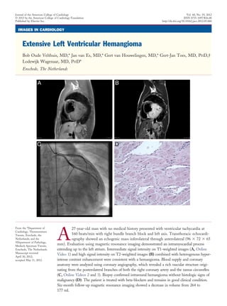
27-year-old man with no medical history presented with ventricular tachycardia at
160 beats/min with right bundle branch block and left axis. Transthoracic echocardi-
ography showed an echogenic mass inferolateral through anterolateral (96 ϫ 72 ϫ 65
mm). Evaluation using magnetic resonance imaging demonstrated an intramyocardial process
extending up to the left atrium. Intermediate signal intensity on T1-weighted images (A, Online
Video 1) and high signal intensity on T2-weighted images (B) combined with heterogenous hyper-
intense contrast enhancement were consistent with a hemangioma. Blood supply and coronary
anatomy were analyzed using coronary angiography, which revealed a rich vascular structure origi-
nating from the posterolateral branches of both the right coronary artery and the ramus circumflex
(C, Online Videos 2 and 3). Biopsy confirmed intramural hemangioma without histologic signs of
malignancy (D). The patient is treated with beta-blockers and remains in good clinical condition.
Six-month follow-up magnetic resonance imaging showed a decrease in volume from 264 to
177 ml.
Journal of the American College of Cardiology Vol. 60, No. 19, 2012
© 2012 by the American College of Cardiology Foundation ISSN 0735-1097/$36.00
Published by Elsevier Inc. http://dx.doi.org/10.1016/j.jacc.2012.05.060