Western blotting
•Download as PPTX, PDF•
3 likes•954 views

A brief Explanation on blotting and western blotting Bishal Panth MSC. Biotechnology

Recommended
More Related Content
What's hot
What's hot (20)
Similar to Western blotting
Similar to Western blotting (20)
More from Bishal Panth
Recently uploaded
Recently uploaded (20)
Western blotting
- 1. Bishal Panth Msc. Biotechnology Second Semester National College 1 Blotting Western
- 2. Content Introduction on blotting Types of Blotting Western Blotting ( Principle, procedure) Reference 2 Bishal Panth || Msc. Biotech
- 3. 3 • Blotting techniques are molecular methods used to identify and measure specific DNA, RNA and protein in complex biological mixtures. • Visualization of specific DNA , RNA & protein among many thousands of contaminating molecules. • It is the technique for transferring DNA , RNA and proteins onto a carrier so they can be separated, and often follows the use of a gel electrophoresis. Introduction on blotting Bishal Panth || Msc. Biotech
- 4. 4 Types of blotting Bishal Panth || Msc. Biotech Blotting Southern Blotting Northern Blotting Western Blotting DNA RNA Protein
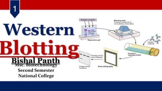
- 5. 5 Western blotting Bishal Panth || Msc. Biotech • Western blotting is a widely used analytical technique in molecular biology to detect specific protein in a complex biological sample or extract. • It is an immunoblotting (protein detection) technique using the separation power of SDS PAGE into an absorbent membrane to assess the presence, amount and molecular- weight of proteins in cellular or tissue extracts using antibodies. • It works on the principle of gel electrophoresis. Proteins are separated based on their size on polyacrylamide gel • The method is characterized by transferring the protein, which was run on a gel by electrophoresis, onto a nitrocellulose membrane. This makes the protein stable on the membrane so that several methods including methods of detection and quantity of the protein content can be detected.
- 6. 6 Bishal Panth || Msc. Biotech Western blotting- Principle Western blot is performed by using polypropylene gel electrophoresis. SDS-PAGE allows protein samples to be separated and transferred to a solid support, such as nitrocellulose (NC) or polyvinylidene difluoride (PVDF) membrane. The solid support called blot absorb the protein and keep its biological activity unchanged and is treated with a protein solution to block the hydrophobic binding site on the membrane. The membrane is treated with the antibody (primary) of the target proteins. Only the proteins to be studied can specifically bind to the primary antibody to form an antigen- antibody complex. The primary antibody-treated membranes are treated with a labeled secondary antibody after washing. After treatment, the labeled secondary antibody that binds to the primary antibody forms an antibody complex that can indicate the location of the primary antibody, both the location of the protein being studied.
- 7. 7 Western blotting- Procedure Bishal Panth || Msc. Biotech The technique consists of three major processes: 1.Separation of proteins by size (Electrophoresis). 2.Transfer to a solid support (Blotting) 3.Marking target protein using a proper primary and secondary antibody to visualize (Detection).
- 8. 8 Bishal Panth || Msc. Biotech Electrophoresis is used to separate proteins according to their electrophoretic mobility which depends on the charge, size of protein molecule, and structure of the proteins. Proteins are moved within the gel onto a membrane made of Nitrocellulose (NC) or Polyvinylidene difluoride (PVDF). The proteins combine with nitrocellulose membrane based on hydrophobic interaction. Fig: SDS Electrophoresis || Source : Research gate Western blotting- Procedure
- 9. 9 Bishal Panth || Msc. Biotech Western blotting- Procedure Western blot uses two types of agarose gel: Stacking gel that is used for concentrate all proteins in one band and Separating gel that allows for separating proteins according to their molecular weight. Fig: SDS Electrophoresis || Source : Proteomics.com
- 10. Bishal Panth || Msc. Biotech 10 Fig: Blotting technique || Source : Proteomics.com Western blotting- Procedure • A foam sponge is taken and laid on the backside, over which goes the filter paper. These should be placed to ensure that both of them are wet and slightly submerged. • The gel is taken out from the tank and placed on the wet filter paper. • The nitrocellulose membrane is wet with the transfer buffer and is placed on top of the gel in a way that there are no bubbles between the gel and the membrane.
- 11. Bishal Panth || Msc. Biotech 11 Fig : Schematic of electrophoretic protein separation in a polyacrylamide gel || Source : bio-rad.com Western blotting- Procedure For detection of the proteins, primary antibody and enzyme-conjugated secondary antibody are used. In addition of substrate, a substrate reacts with the enzyme that is bound to the secondary antibody to generate colored substance, namely, visible protein bands. • Protein bands appeared after protein transfer with buffer in a membrane gives the data for the sample used. • The protein levels can be evaluated by spectrophotometry. • It is used in the clinical diagnosis of different diseases. The confirmatory test for HIV involves a western blot by detecting anti-HIV antibodies in the serum. • used to quantify proteins and other gene products in gene expression studies. • It is also used for the analysis of different biomarkers like growth factors, cytokines, and hormones.
- 12. Western blotting Bishal Panth || Msc. Biotech 12 Fig: Process of Western Blot || Created with BioRender.com || Source : Microbes Notes
- 13. Reference i. Kurien BT, Scofield RH. Western blotting: an introduction. Methods Mol Biol. 2015;1312:17-30. doi: 10.1007/978-1-4939-2694-7_5. PMID: 26043986; PMCID: PMC7304528. ii. Hnasko TS, Hnasko RM. The Western Blot. Methods Mol Biol. 2015;1318:87-96. doi: 10.1007/978-1-4939-2742-5_9. PMID: 26160567. iii. Mahmood, Tahrin, and Ping-Chang Yang. “Western blot: technique, theory, and trouble shooting.” North American journal of medical sciences vol. 4,9 (2012): 429-34. doi:10.4103/1947-2714.100998 iv. Ghosh, Rajeshwary et al. “The necessity of and strategies for improving confidence in the accuracy of western blots.” Expert review of proteomics vol. 11,5 (2014): 549-60. doi:10.1586/14789450.2014.939635 Bishal Panth || Msc. Biotech 13
- 14. Bishal Panth || Msc. Biotech 14