SlideShare a Scribd company logo
1 of 68
Download to read offline
Full Arch Rehabilitation:
All-on-4™ Restorative Steps
Roadmap for Success
Brought to you by:
All-on-4
Restorative
Steps
NobelBiocare: FullArchRehabilitation-All-on-4™RestorativeSteps -RoadmapforSuccess
Full Arch Rehabilitation:
All-on-4™ Restorative Steps
Roadmap for Success
Treatment Planning/Diagnosis
1. Setup meeting with Treatment Team to discuss case – communication is key
2. Qualifying the Patient
When evaluating a candidate for All-on-4™ treatment, the most essential components are the
patients’ current esthetics and function. It is important to identify the patient’s smile line and
vertical occlusal space. If there are noticeable problems with their current esthetics or
function, now is the time to correct these.
Examples of issues that may need to be corrected pre-operatively:
 Poor Fitting Dentures
 Limited Inter-Occlusal Space
 Incorrect Bite
 Insufficient space for prosthesis
3. Treatment Planning Evaluation Questions
Comfort
 Please describe any type of discomfort or awareness of pain associated with your denture
or partial.
 What is your experience sleeping with dentures in your mouth?
 Do you notice a burning sensation in your mouth?
 Do you currently have sore spots with your denture?
Esthetics
 Have you noticed changes in the appearance of your face?
 Are you happy with the appearance of your teeth?
 Are you confident with your smile?
 Are you concerned with signs of aging (appearance of face)?
 Do you have sores at the corners of your mouth?
NobelBiocare: FullArchRehabilitation-All-on-4™RestorativeSteps -RoadmapforSuccess
Function – Chew and Speak
 Can you eat and speak comfortably without pain or looseness of the denture?
 Do you use denture adhesives? Occasional or routine? What is your attitude toward
adhesives?
 Can you easily eat dry food (for example, a granola bar)?
 Do you ever notice dryness of your mouth, including your lips?
 Do you ever feel like your lips stick together?
Dental History
 When was your last reline or adjustment of denture?
 How long have you been using dentures/this denture?
 Did you use a removable partial denture before full?
 What was the reason for tooth loss? (perio/destructive function/caries)
 What food limitation have you experience because of chewing challenges? Any special
requirements for food preparation?
 Can you eat what you would like to eat? Why not?
Examples of Candidates for All-on-4™ Treatment:
NobelBiocare: FullArchRehabilitation-All-on-4™RestorativeSteps -RoadmapforSuccess
Examples of Candidates for All-on-4™ Treatment:
NobelBiocare: FullArchRehabilitation-All-on-4™RestorativeSteps -RoadmapforSuccess
4. Capture Vertical Dimension of Occlusion (VDO) for Restoration/Smile Line
A. Vertical Dimension of Occlusion (VDO), also known as Occlusal Vertical Dimension
(OVD) is a term used to indicate the superior-inferior relationship of the maxilla and the
mandible when the teeth are situated in maximum intercuspation. A VDO is not only
possessed by people who have teeth, however; for completely edentulous individuals who
do not have any teeth with which to position themselves in maximum intercuspation,
VDO can be measured based on the subjective signs related to esthetics and phonetics.
Vertical dimension can be captured using a base plate and bite rim.
 15mm of space (minimum) is required for NobelProcera® Implant Bridge - Fixed
(Restoration (Measure Length of Central Incisor + 6mm)
5. Identify Smile Line and Transition Line of Prosthesis - make sure transition line of
prosthesis is apical to smile line
 If soft tissue is visible while smiling, measure the distance between the gingiva above
central and lateral incisors and the extreme border of the upper lip
Smile Line
Transition Line
of Prosthesis
NobelBiocare: FullArchRehabilitation-All-on-4™RestorativeSteps -RoadmapforSuccess
6. Photographic Evaluation
 Full Face, Lips at Repose
 No animation – with and without Denture/Partial
 Full Face Smiling – with and without Denture/Partial
 Lips Retracted, Teeth apart – with and without Denture/Partial
 Lips Retracted, Teeth together – front, right, and left sides
 Side Profile, Full Face
 Side Profile, Smile
 Intra-oral Alveolar Ridge without Denture
 Denture out of Mouth: Occlusal View and Intaglio View
Example: No Animation with & without Denture/Partial
Example: Smiling with & without Denture/Partial
Example: Retracted with & without Denture/Partial
NobelBiocare: FullArchRehabilitation-All-on-4™RestorativeSteps -RoadmapforSuccess
6. Case Evaluation – Esthetics
 Lip Support
 Naso-Labial Angle
 Facial Midline
 Occlusal Plane
 Lip Size – Flat and Thin
 Tooth Display at rest, and when speaking
 Lip Dynamics
 Smile Line
 Transition Zone
7. Ridge Reduction (Alveolectomy)
– determine how much is necessary
8. Take Accurate Records
A. Assess the condition of the oral mucosa, ridges, facial, cheek, and lip support (contours).
B. Take Impressions of both arches and Bite Registration. The impressions must include the
palate and vestibules in the maxilla and the vestibules and retro molar pads in the
mandible.
C. Make sure to include the Edentulous Saddles
i. Bite Rims are necessary if edentulous saddles cannot be capture on initial
impressions.
ii. If bite will be opened, take a new bite registration with wax rims tried in
NobelBiocare: FullArchRehabilitation-All-on-4™RestorativeSteps -RoadmapforSuccess
9. Shade/Mold Information –
 Consult with patient regarding the color, shape, and size of teeth that will be suitable.
 Send photos, shade/mold information, measurements, and patient’s desires concerning
esthetics of denture to Dental Laboratory. The Laboratory will utilize the impressions
and other information to fabricate immediate denture to be used as provisional prosthesis
on the day of surgery.
10. Evaluate Bone Volume
 With CT Scan and X-rays determine implant/restorative options based on the amount of
bone volume
11. Discuss Patient Symptoms/Issues, Expectations, and Motivation
12. Review steps/coordinate schedules
 Surgical Specialist based on CT Scan determines and orders Implants and Restorative
Components w/alternative sizes/angles:
 Multi-unit Abutments: Straight, 17°, 30° angle correction w/varying tissue depth
 Temporary Copings Multi-unit + Prosthetic Screws (extra)
 Multi-unit Abutment Healing Caps
 Impression Copings – Open Tray – for final prosthesis
 Multi-unit Abutment Lab Analogs – for final prosthesis
NobelBiocare: FullArchRehabilitation-All-on-4™RestorativeSteps -RoadmapforSuccess
Immediate Provisionalization Materials:
Equipment:
 Handheld Light Unit
 Bench Lathe
 Electric Handpiece
 Dust Hood with Vacuum (Handler Bench Top Porta-Vac)
Hand Tools:
 Rubber Dam Punch
 Great White Burs (SSW HP-8)
 #25 Surgical Blades
 #6R Redwood Plaster Knife
 #7R Redwood Plaster Knife
 Small Surgical Scissors
 Instrumentation: Prosthetic Kit (torque wrench, Unigrip drivers 20, 25, 30mm,
multi-unit abutment driver)
 Perio Probe or Explorer
Supplies:
 Blue Mousse Bite Material or Similar
 Rubber Dam
 Aquasil Ultra XLV Fast Set, Teflon Tape, Cavit or Wax for blocking out screw
access holes
 Exothermic Polymer Acrylic - Cold Cure (3M Secure, Dentsply Dual Line,
Unifast Trad, or Quik Set)
 eFiber and Perma Mesh to strengthen the denture (Preat)
 Ivoclar Universal Polishing Paste
 Keystone b12 Brush Wheels
 Lab Pumice and Rag Wheels
 Burs and Brushes for Acrylic Finishing
NobelBiocare: FullArchRehabilitation-All-on-4™RestorativeSteps -RoadmapforSuccess
Dental Laboratory creates Immediate Denture & Surgical Guide:
1. Wax Try-in Denture
 Based upon impression and bite registration a Wax Try-in Denture is created by the
Dental Laboratory. The Wax Try-in for Immediate Denture is designed to verify vertical
dimension, esthetics, phonetics, and facial support. Improvements and modifications to
the denture can be made at this time.
 After modifications are complete, the Dental Laboratory processes an immediate denture
2. Re-mount and Equilibration
3. Cross Mount and Occlusal Guide and Bite Registration
NobelBiocare: FullArchRehabilitation-All-on-4™RestorativeSteps -RoadmapforSuccess
4. Duplicate Denture and Trough out Lingual for Surgical Template
5. Add 30-45 Degree Angulation for Posterior Implant
6. Indicate Amount of Ridge Reduction and Mark on Surgical Template
15mm
NobelBiocare: FullArchRehabilitation-All-on-4™RestorativeSteps -RoadmapforSuccess
Surgery & Immediate Provisionalization:
1. Measure and Mark Vertical Dimension of Occlusion prior to Surgery
 Surgical placement of dental implants is based off the immediate denture in an effort to
maintain the patients’ proper vertical dimension. Before extracting the teeth, the surgical
specialist will mark the chin and nose and measure the distance while the patient is in
occlusion.
2. Extract Teeth and Reduce Ridge (alveolectomy)
 Ridge is reduced based on pre-determined amount indicated in surgical template
(necessary to accommodate prosthesis).
NobelBiocare: FullArchRehabilitation-All-on-4™RestorativeSteps -RoadmapforSuccess
3. Placement of Dental Implants
 Implants are placed in the arch, equally distributed and angulated to avoid the natural
anatomy (sinus and nerve). Implants must achieve initial stability of at least 35Ncm of
torque in order to immediately provisionalize the implants with the patients’ immediate
denture.
 In the case of a dual arch surgery, the upper denture should be provisionalized first,
utilizing the palate as a guide in the maxilla and the retro molar pad and bite registration
in the mandible.
4. Attach Multi-Unit Abutments
 Once implants are placed, multi-unit abutments can be placed - 30° Multi-Unit
Abutments in posterior/tilted implants and Straight or 17° Multi-unit Abutment in
anterior. Hand tighten – take X-ray to verify seated properly. Torque to appropriate
Ncm – Angled (15Ncm)/Straight (35Ncm). Attach Multi-unit Abutment Healing Caps to
top of Multi-unit Abutments prior to suturing.
NobelBiocare: FullArchRehabilitation-All-on-4™RestorativeSteps -RoadmapforSuccess
5. Index position of Multi-Unit Abutments
 Use Blue Mousse to line the intaglio surface of denture to index position of abutments.
6. Hollow-out space in denture for Temporary Coping Multi-unit
7. Attach Temporary Coping Multi-unit to Multi-Unit Abutments
 Place hollowed-out denture over Temporary Coping Multi-unit cylinders (passive fit)
NobelBiocare: FullArchRehabilitation-All-on-4™RestorativeSteps -RoadmapforSuccess
8. Reduce Height of Temporary Coping Multi-Unit
 With Sharpie marker, mark the height the Temporary Coping Multi-unit cylinders need to
be cut down to allow the patient to bite down. Remove Temporary Coping Multi-unit
cylinders and trim to appropriate height. Can be done before or after pick-up with
acrylic.
9. Attach Rubber Dam
 Place Rubber Dam around Temporary Coping Multi-unit cylinders (barrier between
surgical and restorative materials). Place Light Body Impression Material, wax, or
Teflon Tape in top of Temporary Coping Multi-unit cylinder to prevent acrylic from
getting inside. Verify proper seating and alignment of denture with pre-operative Bite
Registration.
10. Pick-up Temporary Coping Multi-unit cylinders
 Use Cold Cure Acrylic (any Exothermic Polymer Acrylic e.g., 3M Secure, Unifast Trad,
or Quik Set). Allow Cold Cure Acrylic to set-up.
NobelBiocare: FullArchRehabilitation-All-on-4™RestorativeSteps -RoadmapforSuccess
11. Remove Prosthesis with Temporary Coping Multi-unit cylinders processed in acrylic
 With UniGrip Driver unscrew prosthesis and remove Prosthetic Screws with a Perio
Probe (wax block-out may make Prosthetic Screws difficult to remove). Attach the white
Healing Cap Multi Units on the Multi Unit Abutments while the provisional prosthesis is
being finished.
12. Convert from Immediate Denture to Fixed Implant Bridge
 Trim palate, borders, phalanges, and remove distal cantilevers beyond 3mm.
13. Polish and Smooth Surface of Fixed Implant Bridge
 Maintain intaglio surface of denture – create ovate pontic contour for ease of
maintenance/hygiene for patient. Remove any sharp angles or edges
NobelBiocare: FullArchRehabilitation-All-on-4™RestorativeSteps -RoadmapforSuccess
14. Attach Provisional Fixed Implant Bridge Prosthesis w/Prosthetic Screws
 Torque Prosthetic Screws with Unigrip Driver to 15Ncm.
 Fill in screw access w/Teflon Tape and composite.
 Adjust occlusion using articulation paper – reducing any high spots to level occlusion.
15. Recommended Diet during Healing Phase
 A semi-solid/soft food diet is recommended while the implants Osseo integrate – 3-4
months. The patients can eat anything they can cut with a fork (cooked vegetables,
canned fruits, well-cooked meat/fish/chicken, etc. Avoid raw vegetables, fruits, and
nuts until the implants have Osseo integrated.)
16. Hygiene and Maintenance
 For the first 14 days following All-on-4™ Surgery & Provisionalization – have the
patient rinse with Peridex Oral Rinse.
 After 2 weeks, use a water pick and soft bristle toothbrush with non-abrasive tooth paste
to clean.
 Regular hygiene visits are recommended every 6 months. Evaluate prosthesis for plaque
build-up or red/inflamed soft tissue. If tissue appears healthy, have hygienist clean
around implants like an ovate pontic on a bridge.
 Remove prosthesis if tissue is red/inflamed or calculus is built up on prosthesis. Use
Unigrip Driver to remove Prosthetic screws and clean in the ultrasonic. Use ProClean
(tartar and stain remover) for cleaning the prosthesis. Soak screws in alcohol or sterile
water. Replace Prosthetic Screws with new screws after removing more than 2 times.
Torque Prosthetic Screws to 15Ncm with Unigrip Driver.
NobelBiocare: FullArchRehabilitation-All-on-4™RestorativeSteps -RoadmapforSuccess
Final Prosthesis – NobelProcera® Implant Bridge:
1. Custom Tray Impression
 Un-screw the provisional implant bridge with Unigrip Driver
 Take an Alginate Impression for custom trays to be manufactured
 Take an impression of the opposing arch, if needed
 Take impressions of temporary dentures, to indicate the patients likes/dislikes of existing
dentures
2. Final Impression
 Un-screw the provisional implant bridge with Unigrip Driver
 Attach Open Tray Multi Unit Impression Copings to Multi Unit Abutments – take X-ray
to verify seated properly. Lute impression copings together with ortho wire and light
cure material or pattern resin – create a rigid frame to ensure accuracy. The Dental
Laboratory will lute the Multi Unit Abutment Replicas together when attaching to the
impression copings and pouring up the master cast to ensure accuracy.
 Make sure the custom tray clears the Open Tray Impression Copings, adjust tray if
needed.
 Use heavy body impression material around the impression copings and a medium body
impression material for the tissue area
NobelBiocare: FullArchRehabilitation-All-on-4™RestorativeSteps -RoadmapforSuccess
3. Verification of Master Cast and Final Records
 Un-screw the provisional implant bridge with Unigrip Driver
 Take a Bite Registration
 Mark Midline, High Lip Line, Incisal Edge and Shade
 Verification Jig Try-in - The Jig MUST sit passively to each implant/abutment. Take an
X-ray to verify seated properly. If the jig does not fit passively, section the jig and re-
take final impression with the custom tray over the jig.
4. Wax Try-in
 Un-screw the provisional implant bridge with Unigrip Driver
 Wax Try-in - If esthetics, phonetics, function, and lip support are acceptable, send to lab
for the NobelProcera® Implant Bridge Titanium framework to be milled. Note: the lip
support is provided by the gingival third of the tooth. There is no denture flange to
provide bulk. If more support is required than what is provided by the wax try-in, then
the necks of the teeth will need to be brought forward.
 Try in Jig (if didn’t fit passively the first time)
NobelBiocare: FullArchRehabilitation-All-on-4™RestorativeSteps -RoadmapforSuccess
5. NobelProcera® Implant Bridge Titanium framework Try-in
 Un-screw the provisional implant bridge with Unigrip Driver
 Try-in the NobelProcera® Implant Bridge Titanium framework, verify passive fit with
each implant/abutment
 Teeth will be set in wax
6. Seat the NobelProcera® Implant Bridge - Final Prosthesis
 Un-screw the provisional implant bridge with Unigrip Driver
 Seat the final prosthesis - Take X-ray to verify seated properly. The final prosthesis
should seat firmly against the soft tissue, like an ovate pontic. The design of the tissue
interface should be such that it causes the tissue to roll over the prosthesis on the buccal
and lingual aspects.
 Torque the Prosthetic Screws with Torque Wrench to 15 Ncm when attaching to Multi-
Unit Abutments. Always use new prosthetic screws to seat the final prosthesis.
 Block out screw access holes to protect screw head with Teflon Tape, Foam, etc. Seal
screw access areas with Acrylic
 A night guard is provided
Full
Arch
Treatment
Options
Full Arch Treatment Options
Option 1: Removable Denture
Advantages
 Relatively inexpensive tooth and gingival replacement
 Provides lip support
 Easy to remove and clean outside of mouth
Disadvantages
 Uncomfortable – may cause sore spots on gum tissue
 Difficult to eat certain foods
 Accelerates bone loss
 Often requires reline to improve fit and comfort as bone deteriorates
 Difficult to speak as the removable denture may move
 May require creams or adhesives to reduce mobility of denture
 Approximately 10% functionality compared to natural teeth
Option 2: 2 or 4 Implant Over Denture (Removable)
Advantages
 Improves stability and functionality to 60% compared to natural teeth
 Relatively inexpensive tooth and gingival replacement
 Provides lip support
 Easy to clean outside of mouth
Disadvantages
 Uncomfortable – may cause sore spots on gum tissue
 Remove and clean denture outside of mouth
 May still move when chewing or speaking
 May require relines to improve fit and comfort as bone deteriorates
Option 3: All-on-4™ Implant Fixed Bridge
Advantages
 Improves functionality to 90% compared to natural teeth
 Eliminates need for bone grafting
 Can provide temporary bridge day of surgery - eat soft foods while healing
 Replaces roots and teeth
 Preserves bone and soft tissue
 Never decay – 95% success rate over 30 years
 Natural-looking esthetics
 Allows you to eat the foods you want
 Able to clean fixed implant bridge like natural teeth
Disadvantages
 Time (healing and restorative)
 Surgical procedure
Natural
Teeth
------------------------100%
No
Teeth,
No
Denture
-----------------
0%
Lower
Denture
----------------------
10%
Implant
Overdenture
-----------------
60%
(Removable
–
Tissue
Borne)
All-on-4™
Fixed
Implant
Bridge-
----
90%
50
40
30%
20
80
70
Relative
Functional
Chewing
Capacity
Upper
Denture
----------------------
Multi-Unit
Abutment
Quick
Guide
Abutment placement instructions
Step 1

– 
Remove the Healing Abutment using the Screwdriver Unigrip
and by rotating it counterclockwise.
Step 2a – Straight Multi-unit Abutment
– 
Use the premounted plastic holder to place the abutment onto
the implant and screw the abutment into the correct position.
If necessary, shorten the holder with a pair of scissors. When
the abutment is seated, remove the plastic holder with a slight
bending movement and hand-tighten with Screwdriver
Machine Multi-unit.
Step 2b – Straight Multi-unit Abutment
– 
Take a radiograph to verify proper seating of the abutment
(radiograph for conical connection is shown). Tighten the
abutment screw to 35 Ncm using the Manual Torque Wrench
Prosthetic and Screwdriver Machine Multi-unit.
Or
Step 2a – Angled Multi-unit Abutment, 17° or 30°
– 
The abutment is placed over the implant by using the
premounted abutment holder. Please note that there are several
possible positions in which to place the abutment based on the
implant connection and abutment angle.
– 
Tighten the abutment screw using a Screwdriver Unigrip
until resistance is felt. The holder is then unscrewed from the
abutment by turning it counterclockwise.
Step 2b – Angled Multi-unit Abutment, 17° or 30°
– 
Take a radiograph to verify proper seating of the abutment
(radiograph for conical connection is shown). Tighten the abutment
screw to 15 Ncm using the Manual Torque Wrench Prosthetic
and Screwdriver Machine Unigrip. Be sure not to exceed 15 Ncm.
See closed or open tray instructions for next steps.
2a
OR
2a
1
2b
2b
Conical connection
Conical connection
Closed tray – Abutment level impression instructions
Step 3
– 
Connect the Impression Coping Closed Tray Multi-unit to
the abutment by rotating it clockwise.
Step 4

– 
Inject a heavy body impression material (polyether material or
polyvinylsiloxane) around the impression coping. Fill the tray
with impression material and record the impression.
Step 5
– 
After setting, remove the impression and disconnect the
impression copings. Attach an Abutment Replica Multi-unit to
each impression coping.
Step 6
– 
Place the impression coping abutment replica assembly into its
corresponding location in the impression.
Step 7
– 
Connect the temporary restoration or healing cap. Send the
impression to the dental laboratory.
6 7
5
3 4
Multi-unit Abutment
Placement and impression techniques
	
Closed tray and open tray Quick Guide
This Quick Guide does not replace the Instructions
for Use. Please review the instructions for use
included with the products.
Indications:
– Multi-unit restorations
– Screw-retained
– 
May be used in combination with framework
design if not all implants benefit from abutments.
– 
Used to elevate seating restoration platform
when restoration-to-implant level is not practical
nor indicated due to depth or angle of implant.
Nobel Biocare USA, LLC. 22715 Savi Ranch Parkway, Yorba Linda, CA 92887
Phone 714 282 4800; Toll free 800 322 5001; Technical services 888 725 7100
©2011 Nobel Biocare USA, LLC. All rights reserved. www.nobelbiocare.com 72194 Rev. 01 (08/11)
Description Item number
NobelReplace® Multi-unit Abutment (internal tri-channel connection)
17° Multi-unit Abut NobelReplace NP 2 mm
17° Multi-unit Abut NobelReplace NP 3 mm
17° Multi-unit Abut NobelReplace RP 2 mm
17° Multi-unit Abut NobelReplace RP 3 mm
17° Multi-unit Abut NobelReplace RP 4 mm
30° Multi-unit Abut NobelReplace RP 4 mm
30° Multi-unit Abut NobelReplace RP 5 mm
30° Multi-unit Abut Non-Engaging NobRpl RP 4 mm
30° Multi-unit Abut Non-Engaging NobRpl RP 5 mm
Multi-unit Abut NobelReplace NP 1 mm
Multi-unit Abut NobelReplace NP 2 mm
Multi-unit Abut NobelReplace NP 3 mm
Multi-unit Abut NobelReplace RP 1 mm
Multi-unit Abut NobelReplace RP 2 mm
Multi-unit Abut NobelReplace RP 3 mm
Multi-unit Abut NobelReplace RP 4 mm
Multi-unit Abut NobelReplace RP 5 mm
Multi-unit Abut NobelReplace WP 1 mm
Multi-unit Abut NobelReplace WP 2 mm
Multi-unit Abut NobelReplace WP 3 mm
29235
29236
29237
29238
29239
29240
29241
33409
33410
29196
29197
29198
29199
29200
29201
29202
29203
29204
29205
29206
Conical Connection Multi-unit Abutment (internal conical connection)
17° Multi-unit Abut CC NP 2.5 mm
17° Multi-unit Abut CC NP 3.5 mm
17° Multi-unit Abut CC RP 2.5 mm
17° Multi-unit Abut CC RP 3.5 mm
30° Multi-unit Abut CC NP 3.5 mm
30° Multi-unit Abut CC NP 4.5 mm
30° Multi-unit Abut CC RP 3.5 mm
30° Multi-unit Abut CC RP 4.5 mm
Multi-unit Abut CC NP 1.5 mm
Multi-unit Abut CC NP 2.5 mm
Multi-unit Abut CC NP 3.5 mm
Multi-unit Abut CC RP 1.5 mm
Multi-unit Abut CC RP 2.5 mm
Multi-unit Abut CC RP 3.5 mm
Multi-unit Abut CC RP 4.5 mm
36614
36615
36618
36619
36620
36621
36622
36623
36611
36613
36624
36616
36617
36625
36626
Multi-unit Abutment // Placement and impression techniques – abutment level // Closed tray and open tray quick guide
Open tray – Abutment level impression instructions
Step 3


– 
Connect the Impression Coping Open Tray Multi-unit on the
abutment and tighten using the Screwdriver Unigrip.
– 
Relieve and perforate the impression tray to allow full seating
of the tray and protrusion of the guide pins. If there is a large
opening, it may be closed off using baseplate wax, with the
guide pins indenting or perforating the wax.
Step 4
– 
Inject a heavy body impression material (polyether material or
polyvinylsiloxane) around the impression coping.
– 
Fill the tray with impression material and seat the impression
tray fully so that the tips of all the guide pins are identifed.
– 
Remove excess impression material from the guide pin
access holes.
Step 5
– 
After setting, unscrew the guide pins and remove the
impression tray and send to the dental laboratory, including
the guide pins. For model fabrication, corresponding implant
replicas should be provided by you or your dental laboratory.
Step 6
– 
Connect the temporary restoration or healing cap.
4
3
6
5
Description Item number
Brånemark System® Multi-unit Abutment (external hex connection)
17º Multi-unit Abutment Brånemark System NP 2 mm
17º Multi-unit Abutment Brånemark System NP 3 mm
17º Multi-unit Abutment Brånemark System RP 2 mm
17º Multi-unit Abutment Brånemark System RP 3 mm
17º Multi-unit Abutment Brånemark System RP 4 mm
30º Multi-unit Abutment Brånemark System RP 4 mm
30º Multi-unit Abutment Brånemark System RP 5 mm
30° Multi-unit Abut Non-Engaging Bmk Syst RP 4 mm
30° Multi-unit Abut Non-Engaging Bmk Syst RP 5 mm
Multi-unit Abutment Brånemark System NP 1 mm
Multi-unit Abutment Brånemark System NP 2 mm
Multi-unit Abutment Brånemark System NP 3 mm
Multi-unit Abutment Brånemark System RP 1 mm
Multi-unit Abutment Brånemark System RP 2 mm
Multi-unit Abutment Brånemark System RP 3 mm
Multi-unit Abutment Brånemark System RP 4 mm
Multi-unit Abutment Brånemark System RP 5 mm
Multi-unit Abutment Brånemark System WP 1 mm
Multi-unit Abutment Brånemark System WP 2 mm
Multi-unit Abutment Brånemark System WP 3 mm
29187
29188
29189
29190
29191
29192
29193
33411
33412
29176
29177
29178
29179
29180
29181
29182
29183
29184
29185
29186
Instruments
Screwdriver Manual Unigrip 20 mm
Screwdriver Manual Unigrip 28 mm
Screwdriver Manual Unigrip 36 mm
Screwdriver Machine Multi-unit 21 mm
Screwdriver Machine Multi-unit Bmk Syst WP 20 mm
Screwdriver Machine Unigrip 20 mm
Screwdriver Machine Unigrip 25 mm
Screwdriver Machine Unigrip 30 mm
Screwdriver Machine Unigrip 35 mm
Manual Torque Wrench Prosthetic
29148
29149
29150
29158
29159
29151
29152
29153
29154
29165
Components
Impression Coping Closed Tray Multi-unit
Impression Coping Closed Tray Multi-unit Bmk Syst WP
Impression Coping Open Tray Multi-unit
Impression Coping Open Tray Multi-unit Bmk Syst WP
Abutment Replica Multi-unit
Abutment Replica Multi-unit Bmk Syst WP
Healing Cap Multi-unit
Healing Cap Multi-unit Bmk Syst WP
Healing Cap Wide Multi-unit
Healing Cap Wide Multi-unit Bmk Syst WP
Temporary Coping Multi-unit
Temporary Coping Multi-unit Bmk Syst WP
Temporary Coping Plastic Multi-unit
29090
29092
29089
29091
31161
31162
31145
29066
31146
29067
29046
29047
DCA 468-0
DCA 705-0
Materials to use for Multi-unit Abutment placement and closed or open tray impression techniques
Product images are not necessarily to scale.
In order to improve readability, Nobel Biocare does not use TM/
®in running text. In so doing,
however, Nobel Biocare does not waive any right to the trademark or registered mark and
nothing herein shall be construed to the contrary.
Case
Study
by
Schroering

Parrish
Clinical Protocol for Rapid,
Graftless, Four-Implant Restoration
of the Fully Edentulous Patient
PracticalPROCEDURES No.29
P R O V I D I N G S O L U T I O N S F O R C L I N I C A L C H A L L E N G E S
Osseointegration has become a predictable biological response to implant placement, enabling
dental professionals to focus on surgical and restorative processes that simplify implant therapy.
This has resulted in techniques that improve the predictability of aesthetic implant placement and
make such treatment options accessible for a greater portion of the patient population. As shown in
the following clinical protocol, the All-on-4™ Technique provides numerous advantages in the
management of the edentulous patient or the soon-to-be edentulous patient.
• Ease of the procedure
• Ability to avoid grafting procedures
• Potential of immediate loading at the
surgical appointment
• Decreased cost to the patient
• Aesthetics
• Can be performed on the partially or
fully edentulous patient
• Ease of maintenance
• Can be flapless when using
NobelGuide™ guided surgery.
*All-on-4, NobelGuide, and Teeth-in-an-Hour are trademarks of Nobel
Biocare, Yorba Linda, CA.
Robert Schroering, DMD* • Ken Parrish, DMD, PhD*
FIGURE. The All-on-4 Technique provides an
immediate fixed restoration to the patient.
As with any implant procedure, the All-on-4™
Technique depends on standard implant
protocols and sound treatment planning. Its
use of the patient’s existing denture or an
immediate denture (for the patient with teeth)
enables simple chairside modification,
relining, and reseating in the patient on the
day of surgery. The primary phases of
treatment—implant placement, abutment
connection, and prosthesis adaptation—are
demonstrated in the following protocol.
Figure 1.5
After implant placement, Multi-Unit Abutments (Nobel
Biocare, Yorba Linda, CA) are placed with an abutment
driver. These abutments help correct the angulation of
the dental implants.
Figure 1.4
The anterior of the sinus is marked with a periodontal
probe in order to identify the mesial wall of the sinus.
This affords the posterior implant the most-posterior
position to create a favorable anterior/posterior spread
without invading the sinus.
Figure 1.1
Preoperative clinical view of partially edentulous patient
who requests a fixed implant restoration.
Figure 1.9
Titanium copings are picked up and the temporary fixed
detachable prosthesis is modified. First molars are eliminated
to reduce cantilever forces during osseointegration.
Figure 1.8
Once the temporary prosthesis is hollowed out chairside,
the surgical field is covered with a rubber dam to protect
the soft tissues during the restorative phase of treatment.
Case 1 — Implant Placement and Prosthesis Conversion
P R A C T I C A L P R O C E D U R E S N o . 2 9
Figure 1.7
Soft tissue closure is accomplished with horizontal mattress
sutures in order to reposition the tissues for optimum healing.
Figure 1.6
Temporary Copings (Nobel Biocare, Yorba Linda, CA)
are placed.
Figure 1.3
All maxillary teeth are extracted and the alveolar bone is
reduced via a “horizontal osteotomy” to a height sufficient
to avoid the smile line.
Figure 1.2
Preoperative panoramic radiograph of the partially
edentulous patient, which permits evaluation of the
bone levels.
Figure 1.11
Postoperative facial view of the temporary fixed
detachable prosthesis delivered that same day utilizing
the All-on-4 Technique.
Figure 1.10
Postoperative panoramic radiograph showing successful
placement of four implants in the maxillary arch.
Additional Evidence of Success
The All-on-4™ Technique represents a viable option for clinicians seeking to bring the
advantages of fixed implant prosthodontics to their patients. In proper clinical situations
with minor prosthetic involvement, the technique utilizes an immediate loading protocol. The
All-on-4 Technique thus constitutes a valuable addition to the practitioner’s armamentarium.
Figure 2.2
Postoperative radiograph following successful placement
of four maxillary implants and the temporary fixed
detachable prosthesis with the All-on-4 Technique.
Figure 2.1
Preoperative view of patient with hopeless dentition in
the maxillary arch.
Figure 3.2
Postoperative radiograph following successful immediate
placement of temporary fixed-detachable dentures using
the All-on-4 Technique. The final fixed detachable
prostheses will be placed four months following surgery.
Figure 3.1
Panoramic radiograph demonstrates a completely
edentulous patient who has worn dentures for 20 years.
*Private practice devoted to advanced implant and periodontal treatment, Louisville, KY.
The authors can be contacted at r.schroering@advancedimplant.com and kenparrish@yahoo.com.
P R A C T I C A L P R O C E D U R E S N o . 2 9
Case 2 — Patient With Hopeless Maxillary Dentition
Case 3 — Completely Edentulous Patient
21243 REV.00 (03/08)
72850 Rev 00 (12/10) Printed in USA. © Nobel Biocare Services, AG. 2010. All rights reserved. www.nobelbiocare.com
Case
Study
by
Babbush
The All-on-Four Immediate Function
Treatment Concept With NobelActive
Implants: A Retrospective Study
Charles A. Babbush, DDS, MScD*
Gary T. Kutsko, DDS
John Brokloff, DDS
The All-on-Four treatment concept provides patients with an immediately loaded fixed
prosthesis supported by 4 implants. This single-center retrospective study evaluated the
concept while using the NobelActive implant (Nobel Biocare, Gothenburg, Sweden). Seven
hundred eight implants placed in 165 subjects demonstrated a cumulative survival rate of
99.6% (99.3% in maxilla and 100% in the mandible) for up to 29 months of loading. The
definitive prosthesis survival rate was 100%.
Key Words: All-on-Four, NobelActive implants
INTRODUCTION
A
common condition in elderly
patients is the occurrence of
edentulism, which can be the
result of many factors such as
poor oral hygiene, dental car-
ies, and periodontal disease. There are also
those patients who face edentulism due to a
terminal nonrestorable dentition. The eden-
tulous condition has been shown to have a
negative impact on oral health–related
quality of life.1
Clinicians are faced with the
growing need to offer solutions to this
population due to an increase in their life
expectancy2,3
and to fabricate prostheses
that provide a replacement for the loss of
natural teeth, allowing optimum satisfaction
and improved quality of life.
The routine treatment for edentulism has
been conventional dentures. National epide-
miological survey data in the United States
suggested that the adult population in need
of 1 or 2 dentures would increase from 35.4
million adults in 2000 to 37.0 million adults
in 2020.4
Clinical studies have reported that
patients with dentures have shown only a
marginal improvement in the quality of life
when compared with implant therapy.5
The
common reasons for dissatisfaction in pa-
tients using dentures are pain, areas of
discomfort, poor denture stability, and diffi-
culties in eating as well as lack of or
compromised retention capability.6
Many
patients wearing complete dentures com-
plain about poor masticatory performance,
loss of function, decreased motor control of
the tongue, reduced bite force, and dimin-
ished oral sensory function.7–10
A review of
the literature noted that prostheses support-
ed by osseointegrated dental implants sig-
nificantly improved the quality of life for
Cleveland ClearChoice Dental Implant Center, Pepper
Pike, Ohio.
*Corresponding author, e-mail: cab@thedentalimplantcenter.
com
DOI: 10.1563/AAID-JOI-D-10-00133
CLINICAL
Journal of Oral Implantology 431
edentulous patients when compared with
conventional dentures.11–15
Immediate loading of implant-supported,
full-arch prostheses for these patients in the
mandible or maxilla has been documented as
a predictable procedure.16–20
Excellent suc-
cess rates for immediately loaded, fixed
prosthetic reconstructions19–26
and long-term
follow-up results have been reported in the
literature.27–29
Immediate loading of implant-
supported fixed full-arch prostheses for these
cases in the edentulous maxilla and mandible
has been associated with a high level of
satisfaction for patients in terms of esthetics,
phonetics, and functionality.17–20,30–34
Dental implants are traditionally placed in
the vertical position. However, in the com-
pletely edentulous jaw as well as in the
postextraction patient, problems such as
minimum bone volume, poor bone quality,
and the need for bone-grafting procedures
prior to implant placement create some
challenging conditions. For these situations,
it has been demonstrated that distal tilting
of implants may be advantageous. Tilting
preserves relevant anatomical structures and
allows for placement of longer implants with
good cortical anchorage in optimal positions
for prosthetic support.35,36
Strain gauge
measurements performed by Krekmanov
reported no significant difference between
tilted and nontilted implants, and theoretical
models showed an increased prosthetic base
due to the inclination of implants, which in
turn can reduce the force acting over the
implants.36
Tilting also increases the inter-
implant space, reduces cantilever length in
jaws,21,31,36,37
and reduces the need for bone
augmentation. Good clinical outcomes have
been reported in various studies using tilted
implants.31,32,35,38–40
The All-on-Four treatment concept pro-
vides edentulous arches and immediate/
postextraction subjects with an immediately
loaded, fixed prosthesis using 4 implants:
2 axially oriented implants in the anterior
region and 2 tilted posterior implants.31,32,37
The principle involves the use of 4 implants
restored with straight and angled multiunit
abutments, which support a provisional,
fixed, immediately loaded, full-arch prosthe-
sis placed on the same day of surgery. The
All-on-Four treatment has been developed
to maximize the use of available bone and
allows immediate function. Overall, pub-
lished data on the All-on-Four concept
reported cumulative survival rates between
92.2% and 100%.31–34,37,40–42
The All-on-Four concept has been report-
ed predominantly in the literature with the
NobelSpeedy or the Branemark System
dental implants. The purpose of this study
was to evaluate the All-on-Four concept
using an implant (NobelActive) with a
tapered body and a variable thread design
for up to 29 months of loading.
MATERIALS AND METHODS
This is a retrospective single-center study.
Subjects with totally edentulous arches and/
or in need of extraction of the remaining
compromised teeth were rehabilitated with
the NobelActive implants. The first implant in
the study was placed on February 21, 2008,
and the last implant was placed on Septem-
ber 12, 2009. Each subject received an
immediately loaded, fixed, complete-arch
provisional prosthesis on the day of implant
placement according to the All-on-Four
technique. The definitive prostheses were
delivered within 6 to 8 months after implant
insertion. An actuarial life table method was
used to determine implant cumulative sur-
vival rate.
Patients treated with the technique and
therefore included in the retrospective anal-
ysis met the following criteria:
N jaw bone profile for the placement of at least
4 implants of at least 10 mm in length in
either healed or immediate extraction sites
432 Vol. XXXVII/No. Four/2011
All-on-Four Immediate Function Treatment
N good general health with acceptable oral
hygiene
N implants achieved stability at insertion
Patients could not be treated according to
the technique if they had insufficient bone
quality and quantity for placement of endos-
seous implants, exhibited severe parafunc-
tional habits, or had a compromised medical
history that would affect implant placement
(eg, bisphophonates, chemotherapy).
Surgical protocol
A cone-beam computerized tomographic
scan (CBCT; I-CAT cone beam CT scan,
Imaging Science Corp, Hatfield, Penn) was
taken prior to surgery, and the bone profile,
which included the bone quality and bone
volume, was assessed43
by 2 experienced
clinicians (C.A.B. and G.T.K.). In the vast
majority of cases, the patient was adminis-
tered intravenous (conscious) sedation using
fentanyl citrate 0.5 mg/mL (fentanyl; Hospira,
Lake Forest, Ill), diazepam 5 mg/mL injection
(Valium; Hospira), as well as nitrous oxide
oxygen inhalation. This was in addition to
articaine hydrochloride 4% and epinephrine
bitartrate 1:100 000 (Septodent, Paris, France),
local anesthesia that was administered in
both block and infiltration technique. A few
of the patients were administered general
anesthesia based their preexisting medical
profile.
Patients were started on a course of
antibiotic (penicillin VK 250 mg, Dispensing
Solutions, Santa Ana, Calif), 4 times a day,
2 days prior to the surgical procedure in
cases in which teeth had to be extracted.
Postoperatively, all patients were given the
same antibiotic 4 times per day over a period
of 10 days. If patients were allergic to
penicillin, clindamycin tablets (clindamycin
HCL 150 mg, Dispensing Solutions) were
given using a similar dosage regimen. In
addition, hydrocodone bitartrate and acet-
aminophen 7.5 mg/750 mg (Vicodin, Dis-
pensing Solutions) were also used as an
analgesic along with anti-inflammatory med-
ication, methylprednisolone, 4-mg dose pack
(Medrol, Dispensing Solutions). At the end of
the procedure, bupivacaine 0.5% with
1:200 000 epinephrine (bupivacaine, Cook-
Waite, Greensboro, NC) was also adminis-
tered for its analgesic-sparing effect.
Implant placement
NobelActive implants were inserted by
(C.A.B.) according to the manufacturer’s
guidelines (manual No. 21279-GB085, Nobel
Biocare Services 2008). Each subject received
2 distally tilted implants in the posterior
region followed by 2 anterior implants in
either the maxilla or the mandible. In the
maxilla, the tilted implants were positioned
just anterior to the maxillary sinus and in the
mandible; the tilted implants were posi-
tioned anterior to the mental foramen.
Implant placement was assisted by the All-
on-Four surgical guide (Nobel Biocare; Fig-
ure 4). The guide was placed into a 2-mm
osteotomy made at the midline of the
mandible and/or maxilla, and the titanium
band was contoured so that the occlusal
centerline of the opposing jaw was followed.
The guide allowed for optimal positioning,
alignment, parallelism, and inclination of the
implants for subsequent anchorage and
prosthetic support. The drill protocol fol-
lowed the manufacturer’s guidelines (All-on-
Four procedures and products, manual
No. 16896 Lot GB 0603, Nobel Biocare
Services, 2006). The implant sites were
usually underprepared avoiding countersink-
ing to engage as maximum cortical support
bone as possible. The recommended drill
sequences for soft bone type IV, medium
type II and type III, and dense type I bone
were followed. A manual surgical torque
wrench (Nobel Biocare) was used to check
the final torque of the implant, which was
carefully documented (Table 1). In cases of
immediate implant placement, the soft
Babbush et al
Journal of Oral Implantology 433
tissues were readapted to obtain a primary
closure around the abutments and fresh
extraction sites and then sutured back into
position with interrupted resorbable 4.0
chromic sutures (Salvin Dental Specialties,
Charlotte, NC). Local bone grafting to cover
exposed threads and/or other osseous de-
fects associated with extraction sockets was
performed at 64% of implant sites with
demineralized bone matrix gel (Dyna graft-
D, Keystone Dental, Boston, Mass), and 1% of
implant sites were grafted with autogenous
bone from the local surgical area.
Straight, 17u multiunit abutment, internal
(Nobel Biocare), and 30u angulated multiunit
abutments, internal (Nobel Biocare), were
used to achieve relative parallelism of the
implants so that a rigid prosthesis would
seat in a passive manner.
Open-tray multiunit impression copings
(Nobel Biocare) were placed on the abut-
ments, and an impression was made with a
custom open tray using precision impression
material (Flexitime, Heraeus Kulzer, Hanau,
Germany). Patients were instructed to avoid
brushing and to use warm water rinses for
the first postoperative week. A cold or room-
temperature soft diet for the first 24 hours
following surgery was recommended, fol-
lowed by a semisolid diet for the next
3 months. Patients were given antibiotics
and analgesics as listed in the surgical
protocol. A CBCT scan was taken immedi-
ately postoperatively to verify the implant
positions and the prosthetic components.
Prosthetic protocol
A provisional denture was prefabricated with
heat-cured acrylic resin (Ivocap high-impact
acrylic, Ivoclar Vivadent, Schaan, Liechten-
stein) prior to the surgical procedure. Imme-
diately following surgery, the denture was
modified to the master model in the
laboratory. Fabrication was completed using
cold-curing material (Probase, Ivoclar Viva-
dent). This provisional all-acrylic resin pros-
thesis was seated within 3 to 4 hours of
completion of surgery on the same day. The
patients were scheduled for routine follow-
up visits after surgery at 1 week, 2 weeks,
4 weeks, and 3 months postoperative and on
a yearly basis. At the 3-month appointment,
fabrication of the definitive prosthesis was
initiated.
Periapical digital radiographs using a
parallel technique were obtained at the 3-
month appointment and thereafter on a
yearly basis from the date of the surgery
(Figures 7, 13, 14, 20, and 29). Implants were
checked by visual observation for plaque
and bleeding on probing at the follow-up
intervals. Periapical radiographs and plaque
and bleeding indices at various follow-up
intervals are part of routine care for patients
at the clinic and not a part of the analysis in
this study.
The definitive prostheses consisted of a
milled titanium frame with a wrap-around
heat-cured acrylic resin (Ivocap high-impact
acrylic). All restorations were performed by 1
clinician (G.T.K.).
Survival criteria
The modified Albrektsson criteria used in this
investigation are the following: an implant
was regarded successful when there was (1)
no radiolucency around the implant; (2) no
signs of infection, pain, or ongoing patholog-
ical processes at the implant site; (3) the
TABLE 1
Torque values (implant insertion)
Ncm n %
,35 14 1.98
35 40 5.65
36–45 53 7.49
46–55 41 5.79
56–65 41 5.79
66–70 471 66.53
Not recorded* 48 6.78
Total 708 100
*Implants achieved primary stability, although
torque was not noted in numerical values.
434 Vol. XXXVII/No. Four/2011
All-on-Four Immediate Function Treatment
implant was restored and functionally loaded;
and (4) the prosthesis was stable for multiple
implants supporting a complete arch prosthe-
sis. An implant was classified surviving when it
remained in the jaw and was functionally
loaded even though all the individual success
criteria were not fulfilled. A failed implant was
an implant that had fractured beyond repair
or could not be classified as a successful or
surviving implant.44
Statistics
A single reviewer abstracted the relevant
data from medical records of the patients
who were treated consecutively with the All-
on-Four technique and entered them into a
spreadsheet (Excel 2007, Microsoft, Red-
mond, Wash). An actuarial life table45
was
used to calculate the cumulative survival
rate. Statistical analysis was done in SPSS
17.0 (SSPS, Chicago, Ill) using the Fisher exact
test to determine the level of significance
(P , .05) comparing the survival rates of the
arches as well as the various implant sizes.
RESULTS
One hundred sixty-five patients (72 men
and 93 women) with a mean age of 59
(SD 611 years) have been included in the
analysis. Seven hundred eight implants re-
storing both jaws (109 maxillae and 68
mandibles) have been placed. Four hundred
thirty-six implants have been placed in the
maxilla and 272 in the mandible. Twelve
patients were treated in both jaws. Each
prosthesis was supported by 4 implants. Most
of the implants were seated with a minimum
of 35-Ncm torque. Two percent (n 5 14) of
the implants were seated at a torque of
,35 Ncm (Table 1). All implants achieved
primary stability at placement. Four hundred
twenty-four implants were placed in extrac-
tion sites immediately after tooth extraction,
and 284 were placed in healed sites. Local
bone grafting was performed at 65% of the
implant sites; no bone grafting was reported
in 35% of the sites. Implant distribution
according to implant type and implant length
is outlined in Tables 2 and 3, respectively.
Implant follow-up occurred up to 29 months.
Acrylic provisional restorations were placed
within 3 to 4 hours of surgery, with occlusal
contact limited to the anterior area only.
Two anterior implants failed in 2 different
patients at the 1-month and 7-month time
points due to mobility. In a third patient, 1
tilted implant failed at the 4-month time
point due to mobility. All 3 implants failed
during the provisional prosthesis phase. All
of the 3 implants have been replaced, and
no further complications have been noted in
these patients. None of the implant failures
compromised the prosthesis function, and
no relation was found between implant
failure and the opposing dentition.
TABLE 2
Implant placement
Position
Maxilla Mandible
Total Number of Implants
Central incisor
Right 11 2
Left 10 7
Lateral incisor
Right 62 37
Left 66 31
Canine
Right 38 28
Left 39 31
First premolar
Right 14 16
Left 14 6
Second premolar
Right 77 45
Left 72 51
First molar
Right 15 7
Left 17 11
Second molar
Right 0 0
Left 1 0
Third molar
Right 0 0
Left 0 0
Total 436 272
Babbush et al
Journal of Oral Implantology 435
Two patients aged 70 and 68 years were
lost to follow-up at the 1-year and less than
3-month visit. These patients passed away
due to natural causes. Two additional
patients were lost to follow-up at the 3-
month follow-up visit. Therefore, a total of 16
implants have been lost to follow-up. One
hundred sixty-two patients in the study have
completed the 6-month follow-up and have
had their definitive prostheses (174) deliv-
ered. Three jaws were lost to follow-up prior
to definitive prosthetic delivery, and 1 jaw
was lost to follow-up after definitive pros-
thetic delivery. One hundred fifty-six patients
have completed the 1-year follow-up.
The overall implant survival rate was
99.6% (1 year; Table 4) with no significant
difference between the maxillae and mandi-
bles (99.3% vs 100%, P 5 .06, Fisher exact
test). The 4.3-mm-diameter implants were
most frequently used, with a survival rate of
100% (99.2% for 3.5 mm and 99.2% for
5.0 mm; Figure 1). No total arch failures have
occurred to date, providing a definitive
prosthesis survival rate of 100%. The life
table analysis demonstrating the cumulative
survival rate is reported in Table 4.
Figures 2 through 8 demonstrate a case in
postextraction sites of the mandible and
maxilla with immediate implant placement,
Figures 9 through 14 show a case in healed
sites of a patient with a severely atrophic
edentulous maxilla. The patient has been
edentulous for 50 years. Figures 15 through
TABLE 3
Implant size (NobelActive TiUnite)*
Implant Diameter Implant Length, mm Maxillae Mandibles
3.5 8.5 — —
10 — —
11.5 12 —
13 31 9
15 28 (1) 42
4.3 8.5 — —
10 2 2
11.5 16 7
13 84 18
15 94 92
18 4 1
5.0 8.5 — —
10 — —
11.5 5 1
13 20 15
15 131 (2) 81
18 9 4
436 (3) 272
*( ) indicates failed implants/replaced.
TABLE 4
Cumulative survival analysis*
Placed/Followed
Implants
Failed
Implants
Time Not
Passed
Lost to
Follow-up CSR
Implant insertion »» 3 months 708 1 0 4 99.9
3 months »» 6 months 703 1 0 8 99.7
6 months »» 12 months 694 1 29 0 99.6
12 months »» 18 months 664 0 336 4 99.6
18 months »» 24 months 324 0 240 0 99.6
»» 24 months 84 0
*CSR indicates cumulative survival rate.
436 Vol. XXXVII/No. Four/2011
All-on-Four Immediate Function Treatment
20 show a case displaying terminal nonrestor-
able dentition of the maxilla and mandible
with extraction and immediate implant recon-
struction, and Figures 21 through 29 show a
case of a patient with an edentulous maxilla
reconstructed with the All-on-Four technique.
No complications were reported during
surgery or immediately after surgery.
DISCUSSION
The overall survival rate was 99.6% (1 year),
with no significant difference between the
maxillae and mandibles. Three implants
failed over a period of 29 months of loading.
The prosthesis survival rate was 100%. This is
in accordance with studies on biomechanical
measurements, which demonstrated that
tilted implants, when part of a prosthetic
support, do not have a negative effect on
the load distribution.16,46,47
In addition, a
biomechanical rationale in tilting distal
implants allows a reduction in cantilever
length due to the more posterior position of
the tilted implants, resulting in a more
favorable stress distribution.47,48
The methodology of using titled implants
maximizing the use of the available bone
without grafting has been reported, leading
to successful clinical outcomes.35,41,42
This is
in comparison to the traditional implant
treatment in which insufficient bone in the
posterior region requires bone-grafting pro-
cedures involving greater chair time for the
patient as well as increased cost and
increased number of procedures.
FIGURE 1. Cumulative survival rate in relation to
implant diameter.
FIGURE 2. Case 1: maxillary and mandibular all-on-Four technique in extraction sites with immediate
implant placement. Preoperative computerized tomography scan.
Babbush et al
Journal of Oral Implantology 437
The results of this study are comparable
with studies of other implant systems using
the All-on-Four concept in the maxilla. Maló et
al32
reported a high survival rate of 97.6% in a
1-year retrospective study in which 128 Brane-
mark implants (Nobel Biocare AB, Goteborg,
Sweden) were immediately loaded in 32
patients. Each jaw received 2 axial and 2 distal
implants (All-on-Four) supported by a fixed all-
acrylic prosthesis in the completely edentu-
FIGURES 3–8. Case 1: maxillary and mandibular All-on-Four technique in extraction sites with immediate
implant placement. FIGURE 3. Extraction sockets in the mandible. FIGURE 4. All-on-Four surgical guide in
situ. FIGURE 5. Implants in final position. FIGURE 6. Postoperative radiographs at 4 months. FIGURE 7.
Clinical photograph of patient at 1 year. FIGURE 8. Postoperative radiographs at 1 year.
438 Vol. XXXVII/No. Four/2011
All-on-Four Immediate Function Treatment
FIGURE 9. Case 2: 50-year history of a severely atrophic edentulous maxilla reconstructed with the All-on-
Four technique. Preoperative computerized tomography scan.
FIGURES 10–14. Case 2: 50-year history of a severely atrophic edentulous maxilla reconstructed with the
All-on-Four technique. FIGURE 10. Implants in final position. FIGURE 11. Fixed implant bridge removed to
demonstrate soft-tissue health at 1 year. FIGURE 12. Clinical view at 1 year. FIGURE 13. Postoperative
radiographs at 1 year demonstrating stable bone levels. FIGURE 14. Postoperative radiographs at 2 years
demonstrating stable bone levels.
Babbush et al
Journal of Oral Implantology 439
lous maxilla.32
Testori et al42
reported a 98.8%
implant survival and a 100% prosthetic
survival rate using a different type of implant
system, angulation of the implant-abutment
connection for the tilted implants, and a
different type of technique for the fabrication
of the final prosthesis. In this prospective,
multicenter center study, 41 patients received
an immediately placed full-arch fixed bridge
supported each by 4 axial and 2 distally tilted
Ossesotite NT implants (3i, Implant Innova-
tions, Palm Beach, Fla) in the edentulous
maxillae.42
Aparacio et al35
reported a survival
rate of 100% for tilted implants, 96.5% survival
for axial implants, and a prosthetic survival
rate of 100% after 5 years when 101 Brane-
mark implants were placed in the severely
resorbed maxilla of 25 patients (59 axially
placed and 42 in a tilted direction). Each
patient received 2 to 5 implants with at least 1
tilted implant.35
Calandriello et al38
reported a
survival rate of 96.7% in a 1-year prospective
clinical study when 60 MKIV and Replace
select implants (Nobel Biocare AB) were
placed in the atrophic maxilla of 18 patients.
In this study, 12 partial- and 7 full-arch, fixed
prostheses were supported by a total of 33
axially placed and 27 tilted implants.38
The results of this study are in accor-
dance with other studies reporting good
FIGURES 15–17. Case 3: All-on-Four technique in maxillary and mandibular (nonrestorable dentition)
extraction sites with immediate implant placement. FIGURE 15. Preoperative computerized tomography
scan. FIGURE 16. (a, b, c) Preoperative clinical photographs of the terminal nonrestorable dentition.
FIGURE 17. (a) Implants placed in maxilla with abutments and healing caps in position. (b) Implants
placed in mandible with abutments and healing caps in position.
440 Vol. XXXVII/No. Four/2011
All-on-Four Immediate Function Treatment
survival rates in the mandible. In a 1-year
retrospective clinical study, Maló et al31
reported an implant survival of 96.7% and
98.2% (2 groups) and a prosthetic survival
rate of 100% when 176 Branemark implants
were placed in 44 patients. An immediately
loaded complete-arch all-acrylic prosthesis
was supported by 4 implants (All-on-Four) in
each completely edentulous mandible.31
Another study reported a 100% implant
and prosthetic survival rate when 96 MKIV
or the NobelSpeedy Groovy implants (Nobel
Biocare AB) were placed in 24 edentulous
patients treated in the mandible according
to the All-on-Four concept.1
In addition, a
100% implant survival and prosthetic surviv-
al rate was reported in a prospective study
when 80 Branemark implants were placed in
20 patients with a extremely atrophic
mandible. Each patient received 2 axially
placed and 2 tilted implants, supporting a
fixed full-arch prosthesis (All-on-Four con-
cept).40
Previously published literature reporting
survival rates using the All-on-Four concept
in both the mandible and maxilla is similar to
the results of this analysis. In a pilot study, a
survival rate of 98.9% was reported in a case
series when 189 NobelSpeedy implants were
placed in 46 patients, supporting 53 full-
arch, all-acrylic prostheses (44 maxillae, 9
mandibles) using the All-on-Four concept.33
Maló et al34
reported a survival rate of 97.2%
and 100% in the maxilla and mandible in a 1-
year prospective study when 92 Nobel-
Speedy implants were placed in 23 consec-
utively treated patients. Each jaw was
restored by a immediate fixed full-arch
prosthesis according to the All-on-Four
concept.34
Pomares41
reported a 96.9%
FIGURES 18–20. FIGURE 18. Case 3: All-on-Four technique in maxillary and mandibular (nonrestorable
dentition) extraction sites with immediate implant placement. Maxillary and mandibular immediate
provisional fixed prosthesis in place. FIGURE 19. Postoperative panoramic radiograph at 4 months.
FIGURE 20. Postoperative radiograph at 15 months demonstrating stable bone levels.
Babbush et al
Journal of Oral Implantology 441
implant survival (96.7% in the maxilla and
97.2% in the mandible) and a 100% pros-
thetic survival rate in a prospective study
when 127 MKIII Groovy implants were placed
in 20 patients (restoring 19 maxillae and
9 mandibles) using the All-on-Four or All-
on-Six concept. A survival rate of 98.4% and
99.7% (maxilla and mandible) at the end of
1 year was reported in another single-cohort
study in which 173 edentulous patients
received 2 distal and 2 anterior axial MKIV
or NobelSpeedy Groovy implants. In this
study, each patient received a full-arch fixed
prosthesis supported by 2 distal and 2 axial
implants (All-on-Four).49
Other long-term studies using the con-
cept are comparable to the data in this
analysis. Implant survival rates from the
follow-up of results of the previously men-
tioned studies from Maló et al31,32
with a
longer follow-up demonstrated a survival rate
of 96.2% in the mandible up to 9 years and
97.7% in the maxilla up to 5 years of follow-
up.37
Citing the published literature, it was
FIGURES 21–25. Case 4: edentulous maxilla reconstructed with the All-on-Four technique. FIGURE 21.
Preoperative clinical photograph. FIGURE 22. Preoperative clinical photograph of the edentulous maxilla.
FIGURE 23. Preoperative panoramic radiograph demonstrating the edentulous maxilla. FIGURE 24. The
abutments and premounted abutment holders adjusting for relative parallelism. FIGURE 25. (a) The final
position of the implants, abutments, and healing caps. (b) The mucoperiosteal flaps repositioned and
sutured with 4-0 chromic interrupted sutures.
442 Vol. XXXVII/No. Four/2011
All-on-Four Immediate Function Treatment
noted that the overall cumulative survival rate
of 99.6% (1 year) in this study for the new
implant system (NobelActive) while using the
All-on-Four concept offers an attractive solu-
tion to clinicians treating edentulous and/or
immediate extraction patients.
CONCLUSION
The overall survival rate using the All-on-
Four immediate function treatment concept
using an implant with a tapered body and a
variable thread design can be considered a
viable treatment concept for patients pre-
FIGURES 26–29. Case 4: edentulous maxilla reconstructed with the All-on-Four technique. FIGURE 26. (a, b)
The tissue and occlusal views of the all-acrylic fixed provisional implant bridge. FIGURE 27. Postoperative
panoramic radiograph taken immediately after implant placement. FIGURE 28. Clinical photograph with
the definitive prosthesis in place. FIGURE 29. Postoperative radiographs at 1 year with the definitive fixed
implant prosthesis in place.
Babbush et al
Journal of Oral Implantology 443
senting with edentulous arches and/or
immediate placement.
NOTE
Dr Babbush has a consulting agreement with
Nobel Biocare AB Sweden for ongoing
clinical studies and continuing education
courses.
REFERENCES
1. Agliardi E, Clericò M, Ciancio P, Massironi D.
Immediate loading of full-arch fixed prostheses sup-
ported by axial and tilted implants for the treatment of
edentulous atrophic mandibles. Quintessence Int.
2010;41:285–293.
2. World Medical Association Declaration of Hel-
sinki. Ethical principles for medical research involving
human subjects. J Am Med Assoc. 2000;284:2043–3045.
3. Salvin B. The business of implant dentistry. In:
Babbush C, Hahn J, Krauser J, Rosenlicht J, eds. Dental
Implants: The Art and Science. 2nd ed. St Louis, Mo:
Saunders Elsevier; 2010:7–13.
4. US Bureau of the Census. Statistical Abstract of
the United States: 1996. 116th ed. Washington, DC: US
Bureau of the Census; 1996:p 15, tables II, No. 16, p 17,
table II, No. 17.
5. Allen PF, McMillan AS. A review of the functional
and psychosocial outcomes of edentulousness treated
with complete replacement dentures. J Can Dent Assoc.
2003;69(10):662.
6. Heath MR. The effect of maximum biting force
and bone loss upon masticatory function and dietary
selection of the elderly. Int Dent J. 1982;32:345–356.
7. Wolff A, Gadre A, Begleiter A, Moskona D,
Cardash H. Correlation between patient satisfaction
with complete dentures and denture quality, oral
condition, and flow rate of submandibular/sublingual
salivary glands. Int J Prosthodont. 2003;16:45–48.
8. Ikebe K, Matsuda K, Morii K, Furuya-Yoshinaka M,
Nokubi T, Renner RP. Association of masticatory
performance with age, posterior occlusal contacts,
occlusal force, and salivary flow in older adults. Int J
Prosthodont. 2006;19:475–481.
9. Koshino H, Hirai T, Ishijima T, Ikeda Y. Tongue
motor skills and masticatory performance in adult
dentates, elderly dentates, and complete denture
wearers. J Prosthet Dent. 1997;77:147–152.
10. Ikebe K, Amemiya M, Morii K, et al. Association
between oral stereognostic ability and masticatory
performance in aged complete denture wearers. Int J
Prosthodont. 2007;20:245–250.
11. Turkyilmaz I, Company AM, McGlumphy EA.
Should edentulous patients be constrained to remov-
able complete dentures? The use of dental implants to
improve the quality of life for edentulous patients.
Gerodontology. 2010;27(1):3–10.
12. Allen F, McMillan A. Food selection and
perceptions of chewing ability following provision of
implant and conventional prostheses in complete
denture wearers. Clin Oral Implants Res. 2002;13:320–
326.
13. Awad MA, Lund JP, Dufresne E, Feine JS.
Comparing the efficacy of mandibular implant-retained
overdentures and conventional dentures among mid-
dle-aged edentulous patients: satisfaction and func-
tional assessment. Int J Prosthodont. 2003;16:117–122.
14. Awad MA, Lund JP, Shapiro SH, et al. Oral
health status and treatment satisfaction with mandib-
ular implant overdentures and conventional dentures: a
randomized clinical trial in a senior population. Int J
Prosthodont. 2003;16:390–396.
15. Heydecke G, Thomason JM, Lund JP, Feine JS.
The impact of conventional and implant supported
prostheses on social and sexual activities in edentulous
adults: results from a randomized trial 2 months after
treatment. J Dent. 2005;33:649–657.
16. Bellini CM, Romeo D, Galbusera F, et al.
Comparison of tilted versus nontilted implant-support-
ed prosthetic designs for the restoration of the
edentuous mandible: a biomechanical study. Int J Oral
Maxillofac Implants. 2009;24:511–517.
17. Malo P, de Araujo Nobre M, Rangert B. Implants
placed in immediate function in periodontally compro-
mised sites: a five-year retrospective and one-year
prospective study [published correction appears in J
Prosthet Dent. 2008;99:167]. J Prosthet Dent. 2007;97(6
suppl):S86–S95.
18. Khatami AH, Smith CR. ‘‘All-on-Four’’ immediate
function concept and clinical report of treatment of an
edentulous mandible with a fixed complete denture
and milled titanium framework. J Prosthodont. 2008;17:
47–51.
19. Ioannidou E, Doufex A. Does loading time
affect implant survival? A meta analysis of 1,266
implants. J Periodontol. 2005;76:1252–1258.
20. Attard NJ, Zarb GA. Immediate and early
implant loading protocols: a literature review of clinical
studies. J Prosthet Dent. 2005;3:242–258.
21. Del Fabbro M, Bellini CM, Romeo D, Francetti L.
Tilted implants for the rehabilitation of edentulous
jaws: a systematic review. Clin Implant Dent Relat Res.
Epub ahead of print, 13 May 2010.
22. Nkenke E, Fenner M. Indications for immediate
loading of implants and implant success. Clin Oral
Implants Res. 2006;17(suppl 2):19–34.
23. Avila G, Galindo P, Rios H, Wang HL. Immediate
implant loading: current status from available literature.
Implant Dent. 2007;16:235–245.
24. Esposito M, Grusovin MG, Willings M, Coulthard
P, Worthington HV. Interventions for replacing missing
teeth: different times for loading dental implants.
Cochrane Database Syst Rev. 2007;(2):CD003878.
25. Jokstad A, Carr AB. What is the effect on
outcomes of time-to-loading of a fixed or removable
prosthesis placed on implant(s)? [published correction
appears in Int J Oral Maxillofac Implants. 2008;23:56]. Int
J Oral Maxillofac Implants. 2007;22(suppl):19–48.
26. Sennerby L, Gottlow J. Clinical outcomes of
immediate/early loading of dental implants: a literature
review of recent controlled prospective clinical studies.
Aust Dent J. 2008;53(suppl 1):S82–S88.
27. Ericsson I, Randow K, Nilner K, Peterson A. Early
functional loading of Brånemark dental implants: 5-year
clinical follow-up study. Clin Implant Dent Relat Res.
2000;2(2):70–77.
444 Vol. XXXVII/No. Four/2011
All-on-Four Immediate Function Treatment
28. Henry P, Rosenberg I. Single-stage surgery for
rehabilitation of the edentulous mandible: preliminary
results. Pract Periodontics Aesthet Dent. 1994;6(9):15–22.
29. Schnitman PA, Wöhrle PS, Rubenstein JE,
DaSilva JD, Wang NH. Ten-year results for Brånemark
implants immediately loaded with fixed prostheses at
implant placement. Int J Oral Maxillofac Implants.
1997;12:495–503.
30. Rosenlicht J, Ward J, Krauser J. Immediate
loading of dental implants In: Babbush C, Hahn J,
Krauser J, Rosenlicht J, eds. Dental Implants: The Art and
Science. 2nd ed. St Louis, Mo: Saunders Elsevier; 2010:
343–345.
31. Maló P, Rangert B, Nobre M. ‘‘All-on-Four’’
immediate-function concept with Brånemark system
implants for completely edentulous mandibles: a
retrospective clinical study. Clin Implant Dent Relat
Res. 2003;5(suppl 1):2–9.
32. Maló P, Rangert B, Nobre M. All-on-4 immedi-
ate-function concept with Brånemark system implants
for completely edentulous maxillae: a 1-year retrospec-
tive clinical study. Clin Implant Dent Relat Res.
2005;7(suppl 1):S88–S94.
33. Maló P, Nobre Mde A, Petersson U, Wigren S. A
pilot study of complete edentulous rehabilitation with
immediate function using a new implant design: case
series. Clin Implant Dent Relat Res. 2006;8:223–232.
34. Maló P, de Araujo Nobre M, Lopes A. The use of
computer-guided flapless implant surgery and four
implants placed in immediate function to support a
fixed denture: preliminary results after a mean follow-
up period of thirteen months [published correction
appears in J Prosthet Dent. 2008;99:167]. J Prosthet Dent.
2007;97(6 suppl):S26–34.
35. Aparicio C, Perales P, Rangert B. Tilted implants
as an alternative to maxillary sinus grafting: a clinical,
radiologic, and periotest study. Clin Implant Dent Relat
Res. 2001;3:39–49.
36. Krekmanov L, Kahn M, Rangert B, Lindstrom H.
Tilting of posterior mandibular and maxillary implants
of improved prosthesis support. Int J Oral Maxillofac
Implants. 2000;15:405–414.
37. Malo P, Lopez I, Nobre M. The All on Four
concept. In: Babbush C, Hahn J, Krauser J, eds. Dental
Implants: The Art and Science. 2nd ed. St Louis, Mo:
Saunders Elsevier; 2010:435.
38. Calandriello R, Tomatis M. Simplified treatment
of the atrophic posterior maxilla via immediate/early
function and tilted implants: a prospective 1-year
clinical study. Clin Implant Dent Relat Res. 2005;7(suppl
1):S1–12.
39. Fortin Y, Sullivan R, Rangert BR. The marius
implant bridge: surgical and prosthetic rehabilitation
for the completely edentulous upper jaw with moder-
ate to severe resorption: a 5-year retrospective clinical
study. Clin Implant Dent Relat Res. 2002;4:69–77.
40. Weinstein R, Agliardi E, Fabbro MD, Romeo D,
Francetti L. Immediate rehabilitation of the extremely
atrophic mandible with fixed full-prosthesis supported
by four implants. Clin Implant Dent Relat Res. Epub
ahead of print, 2010 Feb 11.
41. Pomares C. A retrospective clinical study of
edentulous patients rehabilitated according to the ‘‘all
on four’’ or the ‘‘all on six’’ immediate function concept.
Eur J Oral Implantol. 2009;2(1):55–60.
42. Testori T, Del Fabbro M, Capelli M, Zuffetti F,
Francetti L, Weinstein RL. Immediate occlusal loading
and tilted implants for the rehabilitation of the atrophic
edentulous maxilla: 1-year interim results of a multi-
center prospective study. Clin Oral Implants Res.
2008;19:227–232.
43. Ganz S. Defining new paradigms for assess-
ment of implant receptor sites: the All on Four concept.
In: Babbush C, Hahn J, Krauser J, Rosenlicht J, eds.
Dental Implants: The Art and Science. 2nd ed. St Louis,
Mo: Saunders Elsevier; 2010:148–149.
44. Albrektsson T, Zarb G, Worthington P, Eriksson
AR. The long-term efficacy of currently used dental
implants: a review and proposed criteria of success. Int
J Oral Maxillofac Implants. 1986;1:11–25.
45. Altman D. Practical Statistics for Medical Re-
search. Boca Raton, Fla: CRC Press; 1992.
46. Parel S, Branemark PI, Ohrnell LO, Svensson B.
Remote implant anchorage for the rehabilitation of
maxillary defects. J Prosthet Dent. 2001;86:377–381.
47. Bellini CM, Romeo D, Galbusera F, et al. A finite
element analysis of tilted versus nontilted implant
configurations in edentulous maxilla. Int J Prosthodont.
2009;22:155–157.
48. Zampelis A, Rangert B, Heijl L. Tilting of splinted
implants for improved prosthodontic support: a two-
dimensional finite element analysis [published correc-
tion appears in J Prosthet Dent. 2008;99:167]. J Prosthet
Dent. 2007;97(6 suppl):S35–S43.
49. Agliardi E, Panigatti S, Clericò M, Villa C, Malò P.
Immediate rehabilitation of the edentulous jaws with
full fixed prostheses supported by four implants:
interim results of a single cohort prospective study.
Clin Oral Implants Res. 2010;21:459–465.
Babbush et al
Journal of Oral Implantology 445
Case
Study
by
Khatami
“All-on-Four” Immediate Function Concept and Clinical
Report of Treatment of an Edentulous Mandible
with a Fixed Complete Denture and Milled
Titanium Framework
Amir H. Khatami, DDS1
 Christopher R. Smith, DDS, FACP2
1
Assistant Professor, Department of Restorative Dentistry and Prosthodontics, The Ohio State University, Columbus, OH
2
Associate Professor, Department of Surgery, Section of Dentistry, The University of Chicago Hospitals, Chicago, IL
Keywords
“All-on-Four”; immediate function; conversion
prosthesis; tilting dental implants; milled
titanium framework.
Correspondence
Amir H. Khatami, Department of Restorative
Dentistry and Prosthodontics, The Ohio
State University, College of Dentistry, 305
W. 12th Ave, Columbus, OH 43210.
E-mail: akhatami@msn.com
Presented at the Table Clinic Session,
American College of Prosthodontists 2005
Annual Session, Los Angeles, CA.
Accepted August 2, 2006.
doi: 10.1111/j.1532-849X.2007.00246.x
Abstract
The “All-on-Four” concept—tilting the distal implants in the edentulous arches im-
proves the prosthetic support—increases the inter-implant distance and provides better
implant anchorage in the bone by using longer implants. Computer milling of a solid
block of titanium also provides frameworks with improved fit and fewer technical
challenges than conventional cast or noncast approaches. This clinical report describes
a method of restoring an edentulous mandible with the “All-on-Four” immediate
function concept and a milled titanium framework. The patient in our clinical report
has reported for follow-up visits for 1 year and is satisfied with the outcome of the
treatment. No discernable clinical and radiographic changes were noted around the
dental implants. To date, there have been no prosthetic complications. The patient is
scheduled for quarterly follow-ups to determine the effectiveness of home care.
Immediate loading of implant-supported dental prostheses is
documented in the literature with a high and predictable suc-
cess rate for the edentulous mandible.1–4
The development of
new protocols for immediate loading of dental implants has
switched from placing multiple implants and loading a few to
placing only four implants as an optimal number to restore a
completely edentulous mandible.5
Rehabilitation of the pos-
terior edentulous mandible can at times be hindered by bone
atrophy distal to the mental foramen and bite forces that are
more posterior in the dentition.6
Traditionally, and according
to the original concept of the Branemark system, implants are
placed in a fairly upright position in the anterior edentulous
mandible. Therefore, it is often necessary to fabricate a bi-
lateral cantilever, which is sometimes up to 20-mm long, to
provide the patient with good chewing capacity in the molar
region. Clinical studies have demonstrated that the distal tilting
of implants may be advantageous, with reduction of cantilever
length about 6.5 mm in the mandible and 9.3 mm in the max-
illa.7,8
More recently, a concept was developed to restore the
completely edentulous arches with immediately-loaded, tilted
distal implants and the use of an “All-on-Four” guide (Nobel
Biocare, Yorba Linda, CA) (Fig 1).8,9
This clinical report de-
scribes a method of restoring an edentulous mandible with the
“All-on-Four” immediate function concept and milled titanium
framework.
Clinical report
A 59-year-old African–American female presented to the Sec-
tion of Dentistry at the University of Chicago Hospitals with
the desire to have “new dentures.” Her medical history included
a kidney transplant due to glomerulonephritis, and she was on
a daily dose of corticosteroids. On clinical examination the
patient presented with maxillary and mandibular partial eden-
tulism. She was wearing removable partial dentures (RPDs)
in both arches. The patient was advised to replace the RPDs;
however, she chose extraction of the remaining maxillary and
mandibular teeth and receiving immediate complete denture
(ICD) prostheses instead. Maxillary and mandibular ICDs were
made, and after 6 months were relined for a better prosthetic fit
(Fig 2). Despite all the effort to make her comfortable with the
Journal of Prosthodontics 17 (2008) 47–51 c
 2007 by The American College of Prosthodontists 47
“All-on-four” concept and treatment of edentulous mandible Khatami and Smith
Figure 1 “All-on-Four” guide.
Figure 2 Preoperative view of ICDs.
Figure 3 Maxillary and mandibular edentulous ridges.
mandibular denture, her chief concern was prosthesis stability
in the mouth during function. With the advent of the “All-on-
Four” concept, the patient was presented with the option of
placing four implants in the mandible and immediately loading
the implants with a conversion prosthesis.8
In preparation for
the procedure and due to her medical status, her physician was
consulted. She was instructed to increase the daily dose of her
orally administered corticosteroid (Prednisone, Deltasone
R

,
Kalamazoo, MI) to 20 mg, the day prior to surgery and the day
of surgery. She was also prescribed Penicillin-VK starting 2
days prior to surgery, 2 g per day, for 10 days.
Upon clinical and radiographic evaluation, the mandible was
classified ACP PDI for Complete Edentulism Class I (Figs 3
and 4).10
Figure 4 Preoperative panoramic radiograph.
Figure 5 Surgical template try-in.
Figure 6 Angulated guide pins.
Alginate impressions (Jeltrate, Dentsply International, York,
PA) of the edentulous arches were made, and the patient’s
mandibular denture was duplicated. This duplicate denture
was used as a guide to fabricate the radiographic and sur-
gical templates (Fig 5). A supra-crestal incision was made
from the second mandibular molar area extending to the con-
tralateral side. The mental foramina were located bilaterally
to serve as landmarks for placement of the most distal im-
plants. Implant placement was assisted by the “All-on-Four”
guide. The guide was placed into a 5-mm deep osteotomy site
made at the midline of the mandible, and its titanium band
was adjusted to follow the mandibular arch shape. The guide
is used to find the optimal position and inclination of the im-
plants (Fig 6). Four 4.3 × 13 mm2
implants (Replace Select
Yorba Linda, CA) were placed following the Replace Select
protocol and torqued to 45 Ncm (Fig 7). The platforms of
the most distal implants were angled about 30◦
distally with
the use of the “All-on-Four” guide. Multi-Unit Abutments(tm)
(4-mm height) (Nobel Biocare) were connected to the most
anterior implants, and 17◦
angled Multi-Unit Abutments(tm)
(4-
mm height) were connected to the distal implants to bring the
screw access holes to the occlusal surface of the prosthesis
48 Journal of Prosthodontics 17 (2008) 47–51 c
 2007 by The American College of Prosthodontists
Khatami and Smith “All-on-four” concept and treatment of edentulous mandible
Figure 7 Connected straight and angulated abutments.
Figure 8. Panoramic radiograph after implant placement.
Figure 9 Hollowed mandibular ICD.
(Fig 8). Subsequent to suturing the soft tissue, temporary abut-
ments were connected, and the hollowed-out mandibular den-
ture (Fig 9) was indexed using denture repair resin (Nature-
cryl, GC America, Inc., Alsip, IL). The mandibular denture
was modified to an implant-supported interim fixed prosthe-
sis in the dental laboratory. Following occlusal adjustment,
the prosthesis was inserted with prosthetic retaining screws
(Fig 10).
The patient was given oral hygiene instructions and placed
on Peridex (Zila Professional Pharmaceutical, Phoenix, AZ) for
2 weeks. At the 2-week follow-up appointment, sutures were
removed, and an open tray abutment level impression was made
with transfer copings (Nobel Biocare) and vinylpolysiloxane
(Aquasil, Dentsply International, York, PA). The impression
was poured in die stone (Die-keen, Heraeus-Kulzer, Armonk,
NY), and its accuracy was verified with the passively fitting
existing implant-supported interim fixed prosthesis (Fig 11).
The maxillary denture duplicate cast and the mandibular cast
with the attached interim fixed prosthesis were mounted using
Figure 10 Implant-supported interim fixed prosthesis.
Figure 11 Accuracy verification of the master cast with interim prosthe-
sis.
the arbitrary facebow transfer and interocclusal records. A putty
jig (Lab Putty, Coltene, Cuyahoga Falls, OH) was made to
register the 3D relationship of the interim prosthesis to the
mandibular cast. Suture removal, final mandibular impression,
and the laboratory procedures were completed in one clinic
session.
Using the temporary abutments (Nobel Biocare) and the putty
jig, the resin pattern was fabricated with autopolymerizing resin
(GC Pattern resin, GC America, Inc.) (Fig 12). The pattern was
sent to Nobel Biocare Headquarters in Sweden to fabricate a
milled titanium framework. Once received, the titanium frame-
work was tried intraorally for passivity with the recommended
screw test11–13
(Fig 13). Subsequent to the denture tooth setup,
esthetics, phonetics, and centric relation occlusion were evalu-
ated intraorally.
Necessary adjustments were made, the prosthesis was pro-
cessed with acrylic resin wrap around the framework design,
and occlusion was adjusted intraorally. The finished prosthe-
sis was inserted by torque tightening the prosthesis retaining
screws to 10 Ncm (Figs 14 and 15). The patient was given
oral hygiene instructions and scheduled for follow-up every 3
months. At the 3-, 6-, 9-, and 12-month follow-up appointments,
there were no discernable clinical or radiographic changes
around the dental implants. The patient was instructed on bet-
ter prosthetic care of the gingival and lingual surfaces of the
prosthesis at the 12-month follow-up appointment.
Journal of Prosthodontics 17 (2008) 47–51 c
 2007 by The American College of Prosthodontists 49
“All-on-four” concept and treatment of edentulous mandible Khatami and Smith
Figure 12 Resin pattern guide.
Figure 13. Milled titanium framework try-in.
Figure 14 Final prosthesis.
Discussion
In some completely edentulous patients, implant-supported
prosthetic treatment is almost impossible without complex tech-
niques, such as nerve transposition and grafting in the posterior
mandible. Moreover, upright placement of implants in the an-
terior edentulous mandible necessitates cantilever lengths from
10 to 20 mm to provide the patient with esthetics and function.
When cantilever spans exceeding 7 mm are planned, regardless
of the number of implants, an optimal biomechanical envi-
ronment should exist.14,15
In a biomechanically compromised
environment, such as poor quality bone, the strain transmitted
to the crestal bone can be reduced by increasing the anterior–
posterior spread of the implants, placement of longer implants,
and maximizing the number of implants.16–18
The method of
tilting the distal implants in the edentulous arches represents
an alternative technique, which leads to placement of longer
implants, improved prosthetic support with a shorter cantilever
Figure 15 Occlusal view of final prosthesis.
arm, improved inter-implant distance, and improved anchor-
age in the bone. In vitro studies and theoretical calculations
on single implants have shown that tilted implants may in-
crease the stress to the bone. Tilted single implants may also
be subjected to bending during function, which may lead to
increased marginal bone stress19–21
; however, if such implants
are part of a multiple implant-supported prosthesis, the spread
of the implants and rigidity of the prosthesis will reduce or
change the nature of bending forces.22
In a retrospective clini-
cal study of tilted, immediately-loaded implants of 44 patients,
Malo et al reported 96.7% and 98.2% implant survival rates
for the developmental (more than four implants placed) and the
routine group (four implants placed), respectively.8
They re-
ported a 100% prosthetic survival rate and concluded marginal
bone loss values comparable to values for early loading of the
mandibular full arch prostheses.23
The first patients with fixed complete dentures were provided
with Cr–Co alloy frameworks with resin teeth. This protocol
was modified over time, and gold-alloy casting was introduced
to provide a more stable occlusion in metal and to allow porce-
lain veneering of the framework; however, in many cases, like
severe bone resorption, large amounts of gold alloy had to be
cast. Some of the inherent problems with the conventional lost-
wax technique were distortion related to arch curvature and
the amount of casting alloy. To avoid problems with casting,
a few noncasting approaches, such as premachined gold-alloy
cylinders/bars and laser-welded titanium frameworks, were in-
troduced. These noncasting approaches were technically de-
manding and time consuming.16,24,25
More recently, a new protocol based on using computer
numeric-controlled (CNC) milling of a solid block of titanium
was developed and is free of the technical challenges involved
with the previous approaches. The intraoral precision of the
prosthesis in this method is completely dependent on the ac-
curacy of the master cast and therefore necessitates verifying
impression accuracy with a jig or a well-fitting interim prosthe-
sis.26,27
Ortorp and Jemt, in a 5-year clinical follow-up of 129 eden-
tulous patients, compared the clinical and radiographic perfor-
mance of implant-supported prostheses with milled titanium
frameworks and conventional cast gold-alloy frameworks. They
found lower levels of fracture associated with milled titanium
framework prostheses and also found improved framework fit
compared to that of conventional castings.28
When using gold
50 Journal of Prosthodontics 17 (2008) 47–51 c
 2007 by The American College of Prosthodontists
Khatami and Smith “All-on-four” concept and treatment of edentulous mandible
screws, milled titanium frameworks have preloads similar to
those of gold-alloy frameworks, and the preloads were also
similar before and after veneering the milled titanium frame-
work with acrylic resin or porcelain.29
Conclusion
The patient in our clinical report has been treated with four
dental implants placed with the “All-on-Four” concept in the
mandible and a fixed complete denture with a milled titanium
framework. She was followed up for 12 months and thus far
remains satisfied with the outcome of the treatment. There were
no discernable clinical and radiographic changes around the
dental implants. At the time of this writing, there have been
no prosthetic complications, and the patient is scheduled for
quarterly follow-ups, mainly to determine the effectiveness of
home oral care.
References
1. Schnitman PA, Wohrle PS, Rubenstein JE, et al: Ten-year results
for Branemark implants immediately loaded with fixed
prostheses at implant placement. Int J Oral Maxillofac Implants
1997;12:495-503
2. Schnitman PA, Wohrle PS, Rubenstein JE: Immediate fixed
interim prostheses supported by two-stage threaded implants:
Methodology and results. J Oral Implantol 1990;16:96-105
3. Balshi TJ, Wolfinger GJ: Immediate loading of Branemark
implants in edentulous mandibles: A preliminary report. Implant
Dent 1997;6:83-88
4. Balshi TJ, Wolfinger GJ: Conversion Prosthesis: A transitional
fixed implant-supported prosthesis for an edentulous arch—a
technical note. Int J Oral Maxillofac Implants 1996;11:106-111
5. Duyck J, Van Oosterwyck H, VanderSloten J, et al: Magnitude
and distribution of occlusal forces on oral implants supporting
fixed prostheses: An in vivo study. Clin Oral Implants Res
2000;11:465-475
6. Book K, Karlsson S, Jemt T: Functional adaptation to full-arch
fixed prosthesis supported by osseointegrated implants
in the edentulous mandible. Clin Oral Implants Res 1992;3:17-21
7. Krekmanov L, Khan M, Rangert B, et al: Tilting of posterior
mandibular and maxillary implants for improved prosthesis
support. Int J Oral Maxillofac Implants 2000;15:405-414
8. Malo P, Rangert B, Nobre M: “All-on-Four” immediate-function
concept with Branemark System implants for completely
edentulous mandibles: A retrospective clinical study. Clin
Implant Dent Relat Res 2003;5(Suppl. 1):2-9
9. Malo P, Rangert B, Nobre M: All-on-4 immediate-function
concept with Branemark System implants for completely
edentulous maxillae: A 1-year retrospective clinical study. Clin
Implant Dent Relat Res 2005;7(Suppl. 1):88-94
10. McGarry TJ, Nimmo A, Skiba JF, et al: Classification system for
complete edentulism. J Prosthodont 1999;8:27-39
11. Jemt T: Failures and complications in 391 consecutively inserted
fixed prostheses supported by Branemark implants in edentulous
jaws: A study of treatment from the time of prosthesis placement
to the first annual checkup. Int J Oral Maxillofac Implants
1991;6:270-276
12. Tan KB, Rubenstein JE, Nicholls JI, et al: Three-dimensional
analysis of the casting accuracy of one-piece, osseointegrated
implant-retained prostheses. Int J Prosthodont 1993;6:346-363
13. Kan JY, Rungcharassaeng L, Bohsali K, et al: Clinical methods
for evaluating implant framework fit. J Prosthet Dent
1999;81:7-13
14. Rodriguez AM, Aquilino SA, Lund PS, et al: Evaluation of strain
at the terminal abutment site of a fixed mandibular implant
prosthesis during cantilever loading. J Prosthodont
1993;2:93-102
15. White SN, Caputo AA, Anderkvist T: Effect of cantilever length
on stress transfer by implant-supported prostheses. J Prosthet
Dent 1994;71:493-499
16. Lekholm U, Zarb GA: Patient selection and preparation. In:
Branemark P-I, Zarb, GA Albrektson T (eds): Tissue-Integrated
Prosthesis: Osseointegration in Clinical Dentistry. Chicago, IL,
Quintessence, 1985, pp. 199-209
17. van Steenberghe D, Lekholm U, Bolender C, et al: Applicability
of osseointegrated oral implants in the rehabilitation of partial
edentulism: A prospective multicenter study on 558 fixtures. Int J
Oral Maxillofac Implants 1990;5:272-281
18. Tada S, Stegaroiu R, Kitamura E, et al: Influence of implant
design and bone quality on stress/strain distribution in bone
around implants: A 3-dimensional finite element analysis. Int J
Oral Maxillofac Implants 2003;18:357-368
19. Clelland N, Gilet A, McGlumphy EA, et al: A photoelastic and
strain gauge analysis of angled abutments for an implant system.
Int J Oral Maxillofac Implants 1993;8:541-548
20. Celland N, Lee JK, Bimbenet OC, et al: A three-dimensional
finite element stress analysis of angled abutments for an implant
placed in the anterior maxilla. J Prosthodont 1995;4:95-100
21. Rangert B, Sullivan RM, Jemt T: Load factor control for
implants in the posterior partially edentulous segment. Int J Oral
Maxillofac Implants 1997;12:360-370
22. Daellenbach K, Hurley E, Brusnki JB, et al: Biomechanics of
in-line vs. offset implants supporting a partial prosthesis. J Dent
Res 1996;75:183
23. Petersson A, Rangert B, Randow K, et al: Marginal bone
resorption at different treatment concepts using Branemark
dental implants in anterior mandibles. Clin Implant Dent Relat
Res 2001;3:142-147
24. Branemark PI: Osseointegrated implants in the treatment of the
edentulous jaw. Experience from a 10-year period. Scand J Plast
Reconstr Surg Suppl 1977;16:1-132
25. Lundqvist S, Carlsson GE: Maxillary fixed prostheses on
osseointegrated dental implants. J Prosthet Dent
1983;50:262-270
26. Jemt T, Back T, Petersson A: Precision of CNC-milled titanium
frameworks for implant treatment in the edentulous jaw. Int J
Prosthodont 1999;12:209-215
27. Jemt T, Back T, Petersson A: Photogrammetry—an alternative to
conventional impressions in implant dentistry? A clinical pilot
study. Int J Prosthodont 1999;12:363-368
28. Ortorp A, Jemt T: Clinical experiences of computer numeric
control-milled titanium frameworks supported by implants in the
edentulous jaw: A 5-year prospective study. Clin Implant Dent
Relat Res 2004;6:199-209
29. Ortorp A, Jemt T, Wennerberg A, et al: Screw preloads and
measurements of surface roughness in screw joints: An in vitro
study on implant frameworks. Clin Implant Dent Relat Res
2005;7:141-149
Journal of Prosthodontics 17 (2008) 47–51 c
 2007 by The American College of Prosthodontists 51
Case
Study
by
Bedrossian
DENTAL IMPLANTS
J Oral Maxillofac Surg
66:112-122, 2008
Fixed-Prosthetic Implant Restoration of
the Edentulous Maxilla: A Systematic
Pretreatment Evaluation Method
Edmond Bedrossian, DDS, FACD, FACOMS,*
Richard M. Sullivan, DDS,† Yvan Fortin, DDS,‡
Paulo Malo, DDS,§ and Thomas Indresano, DMD储
Potential candidates for implant restoration of the
completely edentulous maxilla may be interested in
receiving a fixed prosthesis as opposed to a remov-
able overdenture. Multiple surgical approaches are
available in order to provide this type of care. Graft-
less approaches such as the use of tilted implants
including the zygomatic implant, allow the surgeon to
establish adequate support for a fixed prosthesis with-
out bone grafting. Adjunctive procedures such as si-
nus grafting, maxillary osteotomies as well as horizon-
tal augmentations are also available for surgeons who
may prefer the grafting approach for the reconstruc-
tion of this group of patients. The ability to determine
early in the consultation process the type of fixed pros-
theses necessary to provide the best functional and es-
thetic results is advantageous. This current therapy arti-
cle examines 3 critical factors; the nature of the patient’s
dental condition and whether the residual ridge is
visible in both the relaxed lip and smiling state, direct
the choice of fixed dental prostheses. The presence
or the absence of bone in the 3 radiographic zones,
determines whether bone-grafting procedures are
necessary to achieve the desired outcome.
Treatment of the edentulous maxilla poses a num-
ber of challenges. Expectations regarding the esthet-
ics of the definitive prosthesis may be high. Achieving
adequate phonetics and stable masticatory function
are major concerns. Evaluation of the edentulous
maxilla is complicated by the fact that patients may
only be missing clinical crowns, or they may have
experienced a combination of tooth, soft tissue, and
bone loss, with associated changes in facial form.
Bone and soft tissue loss can begin before tooth re-
moval as a result of generalized periodontitis, creating
the appearance of long teeth. The loss of teeth and
use of a removable prosthesis can result in continued
alveolar bone atrophy in both the vertical and hori-
zontal dimensions.1
In a study spanning 25 years,
Tallgren observed that the greatest amount of alveolar
bone atrophy occurs within the first year of edentu-
lism.1
Changes in the jaw relationship as well as facial
musculature also may result in deformation or other
changes in the facial form and morphology.2
A systematic pretreatment approach to evaluating
edentulous patients allows for better communication
between the implant team as well as the patients
leading to a predictable treatment outcome. McGarry
et al 3
developed a classification of complete edentu-
lism that considers the quantity of the residual eden-
tulous ridge, its morphology or topography, and the
relationship of the maxilla to the mandible. Interarch
space, tongue anatomy, and the attachment of the
musculature to the edentulous ridge are considered.
The possible need for preprosthetic surgical proce-
dures prior to the fabrication of complete removable
dentures is also evaluated.
The establishment of evaluation criteria may result
in improved patient care, enhanced communication
between dental professionals, and better screening
and treatment of patients in dental educational cen-
ters.3
Guidelines for the treatment of edentulous pa-
tients with implants should include consistent clinical
*Director of Implant Training, Department of Oral and Maxillo-
facial Surgery, University of the Pacific, San Francisco, CA; and
Private Practice, San Francisco Center for Osseointegration, San
Francisco, CA.
†Clinical Director, Nobel Biocare USA, Yorba Linda, CA.
‡Centre d’Implantologic Dentaire de Quebec, Ste-Foy, Quebec,
Canada.
§Chief Scientist, Nobel Biocare AB, Gothenburg, Sweden.
储Chairman, Department of Oral and Maxillofacial Surgery, Uni-
versity of the Pacific, San Francisco, CA.
Address correspondence and reprint requests to Dr Bedrossian:
University of the Pacific, Oral and Maxillofacial Surgery, 450 Sutter
Street, Suite 2439, San Francisco, CA 94108; e-mail: oms@sfimplants.
com
© 2008 American Association of Oral and Maxillofacial Surgeons
0278-2391/08/6601-0018$32.00/0
doi:10.1016/j.joms.2007.06.687
112
Nobel-Biocare-All-on-4-Restorative-Steps-Roadmap.pdf

More Related Content

Similar to Nobel-Biocare-All-on-4-Restorative-Steps-Roadmap.pdf

Clasp retined partial denture 2
Clasp retined partial denture 2Clasp retined partial denture 2
Clasp retined partial denture 2DrLeenaTomer
 
Common complaints of complete denture wearers
Common complaints of complete denture wearersCommon complaints of complete denture wearers
Common complaints of complete denture wearersArubuola Olawale
 
Mc Cracken chapter 2: Consideration for managing Partial tooth Loss
Mc Cracken chapter 2: Consideration for managing Partial tooth Loss Mc Cracken chapter 2: Consideration for managing Partial tooth Loss
Mc Cracken chapter 2: Consideration for managing Partial tooth Loss Joel Koshy
 
Treatment Planning pt.1-2
Treatment Planning pt.1-2Treatment Planning pt.1-2
Treatment Planning pt.1-2doncurtis
 
Immediate denture
Immediate dentureImmediate denture
Immediate denturedukeheart
 
all-on-4 los algodones.pdf
all-on-4 los algodones.pdfall-on-4 los algodones.pdf
all-on-4 los algodones.pdfAllisonFrazier6
 
Multidisciplinary approaches in dentistry
Multidisciplinary approaches in dentistryMultidisciplinary approaches in dentistry
Multidisciplinary approaches in dentistryAmeera Mostafa
 
Examination, Diagnosis, Treatment Planing
Examination, Diagnosis, Treatment PlaningExamination, Diagnosis, Treatment Planing
Examination, Diagnosis, Treatment PlaningIAU Dent
 
7- Mouth and abutment preparation.pptx
7- Mouth and abutment preparation.pptx7- Mouth and abutment preparation.pptx
7- Mouth and abutment preparation.pptxAmalKaddah1
 
Maxillo-mandibular relation
Maxillo-mandibular relationMaxillo-mandibular relation
Maxillo-mandibular relationMUSTAFANITRO
 
Vertical jaw relation in complete denture
Vertical jaw relation in complete dentureVertical jaw relation in complete denture
Vertical jaw relation in complete denturegujjugullygirl
 
Mouth preparation for removable partial denture/ dental education in india
Mouth preparation for removable partial denture/ dental education in indiaMouth preparation for removable partial denture/ dental education in india
Mouth preparation for removable partial denture/ dental education in indiaIndian dental academy
 
Immediate denture
Immediate denture Immediate denture
Immediate denture memoalawad
 
bracket positioning in orthodontics / dental crown & bridge courses
bracket positioning in orthodontics / dental crown & bridge coursesbracket positioning in orthodontics / dental crown & bridge courses
bracket positioning in orthodontics / dental crown & bridge coursesIndian dental academy
 
full mouth rehabilitation/ academy general dentistry
 full mouth rehabilitation/ academy general dentistry full mouth rehabilitation/ academy general dentistry
full mouth rehabilitation/ academy general dentistryIndian dental academy
 

Similar to Nobel-Biocare-All-on-4-Restorative-Steps-Roadmap.pdf (20)

Clasp retined partial denture 2
Clasp retined partial denture 2Clasp retined partial denture 2
Clasp retined partial denture 2
 
30.insertion and followup
30.insertion and followup30.insertion and followup
30.insertion and followup
 
30.insertion and followup
30.insertion and followup30.insertion and followup
30.insertion and followup
 
Common complaints of complete denture wearers
Common complaints of complete denture wearersCommon complaints of complete denture wearers
Common complaints of complete denture wearers
 
Mc Cracken chapter 2: Consideration for managing Partial tooth Loss
Mc Cracken chapter 2: Consideration for managing Partial tooth Loss Mc Cracken chapter 2: Consideration for managing Partial tooth Loss
Mc Cracken chapter 2: Consideration for managing Partial tooth Loss
 
Implant
ImplantImplant
Implant
 
Treatment Planning pt.1-2
Treatment Planning pt.1-2Treatment Planning pt.1-2
Treatment Planning pt.1-2
 
Immediate denture
Immediate dentureImmediate denture
Immediate denture
 
all-on-4 los algodones.pdf
all-on-4 los algodones.pdfall-on-4 los algodones.pdf
all-on-4 los algodones.pdf
 
Multidisciplinary approaches in dentistry
Multidisciplinary approaches in dentistryMultidisciplinary approaches in dentistry
Multidisciplinary approaches in dentistry
 
Examination, Diagnosis, Treatment Planing
Examination, Diagnosis, Treatment PlaningExamination, Diagnosis, Treatment Planing
Examination, Diagnosis, Treatment Planing
 
7- Mouth and abutment preparation.pptx
7- Mouth and abutment preparation.pptx7- Mouth and abutment preparation.pptx
7- Mouth and abutment preparation.pptx
 
Roth philosophy / dental courses
Roth philosophy / dental coursesRoth philosophy / dental courses
Roth philosophy / dental courses
 
Maxillo-mandibular relation
Maxillo-mandibular relationMaxillo-mandibular relation
Maxillo-mandibular relation
 
Vertical jaw relation in complete denture
Vertical jaw relation in complete dentureVertical jaw relation in complete denture
Vertical jaw relation in complete denture
 
Mouth preparation for removable partial denture/ dental education in india
Mouth preparation for removable partial denture/ dental education in indiaMouth preparation for removable partial denture/ dental education in india
Mouth preparation for removable partial denture/ dental education in india
 
SRC-JDSR-22-142.pdf
SRC-JDSR-22-142.pdfSRC-JDSR-22-142.pdf
SRC-JDSR-22-142.pdf
 
Immediate denture
Immediate denture Immediate denture
Immediate denture
 
bracket positioning in orthodontics / dental crown & bridge courses
bracket positioning in orthodontics / dental crown & bridge coursesbracket positioning in orthodontics / dental crown & bridge courses
bracket positioning in orthodontics / dental crown & bridge courses
 
full mouth rehabilitation/ academy general dentistry
 full mouth rehabilitation/ academy general dentistry full mouth rehabilitation/ academy general dentistry
full mouth rehabilitation/ academy general dentistry
 

Recently uploaded

VIP Mumbai Call Girls Hiranandani Gardens Just Call 9920874524 with A/C Room ...
VIP Mumbai Call Girls Hiranandani Gardens Just Call 9920874524 with A/C Room ...VIP Mumbai Call Girls Hiranandani Gardens Just Call 9920874524 with A/C Room ...
VIP Mumbai Call Girls Hiranandani Gardens Just Call 9920874524 with A/C Room ...Garima Khatri
 
CALL ON ➥9907093804 🔝 Call Girls Hadapsar ( Pune) Girls Service
CALL ON ➥9907093804 🔝 Call Girls Hadapsar ( Pune)  Girls ServiceCALL ON ➥9907093804 🔝 Call Girls Hadapsar ( Pune)  Girls Service
CALL ON ➥9907093804 🔝 Call Girls Hadapsar ( Pune) Girls ServiceMiss joya
 
Russian Call Girls Chickpet - 7001305949 Booking and charges genuine rate for...
Russian Call Girls Chickpet - 7001305949 Booking and charges genuine rate for...Russian Call Girls Chickpet - 7001305949 Booking and charges genuine rate for...
Russian Call Girls Chickpet - 7001305949 Booking and charges genuine rate for...narwatsonia7
 
Call Girls Service in Bommanahalli - 7001305949 with real photos and phone nu...
Call Girls Service in Bommanahalli - 7001305949 with real photos and phone nu...Call Girls Service in Bommanahalli - 7001305949 with real photos and phone nu...
Call Girls Service in Bommanahalli - 7001305949 with real photos and phone nu...narwatsonia7
 
VIP Call Girls Indore Kirti 💚😋 9256729539 🚀 Indore Escorts
VIP Call Girls Indore Kirti 💚😋  9256729539 🚀 Indore EscortsVIP Call Girls Indore Kirti 💚😋  9256729539 🚀 Indore Escorts
VIP Call Girls Indore Kirti 💚😋 9256729539 🚀 Indore Escortsaditipandeya
 
VIP Call Girls Pune Vani 9907093804 Short 1500 Night 6000 Best call girls Ser...
VIP Call Girls Pune Vani 9907093804 Short 1500 Night 6000 Best call girls Ser...VIP Call Girls Pune Vani 9907093804 Short 1500 Night 6000 Best call girls Ser...
VIP Call Girls Pune Vani 9907093804 Short 1500 Night 6000 Best call girls Ser...Miss joya
 
Call Girls Horamavu WhatsApp Number 7001035870 Meeting With Bangalore Escorts
Call Girls Horamavu WhatsApp Number 7001035870 Meeting With Bangalore EscortsCall Girls Horamavu WhatsApp Number 7001035870 Meeting With Bangalore Escorts
Call Girls Horamavu WhatsApp Number 7001035870 Meeting With Bangalore Escortsvidya singh
 
Call Girls Service In Shyam Nagar Whatsapp 8445551418 Independent Escort Service
Call Girls Service In Shyam Nagar Whatsapp 8445551418 Independent Escort ServiceCall Girls Service In Shyam Nagar Whatsapp 8445551418 Independent Escort Service
Call Girls Service In Shyam Nagar Whatsapp 8445551418 Independent Escort Serviceparulsinha
 
VIP Call Girls Tirunelveli Aaradhya 8250192130 Independent Escort Service Tir...
VIP Call Girls Tirunelveli Aaradhya 8250192130 Independent Escort Service Tir...VIP Call Girls Tirunelveli Aaradhya 8250192130 Independent Escort Service Tir...
VIP Call Girls Tirunelveli Aaradhya 8250192130 Independent Escort Service Tir...narwatsonia7
 
Artifacts in Nuclear Medicine with Identifying and resolving artifacts.
Artifacts in Nuclear Medicine with Identifying and resolving artifacts.Artifacts in Nuclear Medicine with Identifying and resolving artifacts.
Artifacts in Nuclear Medicine with Identifying and resolving artifacts.MiadAlsulami
 
Call Girls Service Noida Maya 9711199012 Independent Escort Service Noida
Call Girls Service Noida Maya 9711199012 Independent Escort Service NoidaCall Girls Service Noida Maya 9711199012 Independent Escort Service Noida
Call Girls Service Noida Maya 9711199012 Independent Escort Service NoidaPooja Gupta
 
High Profile Call Girls Jaipur Vani 8445551418 Independent Escort Service Jaipur
High Profile Call Girls Jaipur Vani 8445551418 Independent Escort Service JaipurHigh Profile Call Girls Jaipur Vani 8445551418 Independent Escort Service Jaipur
High Profile Call Girls Jaipur Vani 8445551418 Independent Escort Service Jaipurparulsinha
 
Call Girls Service Jaipur Grishma WhatsApp ❤8445551418 VIP Call Girls Jaipur
Call Girls Service Jaipur Grishma WhatsApp ❤8445551418 VIP Call Girls JaipurCall Girls Service Jaipur Grishma WhatsApp ❤8445551418 VIP Call Girls Jaipur
Call Girls Service Jaipur Grishma WhatsApp ❤8445551418 VIP Call Girls Jaipurparulsinha
 
Russian Call Girls in Bangalore Manisha 7001305949 Independent Escort Service...
Russian Call Girls in Bangalore Manisha 7001305949 Independent Escort Service...Russian Call Girls in Bangalore Manisha 7001305949 Independent Escort Service...
Russian Call Girls in Bangalore Manisha 7001305949 Independent Escort Service...narwatsonia7
 
Hi,Fi Call Girl In Mysore Road - 7001305949 | 24x7 Service Available Near Me
Hi,Fi Call Girl In Mysore Road - 7001305949 | 24x7 Service Available Near MeHi,Fi Call Girl In Mysore Road - 7001305949 | 24x7 Service Available Near Me
Hi,Fi Call Girl In Mysore Road - 7001305949 | 24x7 Service Available Near Menarwatsonia7
 
Call Girl Coimbatore Prisha☎️ 8250192130 Independent Escort Service Coimbatore
Call Girl Coimbatore Prisha☎️  8250192130 Independent Escort Service CoimbatoreCall Girl Coimbatore Prisha☎️  8250192130 Independent Escort Service Coimbatore
Call Girl Coimbatore Prisha☎️ 8250192130 Independent Escort Service Coimbatorenarwatsonia7
 
Russian Call Girls Chennai Madhuri 9907093804 Independent Call Girls Service ...
Russian Call Girls Chennai Madhuri 9907093804 Independent Call Girls Service ...Russian Call Girls Chennai Madhuri 9907093804 Independent Call Girls Service ...
Russian Call Girls Chennai Madhuri 9907093804 Independent Call Girls Service ...Nehru place Escorts
 
Russian Call Girls in Chennai Pallavi 9907093804 Independent Call Girls Servi...
Russian Call Girls in Chennai Pallavi 9907093804 Independent Call Girls Servi...Russian Call Girls in Chennai Pallavi 9907093804 Independent Call Girls Servi...
Russian Call Girls in Chennai Pallavi 9907093804 Independent Call Girls Servi...Nehru place Escorts
 
Call Girls Whitefield Just Call 7001305949 Top Class Call Girl Service Available
Call Girls Whitefield Just Call 7001305949 Top Class Call Girl Service AvailableCall Girls Whitefield Just Call 7001305949 Top Class Call Girl Service Available
Call Girls Whitefield Just Call 7001305949 Top Class Call Girl Service Availablenarwatsonia7
 
Call Girls Service Bellary Road Just Call 7001305949 Enjoy College Girls Service
Call Girls Service Bellary Road Just Call 7001305949 Enjoy College Girls ServiceCall Girls Service Bellary Road Just Call 7001305949 Enjoy College Girls Service
Call Girls Service Bellary Road Just Call 7001305949 Enjoy College Girls Servicenarwatsonia7
 

Recently uploaded (20)

VIP Mumbai Call Girls Hiranandani Gardens Just Call 9920874524 with A/C Room ...
VIP Mumbai Call Girls Hiranandani Gardens Just Call 9920874524 with A/C Room ...VIP Mumbai Call Girls Hiranandani Gardens Just Call 9920874524 with A/C Room ...
VIP Mumbai Call Girls Hiranandani Gardens Just Call 9920874524 with A/C Room ...
 
CALL ON ➥9907093804 🔝 Call Girls Hadapsar ( Pune) Girls Service
CALL ON ➥9907093804 🔝 Call Girls Hadapsar ( Pune)  Girls ServiceCALL ON ➥9907093804 🔝 Call Girls Hadapsar ( Pune)  Girls Service
CALL ON ➥9907093804 🔝 Call Girls Hadapsar ( Pune) Girls Service
 
Russian Call Girls Chickpet - 7001305949 Booking and charges genuine rate for...
Russian Call Girls Chickpet - 7001305949 Booking and charges genuine rate for...Russian Call Girls Chickpet - 7001305949 Booking and charges genuine rate for...
Russian Call Girls Chickpet - 7001305949 Booking and charges genuine rate for...
 
Call Girls Service in Bommanahalli - 7001305949 with real photos and phone nu...
Call Girls Service in Bommanahalli - 7001305949 with real photos and phone nu...Call Girls Service in Bommanahalli - 7001305949 with real photos and phone nu...
Call Girls Service in Bommanahalli - 7001305949 with real photos and phone nu...
 
VIP Call Girls Indore Kirti 💚😋 9256729539 🚀 Indore Escorts
VIP Call Girls Indore Kirti 💚😋  9256729539 🚀 Indore EscortsVIP Call Girls Indore Kirti 💚😋  9256729539 🚀 Indore Escorts
VIP Call Girls Indore Kirti 💚😋 9256729539 🚀 Indore Escorts
 
VIP Call Girls Pune Vani 9907093804 Short 1500 Night 6000 Best call girls Ser...
VIP Call Girls Pune Vani 9907093804 Short 1500 Night 6000 Best call girls Ser...VIP Call Girls Pune Vani 9907093804 Short 1500 Night 6000 Best call girls Ser...
VIP Call Girls Pune Vani 9907093804 Short 1500 Night 6000 Best call girls Ser...
 
Call Girls Horamavu WhatsApp Number 7001035870 Meeting With Bangalore Escorts
Call Girls Horamavu WhatsApp Number 7001035870 Meeting With Bangalore EscortsCall Girls Horamavu WhatsApp Number 7001035870 Meeting With Bangalore Escorts
Call Girls Horamavu WhatsApp Number 7001035870 Meeting With Bangalore Escorts
 
Call Girls Service In Shyam Nagar Whatsapp 8445551418 Independent Escort Service
Call Girls Service In Shyam Nagar Whatsapp 8445551418 Independent Escort ServiceCall Girls Service In Shyam Nagar Whatsapp 8445551418 Independent Escort Service
Call Girls Service In Shyam Nagar Whatsapp 8445551418 Independent Escort Service
 
VIP Call Girls Tirunelveli Aaradhya 8250192130 Independent Escort Service Tir...
VIP Call Girls Tirunelveli Aaradhya 8250192130 Independent Escort Service Tir...VIP Call Girls Tirunelveli Aaradhya 8250192130 Independent Escort Service Tir...
VIP Call Girls Tirunelveli Aaradhya 8250192130 Independent Escort Service Tir...
 
Artifacts in Nuclear Medicine with Identifying and resolving artifacts.
Artifacts in Nuclear Medicine with Identifying and resolving artifacts.Artifacts in Nuclear Medicine with Identifying and resolving artifacts.
Artifacts in Nuclear Medicine with Identifying and resolving artifacts.
 
Call Girls Service Noida Maya 9711199012 Independent Escort Service Noida
Call Girls Service Noida Maya 9711199012 Independent Escort Service NoidaCall Girls Service Noida Maya 9711199012 Independent Escort Service Noida
Call Girls Service Noida Maya 9711199012 Independent Escort Service Noida
 
High Profile Call Girls Jaipur Vani 8445551418 Independent Escort Service Jaipur
High Profile Call Girls Jaipur Vani 8445551418 Independent Escort Service JaipurHigh Profile Call Girls Jaipur Vani 8445551418 Independent Escort Service Jaipur
High Profile Call Girls Jaipur Vani 8445551418 Independent Escort Service Jaipur
 
Call Girls Service Jaipur Grishma WhatsApp ❤8445551418 VIP Call Girls Jaipur
Call Girls Service Jaipur Grishma WhatsApp ❤8445551418 VIP Call Girls JaipurCall Girls Service Jaipur Grishma WhatsApp ❤8445551418 VIP Call Girls Jaipur
Call Girls Service Jaipur Grishma WhatsApp ❤8445551418 VIP Call Girls Jaipur
 
Russian Call Girls in Bangalore Manisha 7001305949 Independent Escort Service...
Russian Call Girls in Bangalore Manisha 7001305949 Independent Escort Service...Russian Call Girls in Bangalore Manisha 7001305949 Independent Escort Service...
Russian Call Girls in Bangalore Manisha 7001305949 Independent Escort Service...
 
Hi,Fi Call Girl In Mysore Road - 7001305949 | 24x7 Service Available Near Me
Hi,Fi Call Girl In Mysore Road - 7001305949 | 24x7 Service Available Near MeHi,Fi Call Girl In Mysore Road - 7001305949 | 24x7 Service Available Near Me
Hi,Fi Call Girl In Mysore Road - 7001305949 | 24x7 Service Available Near Me
 
Call Girl Coimbatore Prisha☎️ 8250192130 Independent Escort Service Coimbatore
Call Girl Coimbatore Prisha☎️  8250192130 Independent Escort Service CoimbatoreCall Girl Coimbatore Prisha☎️  8250192130 Independent Escort Service Coimbatore
Call Girl Coimbatore Prisha☎️ 8250192130 Independent Escort Service Coimbatore
 
Russian Call Girls Chennai Madhuri 9907093804 Independent Call Girls Service ...
Russian Call Girls Chennai Madhuri 9907093804 Independent Call Girls Service ...Russian Call Girls Chennai Madhuri 9907093804 Independent Call Girls Service ...
Russian Call Girls Chennai Madhuri 9907093804 Independent Call Girls Service ...
 
Russian Call Girls in Chennai Pallavi 9907093804 Independent Call Girls Servi...
Russian Call Girls in Chennai Pallavi 9907093804 Independent Call Girls Servi...Russian Call Girls in Chennai Pallavi 9907093804 Independent Call Girls Servi...
Russian Call Girls in Chennai Pallavi 9907093804 Independent Call Girls Servi...
 
Call Girls Whitefield Just Call 7001305949 Top Class Call Girl Service Available
Call Girls Whitefield Just Call 7001305949 Top Class Call Girl Service AvailableCall Girls Whitefield Just Call 7001305949 Top Class Call Girl Service Available
Call Girls Whitefield Just Call 7001305949 Top Class Call Girl Service Available
 
Call Girls Service Bellary Road Just Call 7001305949 Enjoy College Girls Service
Call Girls Service Bellary Road Just Call 7001305949 Enjoy College Girls ServiceCall Girls Service Bellary Road Just Call 7001305949 Enjoy College Girls Service
Call Girls Service Bellary Road Just Call 7001305949 Enjoy College Girls Service
 

Nobel-Biocare-All-on-4-Restorative-Steps-Roadmap.pdf

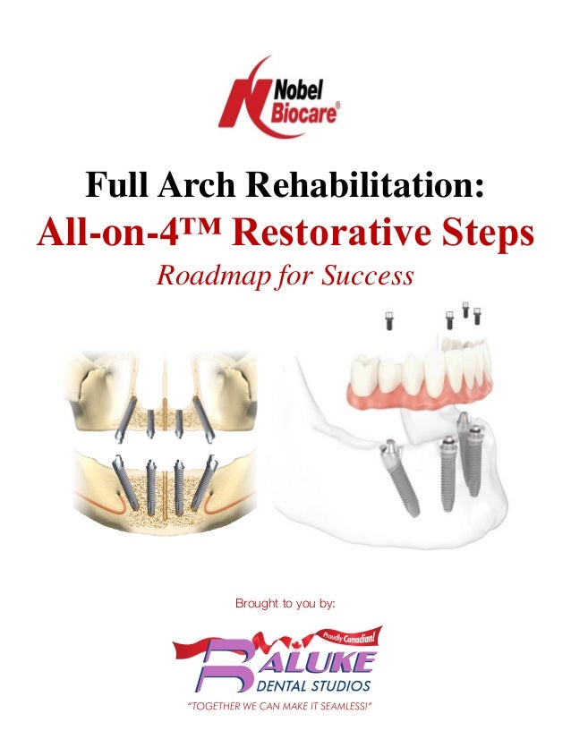
  • 1. Full Arch Rehabilitation: All-on-4™ Restorative Steps Roadmap for Success Brought to you by:
  • 3. NobelBiocare: FullArchRehabilitation-All-on-4™RestorativeSteps -RoadmapforSuccess Full Arch Rehabilitation: All-on-4™ Restorative Steps Roadmap for Success Treatment Planning/Diagnosis 1. Setup meeting with Treatment Team to discuss case – communication is key 2. Qualifying the Patient When evaluating a candidate for All-on-4™ treatment, the most essential components are the patients’ current esthetics and function. It is important to identify the patient’s smile line and vertical occlusal space. If there are noticeable problems with their current esthetics or function, now is the time to correct these. Examples of issues that may need to be corrected pre-operatively:  Poor Fitting Dentures  Limited Inter-Occlusal Space  Incorrect Bite  Insufficient space for prosthesis 3. Treatment Planning Evaluation Questions Comfort  Please describe any type of discomfort or awareness of pain associated with your denture or partial.  What is your experience sleeping with dentures in your mouth?  Do you notice a burning sensation in your mouth?  Do you currently have sore spots with your denture? Esthetics  Have you noticed changes in the appearance of your face?  Are you happy with the appearance of your teeth?  Are you confident with your smile?  Are you concerned with signs of aging (appearance of face)?  Do you have sores at the corners of your mouth?
  • 4. NobelBiocare: FullArchRehabilitation-All-on-4™RestorativeSteps -RoadmapforSuccess Function – Chew and Speak  Can you eat and speak comfortably without pain or looseness of the denture?  Do you use denture adhesives? Occasional or routine? What is your attitude toward adhesives?  Can you easily eat dry food (for example, a granola bar)?  Do you ever notice dryness of your mouth, including your lips?  Do you ever feel like your lips stick together? Dental History  When was your last reline or adjustment of denture?  How long have you been using dentures/this denture?  Did you use a removable partial denture before full?  What was the reason for tooth loss? (perio/destructive function/caries)  What food limitation have you experience because of chewing challenges? Any special requirements for food preparation?  Can you eat what you would like to eat? Why not? Examples of Candidates for All-on-4™ Treatment:
  • 6. NobelBiocare: FullArchRehabilitation-All-on-4™RestorativeSteps -RoadmapforSuccess 4. Capture Vertical Dimension of Occlusion (VDO) for Restoration/Smile Line A. Vertical Dimension of Occlusion (VDO), also known as Occlusal Vertical Dimension (OVD) is a term used to indicate the superior-inferior relationship of the maxilla and the mandible when the teeth are situated in maximum intercuspation. A VDO is not only possessed by people who have teeth, however; for completely edentulous individuals who do not have any teeth with which to position themselves in maximum intercuspation, VDO can be measured based on the subjective signs related to esthetics and phonetics. Vertical dimension can be captured using a base plate and bite rim.  15mm of space (minimum) is required for NobelProcera® Implant Bridge - Fixed (Restoration (Measure Length of Central Incisor + 6mm) 5. Identify Smile Line and Transition Line of Prosthesis - make sure transition line of prosthesis is apical to smile line  If soft tissue is visible while smiling, measure the distance between the gingiva above central and lateral incisors and the extreme border of the upper lip Smile Line Transition Line of Prosthesis
  • 7. NobelBiocare: FullArchRehabilitation-All-on-4™RestorativeSteps -RoadmapforSuccess 6. Photographic Evaluation  Full Face, Lips at Repose  No animation – with and without Denture/Partial  Full Face Smiling – with and without Denture/Partial  Lips Retracted, Teeth apart – with and without Denture/Partial  Lips Retracted, Teeth together – front, right, and left sides  Side Profile, Full Face  Side Profile, Smile  Intra-oral Alveolar Ridge without Denture  Denture out of Mouth: Occlusal View and Intaglio View Example: No Animation with & without Denture/Partial Example: Smiling with & without Denture/Partial Example: Retracted with & without Denture/Partial
  • 8. NobelBiocare: FullArchRehabilitation-All-on-4™RestorativeSteps -RoadmapforSuccess 6. Case Evaluation – Esthetics  Lip Support  Naso-Labial Angle  Facial Midline  Occlusal Plane  Lip Size – Flat and Thin  Tooth Display at rest, and when speaking  Lip Dynamics  Smile Line  Transition Zone 7. Ridge Reduction (Alveolectomy) – determine how much is necessary 8. Take Accurate Records A. Assess the condition of the oral mucosa, ridges, facial, cheek, and lip support (contours). B. Take Impressions of both arches and Bite Registration. The impressions must include the palate and vestibules in the maxilla and the vestibules and retro molar pads in the mandible. C. Make sure to include the Edentulous Saddles i. Bite Rims are necessary if edentulous saddles cannot be capture on initial impressions. ii. If bite will be opened, take a new bite registration with wax rims tried in
  • 9. NobelBiocare: FullArchRehabilitation-All-on-4™RestorativeSteps -RoadmapforSuccess 9. Shade/Mold Information –  Consult with patient regarding the color, shape, and size of teeth that will be suitable.  Send photos, shade/mold information, measurements, and patient’s desires concerning esthetics of denture to Dental Laboratory. The Laboratory will utilize the impressions and other information to fabricate immediate denture to be used as provisional prosthesis on the day of surgery. 10. Evaluate Bone Volume  With CT Scan and X-rays determine implant/restorative options based on the amount of bone volume 11. Discuss Patient Symptoms/Issues, Expectations, and Motivation 12. Review steps/coordinate schedules  Surgical Specialist based on CT Scan determines and orders Implants and Restorative Components w/alternative sizes/angles:  Multi-unit Abutments: Straight, 17°, 30° angle correction w/varying tissue depth  Temporary Copings Multi-unit + Prosthetic Screws (extra)  Multi-unit Abutment Healing Caps  Impression Copings – Open Tray – for final prosthesis  Multi-unit Abutment Lab Analogs – for final prosthesis
  • 10. NobelBiocare: FullArchRehabilitation-All-on-4™RestorativeSteps -RoadmapforSuccess Immediate Provisionalization Materials: Equipment:  Handheld Light Unit  Bench Lathe  Electric Handpiece  Dust Hood with Vacuum (Handler Bench Top Porta-Vac) Hand Tools:  Rubber Dam Punch  Great White Burs (SSW HP-8)  #25 Surgical Blades  #6R Redwood Plaster Knife  #7R Redwood Plaster Knife  Small Surgical Scissors  Instrumentation: Prosthetic Kit (torque wrench, Unigrip drivers 20, 25, 30mm, multi-unit abutment driver)  Perio Probe or Explorer Supplies:  Blue Mousse Bite Material or Similar  Rubber Dam  Aquasil Ultra XLV Fast Set, Teflon Tape, Cavit or Wax for blocking out screw access holes  Exothermic Polymer Acrylic - Cold Cure (3M Secure, Dentsply Dual Line, Unifast Trad, or Quik Set)  eFiber and Perma Mesh to strengthen the denture (Preat)  Ivoclar Universal Polishing Paste  Keystone b12 Brush Wheels  Lab Pumice and Rag Wheels  Burs and Brushes for Acrylic Finishing
  • 11. NobelBiocare: FullArchRehabilitation-All-on-4™RestorativeSteps -RoadmapforSuccess Dental Laboratory creates Immediate Denture & Surgical Guide: 1. Wax Try-in Denture  Based upon impression and bite registration a Wax Try-in Denture is created by the Dental Laboratory. The Wax Try-in for Immediate Denture is designed to verify vertical dimension, esthetics, phonetics, and facial support. Improvements and modifications to the denture can be made at this time.  After modifications are complete, the Dental Laboratory processes an immediate denture 2. Re-mount and Equilibration 3. Cross Mount and Occlusal Guide and Bite Registration
  • 12. NobelBiocare: FullArchRehabilitation-All-on-4™RestorativeSteps -RoadmapforSuccess 4. Duplicate Denture and Trough out Lingual for Surgical Template 5. Add 30-45 Degree Angulation for Posterior Implant 6. Indicate Amount of Ridge Reduction and Mark on Surgical Template 15mm
  • 13. NobelBiocare: FullArchRehabilitation-All-on-4™RestorativeSteps -RoadmapforSuccess Surgery & Immediate Provisionalization: 1. Measure and Mark Vertical Dimension of Occlusion prior to Surgery  Surgical placement of dental implants is based off the immediate denture in an effort to maintain the patients’ proper vertical dimension. Before extracting the teeth, the surgical specialist will mark the chin and nose and measure the distance while the patient is in occlusion. 2. Extract Teeth and Reduce Ridge (alveolectomy)  Ridge is reduced based on pre-determined amount indicated in surgical template (necessary to accommodate prosthesis).
  • 14. NobelBiocare: FullArchRehabilitation-All-on-4™RestorativeSteps -RoadmapforSuccess 3. Placement of Dental Implants  Implants are placed in the arch, equally distributed and angulated to avoid the natural anatomy (sinus and nerve). Implants must achieve initial stability of at least 35Ncm of torque in order to immediately provisionalize the implants with the patients’ immediate denture.  In the case of a dual arch surgery, the upper denture should be provisionalized first, utilizing the palate as a guide in the maxilla and the retro molar pad and bite registration in the mandible. 4. Attach Multi-Unit Abutments  Once implants are placed, multi-unit abutments can be placed - 30° Multi-Unit Abutments in posterior/tilted implants and Straight or 17° Multi-unit Abutment in anterior. Hand tighten – take X-ray to verify seated properly. Torque to appropriate Ncm – Angled (15Ncm)/Straight (35Ncm). Attach Multi-unit Abutment Healing Caps to top of Multi-unit Abutments prior to suturing.
  • 15. NobelBiocare: FullArchRehabilitation-All-on-4™RestorativeSteps -RoadmapforSuccess 5. Index position of Multi-Unit Abutments  Use Blue Mousse to line the intaglio surface of denture to index position of abutments. 6. Hollow-out space in denture for Temporary Coping Multi-unit 7. Attach Temporary Coping Multi-unit to Multi-Unit Abutments  Place hollowed-out denture over Temporary Coping Multi-unit cylinders (passive fit)
  • 16. NobelBiocare: FullArchRehabilitation-All-on-4™RestorativeSteps -RoadmapforSuccess 8. Reduce Height of Temporary Coping Multi-Unit  With Sharpie marker, mark the height the Temporary Coping Multi-unit cylinders need to be cut down to allow the patient to bite down. Remove Temporary Coping Multi-unit cylinders and trim to appropriate height. Can be done before or after pick-up with acrylic. 9. Attach Rubber Dam  Place Rubber Dam around Temporary Coping Multi-unit cylinders (barrier between surgical and restorative materials). Place Light Body Impression Material, wax, or Teflon Tape in top of Temporary Coping Multi-unit cylinder to prevent acrylic from getting inside. Verify proper seating and alignment of denture with pre-operative Bite Registration. 10. Pick-up Temporary Coping Multi-unit cylinders  Use Cold Cure Acrylic (any Exothermic Polymer Acrylic e.g., 3M Secure, Unifast Trad, or Quik Set). Allow Cold Cure Acrylic to set-up.
  • 17. NobelBiocare: FullArchRehabilitation-All-on-4™RestorativeSteps -RoadmapforSuccess 11. Remove Prosthesis with Temporary Coping Multi-unit cylinders processed in acrylic  With UniGrip Driver unscrew prosthesis and remove Prosthetic Screws with a Perio Probe (wax block-out may make Prosthetic Screws difficult to remove). Attach the white Healing Cap Multi Units on the Multi Unit Abutments while the provisional prosthesis is being finished. 12. Convert from Immediate Denture to Fixed Implant Bridge  Trim palate, borders, phalanges, and remove distal cantilevers beyond 3mm. 13. Polish and Smooth Surface of Fixed Implant Bridge  Maintain intaglio surface of denture – create ovate pontic contour for ease of maintenance/hygiene for patient. Remove any sharp angles or edges
  • 18. NobelBiocare: FullArchRehabilitation-All-on-4™RestorativeSteps -RoadmapforSuccess 14. Attach Provisional Fixed Implant Bridge Prosthesis w/Prosthetic Screws  Torque Prosthetic Screws with Unigrip Driver to 15Ncm.  Fill in screw access w/Teflon Tape and composite.  Adjust occlusion using articulation paper – reducing any high spots to level occlusion. 15. Recommended Diet during Healing Phase  A semi-solid/soft food diet is recommended while the implants Osseo integrate – 3-4 months. The patients can eat anything they can cut with a fork (cooked vegetables, canned fruits, well-cooked meat/fish/chicken, etc. Avoid raw vegetables, fruits, and nuts until the implants have Osseo integrated.) 16. Hygiene and Maintenance  For the first 14 days following All-on-4™ Surgery & Provisionalization – have the patient rinse with Peridex Oral Rinse.  After 2 weeks, use a water pick and soft bristle toothbrush with non-abrasive tooth paste to clean.  Regular hygiene visits are recommended every 6 months. Evaluate prosthesis for plaque build-up or red/inflamed soft tissue. If tissue appears healthy, have hygienist clean around implants like an ovate pontic on a bridge.  Remove prosthesis if tissue is red/inflamed or calculus is built up on prosthesis. Use Unigrip Driver to remove Prosthetic screws and clean in the ultrasonic. Use ProClean (tartar and stain remover) for cleaning the prosthesis. Soak screws in alcohol or sterile water. Replace Prosthetic Screws with new screws after removing more than 2 times. Torque Prosthetic Screws to 15Ncm with Unigrip Driver.
  • 19. NobelBiocare: FullArchRehabilitation-All-on-4™RestorativeSteps -RoadmapforSuccess Final Prosthesis – NobelProcera® Implant Bridge: 1. Custom Tray Impression  Un-screw the provisional implant bridge with Unigrip Driver  Take an Alginate Impression for custom trays to be manufactured  Take an impression of the opposing arch, if needed  Take impressions of temporary dentures, to indicate the patients likes/dislikes of existing dentures 2. Final Impression  Un-screw the provisional implant bridge with Unigrip Driver  Attach Open Tray Multi Unit Impression Copings to Multi Unit Abutments – take X-ray to verify seated properly. Lute impression copings together with ortho wire and light cure material or pattern resin – create a rigid frame to ensure accuracy. The Dental Laboratory will lute the Multi Unit Abutment Replicas together when attaching to the impression copings and pouring up the master cast to ensure accuracy.  Make sure the custom tray clears the Open Tray Impression Copings, adjust tray if needed.  Use heavy body impression material around the impression copings and a medium body impression material for the tissue area
  • 20. NobelBiocare: FullArchRehabilitation-All-on-4™RestorativeSteps -RoadmapforSuccess 3. Verification of Master Cast and Final Records  Un-screw the provisional implant bridge with Unigrip Driver  Take a Bite Registration  Mark Midline, High Lip Line, Incisal Edge and Shade  Verification Jig Try-in - The Jig MUST sit passively to each implant/abutment. Take an X-ray to verify seated properly. If the jig does not fit passively, section the jig and re- take final impression with the custom tray over the jig. 4. Wax Try-in  Un-screw the provisional implant bridge with Unigrip Driver  Wax Try-in - If esthetics, phonetics, function, and lip support are acceptable, send to lab for the NobelProcera® Implant Bridge Titanium framework to be milled. Note: the lip support is provided by the gingival third of the tooth. There is no denture flange to provide bulk. If more support is required than what is provided by the wax try-in, then the necks of the teeth will need to be brought forward.  Try in Jig (if didn’t fit passively the first time)
  • 21. NobelBiocare: FullArchRehabilitation-All-on-4™RestorativeSteps -RoadmapforSuccess 5. NobelProcera® Implant Bridge Titanium framework Try-in  Un-screw the provisional implant bridge with Unigrip Driver  Try-in the NobelProcera® Implant Bridge Titanium framework, verify passive fit with each implant/abutment  Teeth will be set in wax 6. Seat the NobelProcera® Implant Bridge - Final Prosthesis  Un-screw the provisional implant bridge with Unigrip Driver  Seat the final prosthesis - Take X-ray to verify seated properly. The final prosthesis should seat firmly against the soft tissue, like an ovate pontic. The design of the tissue interface should be such that it causes the tissue to roll over the prosthesis on the buccal and lingual aspects.  Torque the Prosthetic Screws with Torque Wrench to 15 Ncm when attaching to Multi- Unit Abutments. Always use new prosthetic screws to seat the final prosthesis.  Block out screw access holes to protect screw head with Teflon Tape, Foam, etc. Seal screw access areas with Acrylic  A night guard is provided
  • 23. Full Arch Treatment Options Option 1: Removable Denture Advantages  Relatively inexpensive tooth and gingival replacement  Provides lip support  Easy to remove and clean outside of mouth Disadvantages  Uncomfortable – may cause sore spots on gum tissue  Difficult to eat certain foods  Accelerates bone loss  Often requires reline to improve fit and comfort as bone deteriorates  Difficult to speak as the removable denture may move  May require creams or adhesives to reduce mobility of denture  Approximately 10% functionality compared to natural teeth Option 2: 2 or 4 Implant Over Denture (Removable) Advantages  Improves stability and functionality to 60% compared to natural teeth  Relatively inexpensive tooth and gingival replacement  Provides lip support  Easy to clean outside of mouth Disadvantages  Uncomfortable – may cause sore spots on gum tissue  Remove and clean denture outside of mouth  May still move when chewing or speaking  May require relines to improve fit and comfort as bone deteriorates Option 3: All-on-4™ Implant Fixed Bridge Advantages  Improves functionality to 90% compared to natural teeth  Eliminates need for bone grafting  Can provide temporary bridge day of surgery - eat soft foods while healing  Replaces roots and teeth  Preserves bone and soft tissue  Never decay – 95% success rate over 30 years  Natural-looking esthetics  Allows you to eat the foods you want  Able to clean fixed implant bridge like natural teeth Disadvantages  Time (healing and restorative)  Surgical procedure
  • 26. Abutment placement instructions Step 1 – Remove the Healing Abutment using the Screwdriver Unigrip and by rotating it counterclockwise. Step 2a – Straight Multi-unit Abutment – Use the premounted plastic holder to place the abutment onto the implant and screw the abutment into the correct position. If necessary, shorten the holder with a pair of scissors. When the abutment is seated, remove the plastic holder with a slight bending movement and hand-tighten with Screwdriver Machine Multi-unit. Step 2b – Straight Multi-unit Abutment – Take a radiograph to verify proper seating of the abutment (radiograph for conical connection is shown). Tighten the abutment screw to 35 Ncm using the Manual Torque Wrench Prosthetic and Screwdriver Machine Multi-unit. Or Step 2a – Angled Multi-unit Abutment, 17° or 30° – The abutment is placed over the implant by using the premounted abutment holder. Please note that there are several possible positions in which to place the abutment based on the implant connection and abutment angle. – Tighten the abutment screw using a Screwdriver Unigrip until resistance is felt. The holder is then unscrewed from the abutment by turning it counterclockwise. Step 2b – Angled Multi-unit Abutment, 17° or 30° – Take a radiograph to verify proper seating of the abutment (radiograph for conical connection is shown). Tighten the abutment screw to 15 Ncm using the Manual Torque Wrench Prosthetic and Screwdriver Machine Unigrip. Be sure not to exceed 15 Ncm. See closed or open tray instructions for next steps. 2a OR 2a 1 2b 2b Conical connection Conical connection Closed tray – Abutment level impression instructions Step 3 – Connect the Impression Coping Closed Tray Multi-unit to the abutment by rotating it clockwise. Step 4 – Inject a heavy body impression material (polyether material or polyvinylsiloxane) around the impression coping. Fill the tray with impression material and record the impression. Step 5 – After setting, remove the impression and disconnect the impression copings. Attach an Abutment Replica Multi-unit to each impression coping. Step 6 – Place the impression coping abutment replica assembly into its corresponding location in the impression. Step 7 – Connect the temporary restoration or healing cap. Send the impression to the dental laboratory. 6 7 5 3 4 Multi-unit Abutment Placement and impression techniques Closed tray and open tray Quick Guide This Quick Guide does not replace the Instructions for Use. Please review the instructions for use included with the products. Indications: – Multi-unit restorations – Screw-retained – May be used in combination with framework design if not all implants benefit from abutments. – Used to elevate seating restoration platform when restoration-to-implant level is not practical nor indicated due to depth or angle of implant.
  • 27. Nobel Biocare USA, LLC. 22715 Savi Ranch Parkway, Yorba Linda, CA 92887 Phone 714 282 4800; Toll free 800 322 5001; Technical services 888 725 7100 ©2011 Nobel Biocare USA, LLC. All rights reserved. www.nobelbiocare.com 72194 Rev. 01 (08/11) Description Item number NobelReplace® Multi-unit Abutment (internal tri-channel connection) 17° Multi-unit Abut NobelReplace NP 2 mm 17° Multi-unit Abut NobelReplace NP 3 mm 17° Multi-unit Abut NobelReplace RP 2 mm 17° Multi-unit Abut NobelReplace RP 3 mm 17° Multi-unit Abut NobelReplace RP 4 mm 30° Multi-unit Abut NobelReplace RP 4 mm 30° Multi-unit Abut NobelReplace RP 5 mm 30° Multi-unit Abut Non-Engaging NobRpl RP 4 mm 30° Multi-unit Abut Non-Engaging NobRpl RP 5 mm Multi-unit Abut NobelReplace NP 1 mm Multi-unit Abut NobelReplace NP 2 mm Multi-unit Abut NobelReplace NP 3 mm Multi-unit Abut NobelReplace RP 1 mm Multi-unit Abut NobelReplace RP 2 mm Multi-unit Abut NobelReplace RP 3 mm Multi-unit Abut NobelReplace RP 4 mm Multi-unit Abut NobelReplace RP 5 mm Multi-unit Abut NobelReplace WP 1 mm Multi-unit Abut NobelReplace WP 2 mm Multi-unit Abut NobelReplace WP 3 mm 29235 29236 29237 29238 29239 29240 29241 33409 33410 29196 29197 29198 29199 29200 29201 29202 29203 29204 29205 29206 Conical Connection Multi-unit Abutment (internal conical connection) 17° Multi-unit Abut CC NP 2.5 mm 17° Multi-unit Abut CC NP 3.5 mm 17° Multi-unit Abut CC RP 2.5 mm 17° Multi-unit Abut CC RP 3.5 mm 30° Multi-unit Abut CC NP 3.5 mm 30° Multi-unit Abut CC NP 4.5 mm 30° Multi-unit Abut CC RP 3.5 mm 30° Multi-unit Abut CC RP 4.5 mm Multi-unit Abut CC NP 1.5 mm Multi-unit Abut CC NP 2.5 mm Multi-unit Abut CC NP 3.5 mm Multi-unit Abut CC RP 1.5 mm Multi-unit Abut CC RP 2.5 mm Multi-unit Abut CC RP 3.5 mm Multi-unit Abut CC RP 4.5 mm 36614 36615 36618 36619 36620 36621 36622 36623 36611 36613 36624 36616 36617 36625 36626 Multi-unit Abutment // Placement and impression techniques – abutment level // Closed tray and open tray quick guide Open tray – Abutment level impression instructions Step 3 – Connect the Impression Coping Open Tray Multi-unit on the abutment and tighten using the Screwdriver Unigrip. – Relieve and perforate the impression tray to allow full seating of the tray and protrusion of the guide pins. If there is a large opening, it may be closed off using baseplate wax, with the guide pins indenting or perforating the wax. Step 4 – Inject a heavy body impression material (polyether material or polyvinylsiloxane) around the impression coping. – Fill the tray with impression material and seat the impression tray fully so that the tips of all the guide pins are identifed. – Remove excess impression material from the guide pin access holes. Step 5 – After setting, unscrew the guide pins and remove the impression tray and send to the dental laboratory, including the guide pins. For model fabrication, corresponding implant replicas should be provided by you or your dental laboratory. Step 6 – Connect the temporary restoration or healing cap. 4 3 6 5 Description Item number Brånemark System® Multi-unit Abutment (external hex connection) 17º Multi-unit Abutment Brånemark System NP 2 mm 17º Multi-unit Abutment Brånemark System NP 3 mm 17º Multi-unit Abutment Brånemark System RP 2 mm 17º Multi-unit Abutment Brånemark System RP 3 mm 17º Multi-unit Abutment Brånemark System RP 4 mm 30º Multi-unit Abutment Brånemark System RP 4 mm 30º Multi-unit Abutment Brånemark System RP 5 mm 30° Multi-unit Abut Non-Engaging Bmk Syst RP 4 mm 30° Multi-unit Abut Non-Engaging Bmk Syst RP 5 mm Multi-unit Abutment Brånemark System NP 1 mm Multi-unit Abutment Brånemark System NP 2 mm Multi-unit Abutment Brånemark System NP 3 mm Multi-unit Abutment Brånemark System RP 1 mm Multi-unit Abutment Brånemark System RP 2 mm Multi-unit Abutment Brånemark System RP 3 mm Multi-unit Abutment Brånemark System RP 4 mm Multi-unit Abutment Brånemark System RP 5 mm Multi-unit Abutment Brånemark System WP 1 mm Multi-unit Abutment Brånemark System WP 2 mm Multi-unit Abutment Brånemark System WP 3 mm 29187 29188 29189 29190 29191 29192 29193 33411 33412 29176 29177 29178 29179 29180 29181 29182 29183 29184 29185 29186 Instruments Screwdriver Manual Unigrip 20 mm Screwdriver Manual Unigrip 28 mm Screwdriver Manual Unigrip 36 mm Screwdriver Machine Multi-unit 21 mm Screwdriver Machine Multi-unit Bmk Syst WP 20 mm Screwdriver Machine Unigrip 20 mm Screwdriver Machine Unigrip 25 mm Screwdriver Machine Unigrip 30 mm Screwdriver Machine Unigrip 35 mm Manual Torque Wrench Prosthetic 29148 29149 29150 29158 29159 29151 29152 29153 29154 29165 Components Impression Coping Closed Tray Multi-unit Impression Coping Closed Tray Multi-unit Bmk Syst WP Impression Coping Open Tray Multi-unit Impression Coping Open Tray Multi-unit Bmk Syst WP Abutment Replica Multi-unit Abutment Replica Multi-unit Bmk Syst WP Healing Cap Multi-unit Healing Cap Multi-unit Bmk Syst WP Healing Cap Wide Multi-unit Healing Cap Wide Multi-unit Bmk Syst WP Temporary Coping Multi-unit Temporary Coping Multi-unit Bmk Syst WP Temporary Coping Plastic Multi-unit Temporary Coping Plastic Multi-unit 29090 29092 29089 29091 31161 31162 31145 29066 31146 29067 29046 29047 DCA 468-0 DCA 705-0 Materials to use for Multi-unit Abutment placement and closed or open tray impression techniques Product images are not necessarily to scale. In order to improve readability, Nobel Biocare does not use TM/ ®in running text. In so doing, however, Nobel Biocare does not waive any right to the trademark or registered mark and nothing herein shall be construed to the contrary.
  • 29. Clinical Protocol for Rapid, Graftless, Four-Implant Restoration of the Fully Edentulous Patient PracticalPROCEDURES No.29 P R O V I D I N G S O L U T I O N S F O R C L I N I C A L C H A L L E N G E S Osseointegration has become a predictable biological response to implant placement, enabling dental professionals to focus on surgical and restorative processes that simplify implant therapy. This has resulted in techniques that improve the predictability of aesthetic implant placement and make such treatment options accessible for a greater portion of the patient population. As shown in the following clinical protocol, the All-on-4™ Technique provides numerous advantages in the management of the edentulous patient or the soon-to-be edentulous patient. • Ease of the procedure • Ability to avoid grafting procedures • Potential of immediate loading at the surgical appointment • Decreased cost to the patient • Aesthetics • Can be performed on the partially or fully edentulous patient • Ease of maintenance • Can be flapless when using NobelGuide™ guided surgery. *All-on-4, NobelGuide, and Teeth-in-an-Hour are trademarks of Nobel Biocare, Yorba Linda, CA. Robert Schroering, DMD* • Ken Parrish, DMD, PhD* FIGURE. The All-on-4 Technique provides an immediate fixed restoration to the patient.
  • 30. As with any implant procedure, the All-on-4™ Technique depends on standard implant protocols and sound treatment planning. Its use of the patient’s existing denture or an immediate denture (for the patient with teeth) enables simple chairside modification, relining, and reseating in the patient on the day of surgery. The primary phases of treatment—implant placement, abutment connection, and prosthesis adaptation—are demonstrated in the following protocol. Figure 1.5 After implant placement, Multi-Unit Abutments (Nobel Biocare, Yorba Linda, CA) are placed with an abutment driver. These abutments help correct the angulation of the dental implants. Figure 1.4 The anterior of the sinus is marked with a periodontal probe in order to identify the mesial wall of the sinus. This affords the posterior implant the most-posterior position to create a favorable anterior/posterior spread without invading the sinus. Figure 1.1 Preoperative clinical view of partially edentulous patient who requests a fixed implant restoration. Figure 1.9 Titanium copings are picked up and the temporary fixed detachable prosthesis is modified. First molars are eliminated to reduce cantilever forces during osseointegration. Figure 1.8 Once the temporary prosthesis is hollowed out chairside, the surgical field is covered with a rubber dam to protect the soft tissues during the restorative phase of treatment. Case 1 — Implant Placement and Prosthesis Conversion
  • 31. P R A C T I C A L P R O C E D U R E S N o . 2 9 Figure 1.7 Soft tissue closure is accomplished with horizontal mattress sutures in order to reposition the tissues for optimum healing. Figure 1.6 Temporary Copings (Nobel Biocare, Yorba Linda, CA) are placed. Figure 1.3 All maxillary teeth are extracted and the alveolar bone is reduced via a “horizontal osteotomy” to a height sufficient to avoid the smile line. Figure 1.2 Preoperative panoramic radiograph of the partially edentulous patient, which permits evaluation of the bone levels. Figure 1.11 Postoperative facial view of the temporary fixed detachable prosthesis delivered that same day utilizing the All-on-4 Technique. Figure 1.10 Postoperative panoramic radiograph showing successful placement of four implants in the maxillary arch.
  • 32. Additional Evidence of Success The All-on-4™ Technique represents a viable option for clinicians seeking to bring the advantages of fixed implant prosthodontics to their patients. In proper clinical situations with minor prosthetic involvement, the technique utilizes an immediate loading protocol. The All-on-4 Technique thus constitutes a valuable addition to the practitioner’s armamentarium. Figure 2.2 Postoperative radiograph following successful placement of four maxillary implants and the temporary fixed detachable prosthesis with the All-on-4 Technique. Figure 2.1 Preoperative view of patient with hopeless dentition in the maxillary arch. Figure 3.2 Postoperative radiograph following successful immediate placement of temporary fixed-detachable dentures using the All-on-4 Technique. The final fixed detachable prostheses will be placed four months following surgery. Figure 3.1 Panoramic radiograph demonstrates a completely edentulous patient who has worn dentures for 20 years. *Private practice devoted to advanced implant and periodontal treatment, Louisville, KY. The authors can be contacted at r.schroering@advancedimplant.com and kenparrish@yahoo.com. P R A C T I C A L P R O C E D U R E S N o . 2 9 Case 2 — Patient With Hopeless Maxillary Dentition Case 3 — Completely Edentulous Patient 21243 REV.00 (03/08) 72850 Rev 00 (12/10) Printed in USA. © Nobel Biocare Services, AG. 2010. All rights reserved. www.nobelbiocare.com
  • 34. The All-on-Four Immediate Function Treatment Concept With NobelActive Implants: A Retrospective Study Charles A. Babbush, DDS, MScD* Gary T. Kutsko, DDS John Brokloff, DDS The All-on-Four treatment concept provides patients with an immediately loaded fixed prosthesis supported by 4 implants. This single-center retrospective study evaluated the concept while using the NobelActive implant (Nobel Biocare, Gothenburg, Sweden). Seven hundred eight implants placed in 165 subjects demonstrated a cumulative survival rate of 99.6% (99.3% in maxilla and 100% in the mandible) for up to 29 months of loading. The definitive prosthesis survival rate was 100%. Key Words: All-on-Four, NobelActive implants INTRODUCTION A common condition in elderly patients is the occurrence of edentulism, which can be the result of many factors such as poor oral hygiene, dental car- ies, and periodontal disease. There are also those patients who face edentulism due to a terminal nonrestorable dentition. The eden- tulous condition has been shown to have a negative impact on oral health–related quality of life.1 Clinicians are faced with the growing need to offer solutions to this population due to an increase in their life expectancy2,3 and to fabricate prostheses that provide a replacement for the loss of natural teeth, allowing optimum satisfaction and improved quality of life. The routine treatment for edentulism has been conventional dentures. National epide- miological survey data in the United States suggested that the adult population in need of 1 or 2 dentures would increase from 35.4 million adults in 2000 to 37.0 million adults in 2020.4 Clinical studies have reported that patients with dentures have shown only a marginal improvement in the quality of life when compared with implant therapy.5 The common reasons for dissatisfaction in pa- tients using dentures are pain, areas of discomfort, poor denture stability, and diffi- culties in eating as well as lack of or compromised retention capability.6 Many patients wearing complete dentures com- plain about poor masticatory performance, loss of function, decreased motor control of the tongue, reduced bite force, and dimin- ished oral sensory function.7–10 A review of the literature noted that prostheses support- ed by osseointegrated dental implants sig- nificantly improved the quality of life for Cleveland ClearChoice Dental Implant Center, Pepper Pike, Ohio. *Corresponding author, e-mail: cab@thedentalimplantcenter. com DOI: 10.1563/AAID-JOI-D-10-00133 CLINICAL Journal of Oral Implantology 431
  • 35. edentulous patients when compared with conventional dentures.11–15 Immediate loading of implant-supported, full-arch prostheses for these patients in the mandible or maxilla has been documented as a predictable procedure.16–20 Excellent suc- cess rates for immediately loaded, fixed prosthetic reconstructions19–26 and long-term follow-up results have been reported in the literature.27–29 Immediate loading of implant- supported fixed full-arch prostheses for these cases in the edentulous maxilla and mandible has been associated with a high level of satisfaction for patients in terms of esthetics, phonetics, and functionality.17–20,30–34 Dental implants are traditionally placed in the vertical position. However, in the com- pletely edentulous jaw as well as in the postextraction patient, problems such as minimum bone volume, poor bone quality, and the need for bone-grafting procedures prior to implant placement create some challenging conditions. For these situations, it has been demonstrated that distal tilting of implants may be advantageous. Tilting preserves relevant anatomical structures and allows for placement of longer implants with good cortical anchorage in optimal positions for prosthetic support.35,36 Strain gauge measurements performed by Krekmanov reported no significant difference between tilted and nontilted implants, and theoretical models showed an increased prosthetic base due to the inclination of implants, which in turn can reduce the force acting over the implants.36 Tilting also increases the inter- implant space, reduces cantilever length in jaws,21,31,36,37 and reduces the need for bone augmentation. Good clinical outcomes have been reported in various studies using tilted implants.31,32,35,38–40 The All-on-Four treatment concept pro- vides edentulous arches and immediate/ postextraction subjects with an immediately loaded, fixed prosthesis using 4 implants: 2 axially oriented implants in the anterior region and 2 tilted posterior implants.31,32,37 The principle involves the use of 4 implants restored with straight and angled multiunit abutments, which support a provisional, fixed, immediately loaded, full-arch prosthe- sis placed on the same day of surgery. The All-on-Four treatment has been developed to maximize the use of available bone and allows immediate function. Overall, pub- lished data on the All-on-Four concept reported cumulative survival rates between 92.2% and 100%.31–34,37,40–42 The All-on-Four concept has been report- ed predominantly in the literature with the NobelSpeedy or the Branemark System dental implants. The purpose of this study was to evaluate the All-on-Four concept using an implant (NobelActive) with a tapered body and a variable thread design for up to 29 months of loading. MATERIALS AND METHODS This is a retrospective single-center study. Subjects with totally edentulous arches and/ or in need of extraction of the remaining compromised teeth were rehabilitated with the NobelActive implants. The first implant in the study was placed on February 21, 2008, and the last implant was placed on Septem- ber 12, 2009. Each subject received an immediately loaded, fixed, complete-arch provisional prosthesis on the day of implant placement according to the All-on-Four technique. The definitive prostheses were delivered within 6 to 8 months after implant insertion. An actuarial life table method was used to determine implant cumulative sur- vival rate. Patients treated with the technique and therefore included in the retrospective anal- ysis met the following criteria: N jaw bone profile for the placement of at least 4 implants of at least 10 mm in length in either healed or immediate extraction sites 432 Vol. XXXVII/No. Four/2011 All-on-Four Immediate Function Treatment
  • 36. N good general health with acceptable oral hygiene N implants achieved stability at insertion Patients could not be treated according to the technique if they had insufficient bone quality and quantity for placement of endos- seous implants, exhibited severe parafunc- tional habits, or had a compromised medical history that would affect implant placement (eg, bisphophonates, chemotherapy). Surgical protocol A cone-beam computerized tomographic scan (CBCT; I-CAT cone beam CT scan, Imaging Science Corp, Hatfield, Penn) was taken prior to surgery, and the bone profile, which included the bone quality and bone volume, was assessed43 by 2 experienced clinicians (C.A.B. and G.T.K.). In the vast majority of cases, the patient was adminis- tered intravenous (conscious) sedation using fentanyl citrate 0.5 mg/mL (fentanyl; Hospira, Lake Forest, Ill), diazepam 5 mg/mL injection (Valium; Hospira), as well as nitrous oxide oxygen inhalation. This was in addition to articaine hydrochloride 4% and epinephrine bitartrate 1:100 000 (Septodent, Paris, France), local anesthesia that was administered in both block and infiltration technique. A few of the patients were administered general anesthesia based their preexisting medical profile. Patients were started on a course of antibiotic (penicillin VK 250 mg, Dispensing Solutions, Santa Ana, Calif), 4 times a day, 2 days prior to the surgical procedure in cases in which teeth had to be extracted. Postoperatively, all patients were given the same antibiotic 4 times per day over a period of 10 days. If patients were allergic to penicillin, clindamycin tablets (clindamycin HCL 150 mg, Dispensing Solutions) were given using a similar dosage regimen. In addition, hydrocodone bitartrate and acet- aminophen 7.5 mg/750 mg (Vicodin, Dis- pensing Solutions) were also used as an analgesic along with anti-inflammatory med- ication, methylprednisolone, 4-mg dose pack (Medrol, Dispensing Solutions). At the end of the procedure, bupivacaine 0.5% with 1:200 000 epinephrine (bupivacaine, Cook- Waite, Greensboro, NC) was also adminis- tered for its analgesic-sparing effect. Implant placement NobelActive implants were inserted by (C.A.B.) according to the manufacturer’s guidelines (manual No. 21279-GB085, Nobel Biocare Services 2008). Each subject received 2 distally tilted implants in the posterior region followed by 2 anterior implants in either the maxilla or the mandible. In the maxilla, the tilted implants were positioned just anterior to the maxillary sinus and in the mandible; the tilted implants were posi- tioned anterior to the mental foramen. Implant placement was assisted by the All- on-Four surgical guide (Nobel Biocare; Fig- ure 4). The guide was placed into a 2-mm osteotomy made at the midline of the mandible and/or maxilla, and the titanium band was contoured so that the occlusal centerline of the opposing jaw was followed. The guide allowed for optimal positioning, alignment, parallelism, and inclination of the implants for subsequent anchorage and prosthetic support. The drill protocol fol- lowed the manufacturer’s guidelines (All-on- Four procedures and products, manual No. 16896 Lot GB 0603, Nobel Biocare Services, 2006). The implant sites were usually underprepared avoiding countersink- ing to engage as maximum cortical support bone as possible. The recommended drill sequences for soft bone type IV, medium type II and type III, and dense type I bone were followed. A manual surgical torque wrench (Nobel Biocare) was used to check the final torque of the implant, which was carefully documented (Table 1). In cases of immediate implant placement, the soft Babbush et al Journal of Oral Implantology 433
  • 37. tissues were readapted to obtain a primary closure around the abutments and fresh extraction sites and then sutured back into position with interrupted resorbable 4.0 chromic sutures (Salvin Dental Specialties, Charlotte, NC). Local bone grafting to cover exposed threads and/or other osseous de- fects associated with extraction sockets was performed at 64% of implant sites with demineralized bone matrix gel (Dyna graft- D, Keystone Dental, Boston, Mass), and 1% of implant sites were grafted with autogenous bone from the local surgical area. Straight, 17u multiunit abutment, internal (Nobel Biocare), and 30u angulated multiunit abutments, internal (Nobel Biocare), were used to achieve relative parallelism of the implants so that a rigid prosthesis would seat in a passive manner. Open-tray multiunit impression copings (Nobel Biocare) were placed on the abut- ments, and an impression was made with a custom open tray using precision impression material (Flexitime, Heraeus Kulzer, Hanau, Germany). Patients were instructed to avoid brushing and to use warm water rinses for the first postoperative week. A cold or room- temperature soft diet for the first 24 hours following surgery was recommended, fol- lowed by a semisolid diet for the next 3 months. Patients were given antibiotics and analgesics as listed in the surgical protocol. A CBCT scan was taken immedi- ately postoperatively to verify the implant positions and the prosthetic components. Prosthetic protocol A provisional denture was prefabricated with heat-cured acrylic resin (Ivocap high-impact acrylic, Ivoclar Vivadent, Schaan, Liechten- stein) prior to the surgical procedure. Imme- diately following surgery, the denture was modified to the master model in the laboratory. Fabrication was completed using cold-curing material (Probase, Ivoclar Viva- dent). This provisional all-acrylic resin pros- thesis was seated within 3 to 4 hours of completion of surgery on the same day. The patients were scheduled for routine follow- up visits after surgery at 1 week, 2 weeks, 4 weeks, and 3 months postoperative and on a yearly basis. At the 3-month appointment, fabrication of the definitive prosthesis was initiated. Periapical digital radiographs using a parallel technique were obtained at the 3- month appointment and thereafter on a yearly basis from the date of the surgery (Figures 7, 13, 14, 20, and 29). Implants were checked by visual observation for plaque and bleeding on probing at the follow-up intervals. Periapical radiographs and plaque and bleeding indices at various follow-up intervals are part of routine care for patients at the clinic and not a part of the analysis in this study. The definitive prostheses consisted of a milled titanium frame with a wrap-around heat-cured acrylic resin (Ivocap high-impact acrylic). All restorations were performed by 1 clinician (G.T.K.). Survival criteria The modified Albrektsson criteria used in this investigation are the following: an implant was regarded successful when there was (1) no radiolucency around the implant; (2) no signs of infection, pain, or ongoing patholog- ical processes at the implant site; (3) the TABLE 1 Torque values (implant insertion) Ncm n % ,35 14 1.98 35 40 5.65 36–45 53 7.49 46–55 41 5.79 56–65 41 5.79 66–70 471 66.53 Not recorded* 48 6.78 Total 708 100 *Implants achieved primary stability, although torque was not noted in numerical values. 434 Vol. XXXVII/No. Four/2011 All-on-Four Immediate Function Treatment
  • 38. implant was restored and functionally loaded; and (4) the prosthesis was stable for multiple implants supporting a complete arch prosthe- sis. An implant was classified surviving when it remained in the jaw and was functionally loaded even though all the individual success criteria were not fulfilled. A failed implant was an implant that had fractured beyond repair or could not be classified as a successful or surviving implant.44 Statistics A single reviewer abstracted the relevant data from medical records of the patients who were treated consecutively with the All- on-Four technique and entered them into a spreadsheet (Excel 2007, Microsoft, Red- mond, Wash). An actuarial life table45 was used to calculate the cumulative survival rate. Statistical analysis was done in SPSS 17.0 (SSPS, Chicago, Ill) using the Fisher exact test to determine the level of significance (P , .05) comparing the survival rates of the arches as well as the various implant sizes. RESULTS One hundred sixty-five patients (72 men and 93 women) with a mean age of 59 (SD 611 years) have been included in the analysis. Seven hundred eight implants re- storing both jaws (109 maxillae and 68 mandibles) have been placed. Four hundred thirty-six implants have been placed in the maxilla and 272 in the mandible. Twelve patients were treated in both jaws. Each prosthesis was supported by 4 implants. Most of the implants were seated with a minimum of 35-Ncm torque. Two percent (n 5 14) of the implants were seated at a torque of ,35 Ncm (Table 1). All implants achieved primary stability at placement. Four hundred twenty-four implants were placed in extrac- tion sites immediately after tooth extraction, and 284 were placed in healed sites. Local bone grafting was performed at 65% of the implant sites; no bone grafting was reported in 35% of the sites. Implant distribution according to implant type and implant length is outlined in Tables 2 and 3, respectively. Implant follow-up occurred up to 29 months. Acrylic provisional restorations were placed within 3 to 4 hours of surgery, with occlusal contact limited to the anterior area only. Two anterior implants failed in 2 different patients at the 1-month and 7-month time points due to mobility. In a third patient, 1 tilted implant failed at the 4-month time point due to mobility. All 3 implants failed during the provisional prosthesis phase. All of the 3 implants have been replaced, and no further complications have been noted in these patients. None of the implant failures compromised the prosthesis function, and no relation was found between implant failure and the opposing dentition. TABLE 2 Implant placement Position Maxilla Mandible Total Number of Implants Central incisor Right 11 2 Left 10 7 Lateral incisor Right 62 37 Left 66 31 Canine Right 38 28 Left 39 31 First premolar Right 14 16 Left 14 6 Second premolar Right 77 45 Left 72 51 First molar Right 15 7 Left 17 11 Second molar Right 0 0 Left 1 0 Third molar Right 0 0 Left 0 0 Total 436 272 Babbush et al Journal of Oral Implantology 435
  • 39. Two patients aged 70 and 68 years were lost to follow-up at the 1-year and less than 3-month visit. These patients passed away due to natural causes. Two additional patients were lost to follow-up at the 3- month follow-up visit. Therefore, a total of 16 implants have been lost to follow-up. One hundred sixty-two patients in the study have completed the 6-month follow-up and have had their definitive prostheses (174) deliv- ered. Three jaws were lost to follow-up prior to definitive prosthetic delivery, and 1 jaw was lost to follow-up after definitive pros- thetic delivery. One hundred fifty-six patients have completed the 1-year follow-up. The overall implant survival rate was 99.6% (1 year; Table 4) with no significant difference between the maxillae and mandi- bles (99.3% vs 100%, P 5 .06, Fisher exact test). The 4.3-mm-diameter implants were most frequently used, with a survival rate of 100% (99.2% for 3.5 mm and 99.2% for 5.0 mm; Figure 1). No total arch failures have occurred to date, providing a definitive prosthesis survival rate of 100%. The life table analysis demonstrating the cumulative survival rate is reported in Table 4. Figures 2 through 8 demonstrate a case in postextraction sites of the mandible and maxilla with immediate implant placement, Figures 9 through 14 show a case in healed sites of a patient with a severely atrophic edentulous maxilla. The patient has been edentulous for 50 years. Figures 15 through TABLE 3 Implant size (NobelActive TiUnite)* Implant Diameter Implant Length, mm Maxillae Mandibles 3.5 8.5 — — 10 — — 11.5 12 — 13 31 9 15 28 (1) 42 4.3 8.5 — — 10 2 2 11.5 16 7 13 84 18 15 94 92 18 4 1 5.0 8.5 — — 10 — — 11.5 5 1 13 20 15 15 131 (2) 81 18 9 4 436 (3) 272 *( ) indicates failed implants/replaced. TABLE 4 Cumulative survival analysis* Placed/Followed Implants Failed Implants Time Not Passed Lost to Follow-up CSR Implant insertion »» 3 months 708 1 0 4 99.9 3 months »» 6 months 703 1 0 8 99.7 6 months »» 12 months 694 1 29 0 99.6 12 months »» 18 months 664 0 336 4 99.6 18 months »» 24 months 324 0 240 0 99.6 »» 24 months 84 0 *CSR indicates cumulative survival rate. 436 Vol. XXXVII/No. Four/2011 All-on-Four Immediate Function Treatment
  • 40. 20 show a case displaying terminal nonrestor- able dentition of the maxilla and mandible with extraction and immediate implant recon- struction, and Figures 21 through 29 show a case of a patient with an edentulous maxilla reconstructed with the All-on-Four technique. No complications were reported during surgery or immediately after surgery. DISCUSSION The overall survival rate was 99.6% (1 year), with no significant difference between the maxillae and mandibles. Three implants failed over a period of 29 months of loading. The prosthesis survival rate was 100%. This is in accordance with studies on biomechanical measurements, which demonstrated that tilted implants, when part of a prosthetic support, do not have a negative effect on the load distribution.16,46,47 In addition, a biomechanical rationale in tilting distal implants allows a reduction in cantilever length due to the more posterior position of the tilted implants, resulting in a more favorable stress distribution.47,48 The methodology of using titled implants maximizing the use of the available bone without grafting has been reported, leading to successful clinical outcomes.35,41,42 This is in comparison to the traditional implant treatment in which insufficient bone in the posterior region requires bone-grafting pro- cedures involving greater chair time for the patient as well as increased cost and increased number of procedures. FIGURE 1. Cumulative survival rate in relation to implant diameter. FIGURE 2. Case 1: maxillary and mandibular all-on-Four technique in extraction sites with immediate implant placement. Preoperative computerized tomography scan. Babbush et al Journal of Oral Implantology 437
  • 41. The results of this study are comparable with studies of other implant systems using the All-on-Four concept in the maxilla. Maló et al32 reported a high survival rate of 97.6% in a 1-year retrospective study in which 128 Brane- mark implants (Nobel Biocare AB, Goteborg, Sweden) were immediately loaded in 32 patients. Each jaw received 2 axial and 2 distal implants (All-on-Four) supported by a fixed all- acrylic prosthesis in the completely edentu- FIGURES 3–8. Case 1: maxillary and mandibular All-on-Four technique in extraction sites with immediate implant placement. FIGURE 3. Extraction sockets in the mandible. FIGURE 4. All-on-Four surgical guide in situ. FIGURE 5. Implants in final position. FIGURE 6. Postoperative radiographs at 4 months. FIGURE 7. Clinical photograph of patient at 1 year. FIGURE 8. Postoperative radiographs at 1 year. 438 Vol. XXXVII/No. Four/2011 All-on-Four Immediate Function Treatment
  • 42. FIGURE 9. Case 2: 50-year history of a severely atrophic edentulous maxilla reconstructed with the All-on- Four technique. Preoperative computerized tomography scan. FIGURES 10–14. Case 2: 50-year history of a severely atrophic edentulous maxilla reconstructed with the All-on-Four technique. FIGURE 10. Implants in final position. FIGURE 11. Fixed implant bridge removed to demonstrate soft-tissue health at 1 year. FIGURE 12. Clinical view at 1 year. FIGURE 13. Postoperative radiographs at 1 year demonstrating stable bone levels. FIGURE 14. Postoperative radiographs at 2 years demonstrating stable bone levels. Babbush et al Journal of Oral Implantology 439
  • 43. lous maxilla.32 Testori et al42 reported a 98.8% implant survival and a 100% prosthetic survival rate using a different type of implant system, angulation of the implant-abutment connection for the tilted implants, and a different type of technique for the fabrication of the final prosthesis. In this prospective, multicenter center study, 41 patients received an immediately placed full-arch fixed bridge supported each by 4 axial and 2 distally tilted Ossesotite NT implants (3i, Implant Innova- tions, Palm Beach, Fla) in the edentulous maxillae.42 Aparacio et al35 reported a survival rate of 100% for tilted implants, 96.5% survival for axial implants, and a prosthetic survival rate of 100% after 5 years when 101 Brane- mark implants were placed in the severely resorbed maxilla of 25 patients (59 axially placed and 42 in a tilted direction). Each patient received 2 to 5 implants with at least 1 tilted implant.35 Calandriello et al38 reported a survival rate of 96.7% in a 1-year prospective clinical study when 60 MKIV and Replace select implants (Nobel Biocare AB) were placed in the atrophic maxilla of 18 patients. In this study, 12 partial- and 7 full-arch, fixed prostheses were supported by a total of 33 axially placed and 27 tilted implants.38 The results of this study are in accor- dance with other studies reporting good FIGURES 15–17. Case 3: All-on-Four technique in maxillary and mandibular (nonrestorable dentition) extraction sites with immediate implant placement. FIGURE 15. Preoperative computerized tomography scan. FIGURE 16. (a, b, c) Preoperative clinical photographs of the terminal nonrestorable dentition. FIGURE 17. (a) Implants placed in maxilla with abutments and healing caps in position. (b) Implants placed in mandible with abutments and healing caps in position. 440 Vol. XXXVII/No. Four/2011 All-on-Four Immediate Function Treatment
  • 44. survival rates in the mandible. In a 1-year retrospective clinical study, Maló et al31 reported an implant survival of 96.7% and 98.2% (2 groups) and a prosthetic survival rate of 100% when 176 Branemark implants were placed in 44 patients. An immediately loaded complete-arch all-acrylic prosthesis was supported by 4 implants (All-on-Four) in each completely edentulous mandible.31 Another study reported a 100% implant and prosthetic survival rate when 96 MKIV or the NobelSpeedy Groovy implants (Nobel Biocare AB) were placed in 24 edentulous patients treated in the mandible according to the All-on-Four concept.1 In addition, a 100% implant survival and prosthetic surviv- al rate was reported in a prospective study when 80 Branemark implants were placed in 20 patients with a extremely atrophic mandible. Each patient received 2 axially placed and 2 tilted implants, supporting a fixed full-arch prosthesis (All-on-Four con- cept).40 Previously published literature reporting survival rates using the All-on-Four concept in both the mandible and maxilla is similar to the results of this analysis. In a pilot study, a survival rate of 98.9% was reported in a case series when 189 NobelSpeedy implants were placed in 46 patients, supporting 53 full- arch, all-acrylic prostheses (44 maxillae, 9 mandibles) using the All-on-Four concept.33 Maló et al34 reported a survival rate of 97.2% and 100% in the maxilla and mandible in a 1- year prospective study when 92 Nobel- Speedy implants were placed in 23 consec- utively treated patients. Each jaw was restored by a immediate fixed full-arch prosthesis according to the All-on-Four concept.34 Pomares41 reported a 96.9% FIGURES 18–20. FIGURE 18. Case 3: All-on-Four technique in maxillary and mandibular (nonrestorable dentition) extraction sites with immediate implant placement. Maxillary and mandibular immediate provisional fixed prosthesis in place. FIGURE 19. Postoperative panoramic radiograph at 4 months. FIGURE 20. Postoperative radiograph at 15 months demonstrating stable bone levels. Babbush et al Journal of Oral Implantology 441
  • 45. implant survival (96.7% in the maxilla and 97.2% in the mandible) and a 100% pros- thetic survival rate in a prospective study when 127 MKIII Groovy implants were placed in 20 patients (restoring 19 maxillae and 9 mandibles) using the All-on-Four or All- on-Six concept. A survival rate of 98.4% and 99.7% (maxilla and mandible) at the end of 1 year was reported in another single-cohort study in which 173 edentulous patients received 2 distal and 2 anterior axial MKIV or NobelSpeedy Groovy implants. In this study, each patient received a full-arch fixed prosthesis supported by 2 distal and 2 axial implants (All-on-Four).49 Other long-term studies using the con- cept are comparable to the data in this analysis. Implant survival rates from the follow-up of results of the previously men- tioned studies from Maló et al31,32 with a longer follow-up demonstrated a survival rate of 96.2% in the mandible up to 9 years and 97.7% in the maxilla up to 5 years of follow- up.37 Citing the published literature, it was FIGURES 21–25. Case 4: edentulous maxilla reconstructed with the All-on-Four technique. FIGURE 21. Preoperative clinical photograph. FIGURE 22. Preoperative clinical photograph of the edentulous maxilla. FIGURE 23. Preoperative panoramic radiograph demonstrating the edentulous maxilla. FIGURE 24. The abutments and premounted abutment holders adjusting for relative parallelism. FIGURE 25. (a) The final position of the implants, abutments, and healing caps. (b) The mucoperiosteal flaps repositioned and sutured with 4-0 chromic interrupted sutures. 442 Vol. XXXVII/No. Four/2011 All-on-Four Immediate Function Treatment
  • 46. noted that the overall cumulative survival rate of 99.6% (1 year) in this study for the new implant system (NobelActive) while using the All-on-Four concept offers an attractive solu- tion to clinicians treating edentulous and/or immediate extraction patients. CONCLUSION The overall survival rate using the All-on- Four immediate function treatment concept using an implant with a tapered body and a variable thread design can be considered a viable treatment concept for patients pre- FIGURES 26–29. Case 4: edentulous maxilla reconstructed with the All-on-Four technique. FIGURE 26. (a, b) The tissue and occlusal views of the all-acrylic fixed provisional implant bridge. FIGURE 27. Postoperative panoramic radiograph taken immediately after implant placement. FIGURE 28. Clinical photograph with the definitive prosthesis in place. FIGURE 29. Postoperative radiographs at 1 year with the definitive fixed implant prosthesis in place. Babbush et al Journal of Oral Implantology 443
  • 47. senting with edentulous arches and/or immediate placement. NOTE Dr Babbush has a consulting agreement with Nobel Biocare AB Sweden for ongoing clinical studies and continuing education courses. REFERENCES 1. Agliardi E, Clericò M, Ciancio P, Massironi D. Immediate loading of full-arch fixed prostheses sup- ported by axial and tilted implants for the treatment of edentulous atrophic mandibles. Quintessence Int. 2010;41:285–293. 2. World Medical Association Declaration of Hel- sinki. Ethical principles for medical research involving human subjects. J Am Med Assoc. 2000;284:2043–3045. 3. Salvin B. The business of implant dentistry. In: Babbush C, Hahn J, Krauser J, Rosenlicht J, eds. Dental Implants: The Art and Science. 2nd ed. St Louis, Mo: Saunders Elsevier; 2010:7–13. 4. US Bureau of the Census. Statistical Abstract of the United States: 1996. 116th ed. Washington, DC: US Bureau of the Census; 1996:p 15, tables II, No. 16, p 17, table II, No. 17. 5. Allen PF, McMillan AS. A review of the functional and psychosocial outcomes of edentulousness treated with complete replacement dentures. J Can Dent Assoc. 2003;69(10):662. 6. Heath MR. The effect of maximum biting force and bone loss upon masticatory function and dietary selection of the elderly. Int Dent J. 1982;32:345–356. 7. Wolff A, Gadre A, Begleiter A, Moskona D, Cardash H. Correlation between patient satisfaction with complete dentures and denture quality, oral condition, and flow rate of submandibular/sublingual salivary glands. Int J Prosthodont. 2003;16:45–48. 8. Ikebe K, Matsuda K, Morii K, Furuya-Yoshinaka M, Nokubi T, Renner RP. Association of masticatory performance with age, posterior occlusal contacts, occlusal force, and salivary flow in older adults. Int J Prosthodont. 2006;19:475–481. 9. Koshino H, Hirai T, Ishijima T, Ikeda Y. Tongue motor skills and masticatory performance in adult dentates, elderly dentates, and complete denture wearers. J Prosthet Dent. 1997;77:147–152. 10. Ikebe K, Amemiya M, Morii K, et al. Association between oral stereognostic ability and masticatory performance in aged complete denture wearers. Int J Prosthodont. 2007;20:245–250. 11. Turkyilmaz I, Company AM, McGlumphy EA. Should edentulous patients be constrained to remov- able complete dentures? The use of dental implants to improve the quality of life for edentulous patients. Gerodontology. 2010;27(1):3–10. 12. Allen F, McMillan A. Food selection and perceptions of chewing ability following provision of implant and conventional prostheses in complete denture wearers. Clin Oral Implants Res. 2002;13:320– 326. 13. Awad MA, Lund JP, Dufresne E, Feine JS. Comparing the efficacy of mandibular implant-retained overdentures and conventional dentures among mid- dle-aged edentulous patients: satisfaction and func- tional assessment. Int J Prosthodont. 2003;16:117–122. 14. Awad MA, Lund JP, Shapiro SH, et al. Oral health status and treatment satisfaction with mandib- ular implant overdentures and conventional dentures: a randomized clinical trial in a senior population. Int J Prosthodont. 2003;16:390–396. 15. Heydecke G, Thomason JM, Lund JP, Feine JS. The impact of conventional and implant supported prostheses on social and sexual activities in edentulous adults: results from a randomized trial 2 months after treatment. J Dent. 2005;33:649–657. 16. Bellini CM, Romeo D, Galbusera F, et al. Comparison of tilted versus nontilted implant-support- ed prosthetic designs for the restoration of the edentuous mandible: a biomechanical study. Int J Oral Maxillofac Implants. 2009;24:511–517. 17. Malo P, de Araujo Nobre M, Rangert B. Implants placed in immediate function in periodontally compro- mised sites: a five-year retrospective and one-year prospective study [published correction appears in J Prosthet Dent. 2008;99:167]. J Prosthet Dent. 2007;97(6 suppl):S86–S95. 18. Khatami AH, Smith CR. ‘‘All-on-Four’’ immediate function concept and clinical report of treatment of an edentulous mandible with a fixed complete denture and milled titanium framework. J Prosthodont. 2008;17: 47–51. 19. Ioannidou E, Doufex A. Does loading time affect implant survival? A meta analysis of 1,266 implants. J Periodontol. 2005;76:1252–1258. 20. Attard NJ, Zarb GA. Immediate and early implant loading protocols: a literature review of clinical studies. J Prosthet Dent. 2005;3:242–258. 21. Del Fabbro M, Bellini CM, Romeo D, Francetti L. Tilted implants for the rehabilitation of edentulous jaws: a systematic review. Clin Implant Dent Relat Res. Epub ahead of print, 13 May 2010. 22. Nkenke E, Fenner M. Indications for immediate loading of implants and implant success. Clin Oral Implants Res. 2006;17(suppl 2):19–34. 23. Avila G, Galindo P, Rios H, Wang HL. Immediate implant loading: current status from available literature. Implant Dent. 2007;16:235–245. 24. Esposito M, Grusovin MG, Willings M, Coulthard P, Worthington HV. Interventions for replacing missing teeth: different times for loading dental implants. Cochrane Database Syst Rev. 2007;(2):CD003878. 25. Jokstad A, Carr AB. What is the effect on outcomes of time-to-loading of a fixed or removable prosthesis placed on implant(s)? [published correction appears in Int J Oral Maxillofac Implants. 2008;23:56]. Int J Oral Maxillofac Implants. 2007;22(suppl):19–48. 26. Sennerby L, Gottlow J. Clinical outcomes of immediate/early loading of dental implants: a literature review of recent controlled prospective clinical studies. Aust Dent J. 2008;53(suppl 1):S82–S88. 27. Ericsson I, Randow K, Nilner K, Peterson A. Early functional loading of Brånemark dental implants: 5-year clinical follow-up study. Clin Implant Dent Relat Res. 2000;2(2):70–77. 444 Vol. XXXVII/No. Four/2011 All-on-Four Immediate Function Treatment
  • 48. 28. Henry P, Rosenberg I. Single-stage surgery for rehabilitation of the edentulous mandible: preliminary results. Pract Periodontics Aesthet Dent. 1994;6(9):15–22. 29. Schnitman PA, Wöhrle PS, Rubenstein JE, DaSilva JD, Wang NH. Ten-year results for Brånemark implants immediately loaded with fixed prostheses at implant placement. Int J Oral Maxillofac Implants. 1997;12:495–503. 30. Rosenlicht J, Ward J, Krauser J. Immediate loading of dental implants In: Babbush C, Hahn J, Krauser J, Rosenlicht J, eds. Dental Implants: The Art and Science. 2nd ed. St Louis, Mo: Saunders Elsevier; 2010: 343–345. 31. Maló P, Rangert B, Nobre M. ‘‘All-on-Four’’ immediate-function concept with Brånemark system implants for completely edentulous mandibles: a retrospective clinical study. Clin Implant Dent Relat Res. 2003;5(suppl 1):2–9. 32. Maló P, Rangert B, Nobre M. All-on-4 immedi- ate-function concept with Brånemark system implants for completely edentulous maxillae: a 1-year retrospec- tive clinical study. Clin Implant Dent Relat Res. 2005;7(suppl 1):S88–S94. 33. Maló P, Nobre Mde A, Petersson U, Wigren S. A pilot study of complete edentulous rehabilitation with immediate function using a new implant design: case series. Clin Implant Dent Relat Res. 2006;8:223–232. 34. Maló P, de Araujo Nobre M, Lopes A. The use of computer-guided flapless implant surgery and four implants placed in immediate function to support a fixed denture: preliminary results after a mean follow- up period of thirteen months [published correction appears in J Prosthet Dent. 2008;99:167]. J Prosthet Dent. 2007;97(6 suppl):S26–34. 35. Aparicio C, Perales P, Rangert B. Tilted implants as an alternative to maxillary sinus grafting: a clinical, radiologic, and periotest study. Clin Implant Dent Relat Res. 2001;3:39–49. 36. Krekmanov L, Kahn M, Rangert B, Lindstrom H. Tilting of posterior mandibular and maxillary implants of improved prosthesis support. Int J Oral Maxillofac Implants. 2000;15:405–414. 37. Malo P, Lopez I, Nobre M. The All on Four concept. In: Babbush C, Hahn J, Krauser J, eds. Dental Implants: The Art and Science. 2nd ed. St Louis, Mo: Saunders Elsevier; 2010:435. 38. Calandriello R, Tomatis M. Simplified treatment of the atrophic posterior maxilla via immediate/early function and tilted implants: a prospective 1-year clinical study. Clin Implant Dent Relat Res. 2005;7(suppl 1):S1–12. 39. Fortin Y, Sullivan R, Rangert BR. The marius implant bridge: surgical and prosthetic rehabilitation for the completely edentulous upper jaw with moder- ate to severe resorption: a 5-year retrospective clinical study. Clin Implant Dent Relat Res. 2002;4:69–77. 40. Weinstein R, Agliardi E, Fabbro MD, Romeo D, Francetti L. Immediate rehabilitation of the extremely atrophic mandible with fixed full-prosthesis supported by four implants. Clin Implant Dent Relat Res. Epub ahead of print, 2010 Feb 11. 41. Pomares C. A retrospective clinical study of edentulous patients rehabilitated according to the ‘‘all on four’’ or the ‘‘all on six’’ immediate function concept. Eur J Oral Implantol. 2009;2(1):55–60. 42. Testori T, Del Fabbro M, Capelli M, Zuffetti F, Francetti L, Weinstein RL. Immediate occlusal loading and tilted implants for the rehabilitation of the atrophic edentulous maxilla: 1-year interim results of a multi- center prospective study. Clin Oral Implants Res. 2008;19:227–232. 43. Ganz S. Defining new paradigms for assess- ment of implant receptor sites: the All on Four concept. In: Babbush C, Hahn J, Krauser J, Rosenlicht J, eds. Dental Implants: The Art and Science. 2nd ed. St Louis, Mo: Saunders Elsevier; 2010:148–149. 44. Albrektsson T, Zarb G, Worthington P, Eriksson AR. The long-term efficacy of currently used dental implants: a review and proposed criteria of success. Int J Oral Maxillofac Implants. 1986;1:11–25. 45. Altman D. Practical Statistics for Medical Re- search. Boca Raton, Fla: CRC Press; 1992. 46. Parel S, Branemark PI, Ohrnell LO, Svensson B. Remote implant anchorage for the rehabilitation of maxillary defects. J Prosthet Dent. 2001;86:377–381. 47. Bellini CM, Romeo D, Galbusera F, et al. A finite element analysis of tilted versus nontilted implant configurations in edentulous maxilla. Int J Prosthodont. 2009;22:155–157. 48. Zampelis A, Rangert B, Heijl L. Tilting of splinted implants for improved prosthodontic support: a two- dimensional finite element analysis [published correc- tion appears in J Prosthet Dent. 2008;99:167]. J Prosthet Dent. 2007;97(6 suppl):S35–S43. 49. Agliardi E, Panigatti S, Clericò M, Villa C, Malò P. Immediate rehabilitation of the edentulous jaws with full fixed prostheses supported by four implants: interim results of a single cohort prospective study. Clin Oral Implants Res. 2010;21:459–465. Babbush et al Journal of Oral Implantology 445
  • 50. “All-on-Four” Immediate Function Concept and Clinical Report of Treatment of an Edentulous Mandible with a Fixed Complete Denture and Milled Titanium Framework Amir H. Khatami, DDS1 Christopher R. Smith, DDS, FACP2 1 Assistant Professor, Department of Restorative Dentistry and Prosthodontics, The Ohio State University, Columbus, OH 2 Associate Professor, Department of Surgery, Section of Dentistry, The University of Chicago Hospitals, Chicago, IL Keywords “All-on-Four”; immediate function; conversion prosthesis; tilting dental implants; milled titanium framework. Correspondence Amir H. Khatami, Department of Restorative Dentistry and Prosthodontics, The Ohio State University, College of Dentistry, 305 W. 12th Ave, Columbus, OH 43210. E-mail: akhatami@msn.com Presented at the Table Clinic Session, American College of Prosthodontists 2005 Annual Session, Los Angeles, CA. Accepted August 2, 2006. doi: 10.1111/j.1532-849X.2007.00246.x Abstract The “All-on-Four” concept—tilting the distal implants in the edentulous arches im- proves the prosthetic support—increases the inter-implant distance and provides better implant anchorage in the bone by using longer implants. Computer milling of a solid block of titanium also provides frameworks with improved fit and fewer technical challenges than conventional cast or noncast approaches. This clinical report describes a method of restoring an edentulous mandible with the “All-on-Four” immediate function concept and a milled titanium framework. The patient in our clinical report has reported for follow-up visits for 1 year and is satisfied with the outcome of the treatment. No discernable clinical and radiographic changes were noted around the dental implants. To date, there have been no prosthetic complications. The patient is scheduled for quarterly follow-ups to determine the effectiveness of home care. Immediate loading of implant-supported dental prostheses is documented in the literature with a high and predictable suc- cess rate for the edentulous mandible.1–4 The development of new protocols for immediate loading of dental implants has switched from placing multiple implants and loading a few to placing only four implants as an optimal number to restore a completely edentulous mandible.5 Rehabilitation of the pos- terior edentulous mandible can at times be hindered by bone atrophy distal to the mental foramen and bite forces that are more posterior in the dentition.6 Traditionally, and according to the original concept of the Branemark system, implants are placed in a fairly upright position in the anterior edentulous mandible. Therefore, it is often necessary to fabricate a bi- lateral cantilever, which is sometimes up to 20-mm long, to provide the patient with good chewing capacity in the molar region. Clinical studies have demonstrated that the distal tilting of implants may be advantageous, with reduction of cantilever length about 6.5 mm in the mandible and 9.3 mm in the max- illa.7,8 More recently, a concept was developed to restore the completely edentulous arches with immediately-loaded, tilted distal implants and the use of an “All-on-Four” guide (Nobel Biocare, Yorba Linda, CA) (Fig 1).8,9 This clinical report de- scribes a method of restoring an edentulous mandible with the “All-on-Four” immediate function concept and milled titanium framework. Clinical report A 59-year-old African–American female presented to the Sec- tion of Dentistry at the University of Chicago Hospitals with the desire to have “new dentures.” Her medical history included a kidney transplant due to glomerulonephritis, and she was on a daily dose of corticosteroids. On clinical examination the patient presented with maxillary and mandibular partial eden- tulism. She was wearing removable partial dentures (RPDs) in both arches. The patient was advised to replace the RPDs; however, she chose extraction of the remaining maxillary and mandibular teeth and receiving immediate complete denture (ICD) prostheses instead. Maxillary and mandibular ICDs were made, and after 6 months were relined for a better prosthetic fit (Fig 2). Despite all the effort to make her comfortable with the Journal of Prosthodontics 17 (2008) 47–51 c 2007 by The American College of Prosthodontists 47
  • 51. “All-on-four” concept and treatment of edentulous mandible Khatami and Smith Figure 1 “All-on-Four” guide. Figure 2 Preoperative view of ICDs. Figure 3 Maxillary and mandibular edentulous ridges. mandibular denture, her chief concern was prosthesis stability in the mouth during function. With the advent of the “All-on- Four” concept, the patient was presented with the option of placing four implants in the mandible and immediately loading the implants with a conversion prosthesis.8 In preparation for the procedure and due to her medical status, her physician was consulted. She was instructed to increase the daily dose of her orally administered corticosteroid (Prednisone, Deltasone R , Kalamazoo, MI) to 20 mg, the day prior to surgery and the day of surgery. She was also prescribed Penicillin-VK starting 2 days prior to surgery, 2 g per day, for 10 days. Upon clinical and radiographic evaluation, the mandible was classified ACP PDI for Complete Edentulism Class I (Figs 3 and 4).10 Figure 4 Preoperative panoramic radiograph. Figure 5 Surgical template try-in. Figure 6 Angulated guide pins. Alginate impressions (Jeltrate, Dentsply International, York, PA) of the edentulous arches were made, and the patient’s mandibular denture was duplicated. This duplicate denture was used as a guide to fabricate the radiographic and sur- gical templates (Fig 5). A supra-crestal incision was made from the second mandibular molar area extending to the con- tralateral side. The mental foramina were located bilaterally to serve as landmarks for placement of the most distal im- plants. Implant placement was assisted by the “All-on-Four” guide. The guide was placed into a 5-mm deep osteotomy site made at the midline of the mandible, and its titanium band was adjusted to follow the mandibular arch shape. The guide is used to find the optimal position and inclination of the im- plants (Fig 6). Four 4.3 × 13 mm2 implants (Replace Select Yorba Linda, CA) were placed following the Replace Select protocol and torqued to 45 Ncm (Fig 7). The platforms of the most distal implants were angled about 30◦ distally with the use of the “All-on-Four” guide. Multi-Unit Abutments(tm) (4-mm height) (Nobel Biocare) were connected to the most anterior implants, and 17◦ angled Multi-Unit Abutments(tm) (4- mm height) were connected to the distal implants to bring the screw access holes to the occlusal surface of the prosthesis 48 Journal of Prosthodontics 17 (2008) 47–51 c 2007 by The American College of Prosthodontists
  • 52. Khatami and Smith “All-on-four” concept and treatment of edentulous mandible Figure 7 Connected straight and angulated abutments. Figure 8. Panoramic radiograph after implant placement. Figure 9 Hollowed mandibular ICD. (Fig 8). Subsequent to suturing the soft tissue, temporary abut- ments were connected, and the hollowed-out mandibular den- ture (Fig 9) was indexed using denture repair resin (Nature- cryl, GC America, Inc., Alsip, IL). The mandibular denture was modified to an implant-supported interim fixed prosthe- sis in the dental laboratory. Following occlusal adjustment, the prosthesis was inserted with prosthetic retaining screws (Fig 10). The patient was given oral hygiene instructions and placed on Peridex (Zila Professional Pharmaceutical, Phoenix, AZ) for 2 weeks. At the 2-week follow-up appointment, sutures were removed, and an open tray abutment level impression was made with transfer copings (Nobel Biocare) and vinylpolysiloxane (Aquasil, Dentsply International, York, PA). The impression was poured in die stone (Die-keen, Heraeus-Kulzer, Armonk, NY), and its accuracy was verified with the passively fitting existing implant-supported interim fixed prosthesis (Fig 11). The maxillary denture duplicate cast and the mandibular cast with the attached interim fixed prosthesis were mounted using Figure 10 Implant-supported interim fixed prosthesis. Figure 11 Accuracy verification of the master cast with interim prosthe- sis. the arbitrary facebow transfer and interocclusal records. A putty jig (Lab Putty, Coltene, Cuyahoga Falls, OH) was made to register the 3D relationship of the interim prosthesis to the mandibular cast. Suture removal, final mandibular impression, and the laboratory procedures were completed in one clinic session. Using the temporary abutments (Nobel Biocare) and the putty jig, the resin pattern was fabricated with autopolymerizing resin (GC Pattern resin, GC America, Inc.) (Fig 12). The pattern was sent to Nobel Biocare Headquarters in Sweden to fabricate a milled titanium framework. Once received, the titanium frame- work was tried intraorally for passivity with the recommended screw test11–13 (Fig 13). Subsequent to the denture tooth setup, esthetics, phonetics, and centric relation occlusion were evalu- ated intraorally. Necessary adjustments were made, the prosthesis was pro- cessed with acrylic resin wrap around the framework design, and occlusion was adjusted intraorally. The finished prosthe- sis was inserted by torque tightening the prosthesis retaining screws to 10 Ncm (Figs 14 and 15). The patient was given oral hygiene instructions and scheduled for follow-up every 3 months. At the 3-, 6-, 9-, and 12-month follow-up appointments, there were no discernable clinical or radiographic changes around the dental implants. The patient was instructed on bet- ter prosthetic care of the gingival and lingual surfaces of the prosthesis at the 12-month follow-up appointment. Journal of Prosthodontics 17 (2008) 47–51 c 2007 by The American College of Prosthodontists 49
  • 53. “All-on-four” concept and treatment of edentulous mandible Khatami and Smith Figure 12 Resin pattern guide. Figure 13. Milled titanium framework try-in. Figure 14 Final prosthesis. Discussion In some completely edentulous patients, implant-supported prosthetic treatment is almost impossible without complex tech- niques, such as nerve transposition and grafting in the posterior mandible. Moreover, upright placement of implants in the an- terior edentulous mandible necessitates cantilever lengths from 10 to 20 mm to provide the patient with esthetics and function. When cantilever spans exceeding 7 mm are planned, regardless of the number of implants, an optimal biomechanical envi- ronment should exist.14,15 In a biomechanically compromised environment, such as poor quality bone, the strain transmitted to the crestal bone can be reduced by increasing the anterior– posterior spread of the implants, placement of longer implants, and maximizing the number of implants.16–18 The method of tilting the distal implants in the edentulous arches represents an alternative technique, which leads to placement of longer implants, improved prosthetic support with a shorter cantilever Figure 15 Occlusal view of final prosthesis. arm, improved inter-implant distance, and improved anchor- age in the bone. In vitro studies and theoretical calculations on single implants have shown that tilted implants may in- crease the stress to the bone. Tilted single implants may also be subjected to bending during function, which may lead to increased marginal bone stress19–21 ; however, if such implants are part of a multiple implant-supported prosthesis, the spread of the implants and rigidity of the prosthesis will reduce or change the nature of bending forces.22 In a retrospective clini- cal study of tilted, immediately-loaded implants of 44 patients, Malo et al reported 96.7% and 98.2% implant survival rates for the developmental (more than four implants placed) and the routine group (four implants placed), respectively.8 They re- ported a 100% prosthetic survival rate and concluded marginal bone loss values comparable to values for early loading of the mandibular full arch prostheses.23 The first patients with fixed complete dentures were provided with Cr–Co alloy frameworks with resin teeth. This protocol was modified over time, and gold-alloy casting was introduced to provide a more stable occlusion in metal and to allow porce- lain veneering of the framework; however, in many cases, like severe bone resorption, large amounts of gold alloy had to be cast. Some of the inherent problems with the conventional lost- wax technique were distortion related to arch curvature and the amount of casting alloy. To avoid problems with casting, a few noncasting approaches, such as premachined gold-alloy cylinders/bars and laser-welded titanium frameworks, were in- troduced. These noncasting approaches were technically de- manding and time consuming.16,24,25 More recently, a new protocol based on using computer numeric-controlled (CNC) milling of a solid block of titanium was developed and is free of the technical challenges involved with the previous approaches. The intraoral precision of the prosthesis in this method is completely dependent on the ac- curacy of the master cast and therefore necessitates verifying impression accuracy with a jig or a well-fitting interim prosthe- sis.26,27 Ortorp and Jemt, in a 5-year clinical follow-up of 129 eden- tulous patients, compared the clinical and radiographic perfor- mance of implant-supported prostheses with milled titanium frameworks and conventional cast gold-alloy frameworks. They found lower levels of fracture associated with milled titanium framework prostheses and also found improved framework fit compared to that of conventional castings.28 When using gold 50 Journal of Prosthodontics 17 (2008) 47–51 c 2007 by The American College of Prosthodontists
  • 54. Khatami and Smith “All-on-four” concept and treatment of edentulous mandible screws, milled titanium frameworks have preloads similar to those of gold-alloy frameworks, and the preloads were also similar before and after veneering the milled titanium frame- work with acrylic resin or porcelain.29 Conclusion The patient in our clinical report has been treated with four dental implants placed with the “All-on-Four” concept in the mandible and a fixed complete denture with a milled titanium framework. She was followed up for 12 months and thus far remains satisfied with the outcome of the treatment. There were no discernable clinical and radiographic changes around the dental implants. At the time of this writing, there have been no prosthetic complications, and the patient is scheduled for quarterly follow-ups, mainly to determine the effectiveness of home oral care. References 1. Schnitman PA, Wohrle PS, Rubenstein JE, et al: Ten-year results for Branemark implants immediately loaded with fixed prostheses at implant placement. Int J Oral Maxillofac Implants 1997;12:495-503 2. Schnitman PA, Wohrle PS, Rubenstein JE: Immediate fixed interim prostheses supported by two-stage threaded implants: Methodology and results. J Oral Implantol 1990;16:96-105 3. Balshi TJ, Wolfinger GJ: Immediate loading of Branemark implants in edentulous mandibles: A preliminary report. Implant Dent 1997;6:83-88 4. Balshi TJ, Wolfinger GJ: Conversion Prosthesis: A transitional fixed implant-supported prosthesis for an edentulous arch—a technical note. Int J Oral Maxillofac Implants 1996;11:106-111 5. Duyck J, Van Oosterwyck H, VanderSloten J, et al: Magnitude and distribution of occlusal forces on oral implants supporting fixed prostheses: An in vivo study. Clin Oral Implants Res 2000;11:465-475 6. Book K, Karlsson S, Jemt T: Functional adaptation to full-arch fixed prosthesis supported by osseointegrated implants in the edentulous mandible. Clin Oral Implants Res 1992;3:17-21 7. Krekmanov L, Khan M, Rangert B, et al: Tilting of posterior mandibular and maxillary implants for improved prosthesis support. Int J Oral Maxillofac Implants 2000;15:405-414 8. Malo P, Rangert B, Nobre M: “All-on-Four” immediate-function concept with Branemark System implants for completely edentulous mandibles: A retrospective clinical study. Clin Implant Dent Relat Res 2003;5(Suppl. 1):2-9 9. Malo P, Rangert B, Nobre M: All-on-4 immediate-function concept with Branemark System implants for completely edentulous maxillae: A 1-year retrospective clinical study. Clin Implant Dent Relat Res 2005;7(Suppl. 1):88-94 10. McGarry TJ, Nimmo A, Skiba JF, et al: Classification system for complete edentulism. J Prosthodont 1999;8:27-39 11. Jemt T: Failures and complications in 391 consecutively inserted fixed prostheses supported by Branemark implants in edentulous jaws: A study of treatment from the time of prosthesis placement to the first annual checkup. Int J Oral Maxillofac Implants 1991;6:270-276 12. Tan KB, Rubenstein JE, Nicholls JI, et al: Three-dimensional analysis of the casting accuracy of one-piece, osseointegrated implant-retained prostheses. Int J Prosthodont 1993;6:346-363 13. Kan JY, Rungcharassaeng L, Bohsali K, et al: Clinical methods for evaluating implant framework fit. J Prosthet Dent 1999;81:7-13 14. Rodriguez AM, Aquilino SA, Lund PS, et al: Evaluation of strain at the terminal abutment site of a fixed mandibular implant prosthesis during cantilever loading. J Prosthodont 1993;2:93-102 15. White SN, Caputo AA, Anderkvist T: Effect of cantilever length on stress transfer by implant-supported prostheses. J Prosthet Dent 1994;71:493-499 16. Lekholm U, Zarb GA: Patient selection and preparation. In: Branemark P-I, Zarb, GA Albrektson T (eds): Tissue-Integrated Prosthesis: Osseointegration in Clinical Dentistry. Chicago, IL, Quintessence, 1985, pp. 199-209 17. van Steenberghe D, Lekholm U, Bolender C, et al: Applicability of osseointegrated oral implants in the rehabilitation of partial edentulism: A prospective multicenter study on 558 fixtures. Int J Oral Maxillofac Implants 1990;5:272-281 18. Tada S, Stegaroiu R, Kitamura E, et al: Influence of implant design and bone quality on stress/strain distribution in bone around implants: A 3-dimensional finite element analysis. Int J Oral Maxillofac Implants 2003;18:357-368 19. Clelland N, Gilet A, McGlumphy EA, et al: A photoelastic and strain gauge analysis of angled abutments for an implant system. Int J Oral Maxillofac Implants 1993;8:541-548 20. Celland N, Lee JK, Bimbenet OC, et al: A three-dimensional finite element stress analysis of angled abutments for an implant placed in the anterior maxilla. J Prosthodont 1995;4:95-100 21. Rangert B, Sullivan RM, Jemt T: Load factor control for implants in the posterior partially edentulous segment. Int J Oral Maxillofac Implants 1997;12:360-370 22. Daellenbach K, Hurley E, Brusnki JB, et al: Biomechanics of in-line vs. offset implants supporting a partial prosthesis. J Dent Res 1996;75:183 23. Petersson A, Rangert B, Randow K, et al: Marginal bone resorption at different treatment concepts using Branemark dental implants in anterior mandibles. Clin Implant Dent Relat Res 2001;3:142-147 24. Branemark PI: Osseointegrated implants in the treatment of the edentulous jaw. Experience from a 10-year period. Scand J Plast Reconstr Surg Suppl 1977;16:1-132 25. Lundqvist S, Carlsson GE: Maxillary fixed prostheses on osseointegrated dental implants. J Prosthet Dent 1983;50:262-270 26. Jemt T, Back T, Petersson A: Precision of CNC-milled titanium frameworks for implant treatment in the edentulous jaw. Int J Prosthodont 1999;12:209-215 27. Jemt T, Back T, Petersson A: Photogrammetry—an alternative to conventional impressions in implant dentistry? A clinical pilot study. Int J Prosthodont 1999;12:363-368 28. Ortorp A, Jemt T: Clinical experiences of computer numeric control-milled titanium frameworks supported by implants in the edentulous jaw: A 5-year prospective study. Clin Implant Dent Relat Res 2004;6:199-209 29. Ortorp A, Jemt T, Wennerberg A, et al: Screw preloads and measurements of surface roughness in screw joints: An in vitro study on implant frameworks. Clin Implant Dent Relat Res 2005;7:141-149 Journal of Prosthodontics 17 (2008) 47–51 c 2007 by The American College of Prosthodontists 51
  • 56. DENTAL IMPLANTS J Oral Maxillofac Surg 66:112-122, 2008 Fixed-Prosthetic Implant Restoration of the Edentulous Maxilla: A Systematic Pretreatment Evaluation Method Edmond Bedrossian, DDS, FACD, FACOMS,* Richard M. Sullivan, DDS,† Yvan Fortin, DDS,‡ Paulo Malo, DDS,§ and Thomas Indresano, DMD储 Potential candidates for implant restoration of the completely edentulous maxilla may be interested in receiving a fixed prosthesis as opposed to a remov- able overdenture. Multiple surgical approaches are available in order to provide this type of care. Graft- less approaches such as the use of tilted implants including the zygomatic implant, allow the surgeon to establish adequate support for a fixed prosthesis with- out bone grafting. Adjunctive procedures such as si- nus grafting, maxillary osteotomies as well as horizon- tal augmentations are also available for surgeons who may prefer the grafting approach for the reconstruc- tion of this group of patients. The ability to determine early in the consultation process the type of fixed pros- theses necessary to provide the best functional and es- thetic results is advantageous. This current therapy arti- cle examines 3 critical factors; the nature of the patient’s dental condition and whether the residual ridge is visible in both the relaxed lip and smiling state, direct the choice of fixed dental prostheses. The presence or the absence of bone in the 3 radiographic zones, determines whether bone-grafting procedures are necessary to achieve the desired outcome. Treatment of the edentulous maxilla poses a num- ber of challenges. Expectations regarding the esthet- ics of the definitive prosthesis may be high. Achieving adequate phonetics and stable masticatory function are major concerns. Evaluation of the edentulous maxilla is complicated by the fact that patients may only be missing clinical crowns, or they may have experienced a combination of tooth, soft tissue, and bone loss, with associated changes in facial form. Bone and soft tissue loss can begin before tooth re- moval as a result of generalized periodontitis, creating the appearance of long teeth. The loss of teeth and use of a removable prosthesis can result in continued alveolar bone atrophy in both the vertical and hori- zontal dimensions.1 In a study spanning 25 years, Tallgren observed that the greatest amount of alveolar bone atrophy occurs within the first year of edentu- lism.1 Changes in the jaw relationship as well as facial musculature also may result in deformation or other changes in the facial form and morphology.2 A systematic pretreatment approach to evaluating edentulous patients allows for better communication between the implant team as well as the patients leading to a predictable treatment outcome. McGarry et al 3 developed a classification of complete edentu- lism that considers the quantity of the residual eden- tulous ridge, its morphology or topography, and the relationship of the maxilla to the mandible. Interarch space, tongue anatomy, and the attachment of the musculature to the edentulous ridge are considered. The possible need for preprosthetic surgical proce- dures prior to the fabrication of complete removable dentures is also evaluated. The establishment of evaluation criteria may result in improved patient care, enhanced communication between dental professionals, and better screening and treatment of patients in dental educational cen- ters.3 Guidelines for the treatment of edentulous pa- tients with implants should include consistent clinical *Director of Implant Training, Department of Oral and Maxillo- facial Surgery, University of the Pacific, San Francisco, CA; and Private Practice, San Francisco Center for Osseointegration, San Francisco, CA. †Clinical Director, Nobel Biocare USA, Yorba Linda, CA. ‡Centre d’Implantologic Dentaire de Quebec, Ste-Foy, Quebec, Canada. §Chief Scientist, Nobel Biocare AB, Gothenburg, Sweden. 储Chairman, Department of Oral and Maxillofacial Surgery, Uni- versity of the Pacific, San Francisco, CA. Address correspondence and reprint requests to Dr Bedrossian: University of the Pacific, Oral and Maxillofacial Surgery, 450 Sutter Street, Suite 2439, San Francisco, CA 94108; e-mail: oms@sfimplants. com © 2008 American Association of Oral and Maxillofacial Surgeons 0278-2391/08/6601-0018$32.00/0 doi:10.1016/j.joms.2007.06.687 112