3. INTRODUCTION
• Humans have known about breast cancer for a
long time.
• For example, the Edwin Smith Surgical
Papyrus describes cases of breast cancer. This
medical text dates back to 3,000–2,500 B.C.E
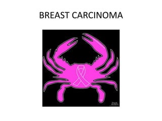
• We see breast cancer resembling a crab , just
as the crab has legs on both sides of body so
as the disease which spreads in all direction –
clinical observation of GALEN in 2 nd century .
4. Anatomy of breast
• Modified sweat gland / apocrine gland /
Mammary gland
• Situated within the superficial pectoral fascia.
- The superficial fascia splits to enclose the
breast to form the anterior and posterior
lamellae. •
5.
6. • The floor is formed by
the deep pectoral
fascia.
• This overlies pectoralis
major and serratus
anterior superiorly and
external oblique and its
aponeurosis inferiorly.
28. EPIDEMOLOGY
• BREAST CANCER IS SECOND LEADING CAUSE
OF CANCER RELATED DEATHS , SECOND TO
LUNG CANCER .
• 40000 PEOPLE DIE OF THIS DISEASE EVERY
YEAR.
• 1 MILLION CASES ARE DIAGNOSED EACH
YEAR.
29. Risk factors for breast cancers
• Non modifiable factors
• Increasing age
• Female sex
• Early age at menarche before age of 12 year
• Older age at menopause beyond age of 55 years .
• Nulliparity
• Family history
• BRCA 1, BRCA 2
• Personal history of breast cancer
• History of radiation exposure
30. Modifiable risk factors
• Reprodutive factors
• Age at first live birth
• Parity
• Lack of breast feeding
• Obesity
• Alcohol consumption
• Tobacoo smoking
• HRT
• Night shifts
• Decrease physical activity
40. Patient history
• Patient age
• Reproductive history
• Previous history of any breast biopsies
• Any hystrectomy
• HRT , OCP .
• Family history
• History of mass,breast pain ,nipple discharge,skin
changes, relation with mensuration
41. CLINICAL PRESENTATION
• Paget’s Disease of the
Nipple
• Skin
Tethering/dimpling/puckeri
ng
• Peau d’Orange
• Skin Ulceration / Fungation
• Visible / Palpable Lump
Hard Consistency
• Non Tender
• Low mobility
• Axillary Lymphnodes+
• Nipple Retraction
• Nipple Discharge
42.
43.
44. The location of breast cancer is as
follows:
• Upper outer quadrant:
50%
• Central area: 18%
• Lower outer quadrant:
11%
• Upper inner quadrant:
15%
• Lower inner quadrant:
6%
45. Physical examination
• Obvious mass
• Asymemetries
• Skin changes
• Nipple retraction and discharge
• Paeu de orange
• Skin fixity
• Tenderness warmth
• Dimpling
122. STAGING WORK UP
• Mamography of the same breast a nd
contralateral breast .
• Ultrasound of abdomen to rule out metastasis
• Xray chest to rule out metastasis of lungs
• Liver function test
125. • Stage 1 and 2 : Are called as early breast
cancer (EBC)
• Stage 3 : are called as locally advanced breast
cancer (LABC)
• 3a –T3N1 or 2
• 3b- T4 any N
• 3c - N3 any T
• Stage 4 : is a metastatic cancer .
175. NOTTINGHAM PROGNOSTIC INDEX- NPI
The index is calculated using the formula: NPI = [0.2 x S] + N + G
Where:
S is the size of the index lesion in centimetres
N is the node status: 0 nodes = 1, 1-4 nodes = 2, >4 nodes = 3
G is the grade of tumour: Grade I =1, Grade II =2, Grade III =3
NPI Score Prognosis 5yr survival :
2 to 2.4 Excellent 93%
2.4 to 3.4 Good 85%
3.4 to 5.4 Moderate 70%
> 5.4 Poor 50%
176. Followup
Monthly self examination of the breast
• Regular physical examination following mastectomy is necessary
• Every 4 months for years 1 and 2,
• Every 6 months for years 3 through 5,
• Every 12 months thereafter
• Contralateral mammogram yearly
• Routine bone scans, skeletal surveys, CT of abdomen and brain- Not
necessary, Yield is low