
Recommended
More Related Content
Similar to L20-Thalamus & Limbic System.ppt
Similar to L20-Thalamus & Limbic System.ppt (20)
More from AzmiNizar1
More from AzmiNizar1 (10)
Recently uploaded
Recently uploaded (20)
L20-Thalamus & Limbic System.ppt
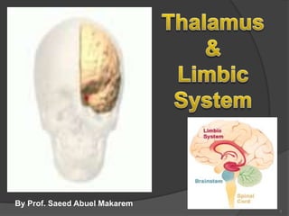
- 1. 1 By Prof. Saeed Abuel Makarem
- 2. By the end of the lecture, the student should be able to: Describe the anatomy and main functions of the thalamus. Name and identify different nuclei of the thalamus. Describe the main connections and functions of thalamic nuclei. Name and identify different parts of the limbic system. Describe main functions of the limbic system. Describe the effects of lesions of the limbic system. Objectives
- 3. 3 THALAMUS It is the largest part of the diencephalon It is the largest nuclear mass of the whole body. It is formed of two oval masses of grey matter. It is the gateway to the sensory cortex. It resemble a small hen. Together with the hypothalamus they form the lateral wall of the 3rd ventricle. PONS THALAMUS Midbrain
- 4. 4 It relays and sends received information to the cerebral cortex. Axons from every sensory system (except olfaction) synapse in the thalamus as the last relay site 'last stop' before the information reaches the cerebral cortex. There are some thalamic nuclei that receive input from: Cerebellar nuclei, Basal ganglia- and Limbic brain regions. THALAMUS
- 5. It has 4 surfaces & 2 ends. Surfaces Lateral: Posterior limb of the internal capsule (L). Medial: The 3rd ventricle. It is connected to the thalamus of the opposite side by the interthalamic connexus, or interthalamic adhesion or Massa intermedia. Superior: Fornix and lateral ventricle.(s). Inferior: Anteriorly: Hypothalamus, & Posteriorly : Subthalamus. 5 Relations I S L 3rd
- 6. Anterior end: Forms a projection, called the anterior tubercle of thalamus It lies just behind the interventricular foramen. Posterior end: Forms a projection called Pulvinar which lies above the superior colliculus and the lateral & medial Geniculate bodies. 6
- 7. Internal Structure White matter: External medullary lamina: Covers the lateral surface of the thalamus. It consists of thalamocortical & corticothalamic fibers. Internal medullary lamina: Bundle of Y- shaped myelinated (afferent & efferent) fibers. It divides the thalamus into: anterior , medial, lateral nuclear groups. Each of these group is subdivided into a number of named nuclei. 7
- 8. 8 It is divided into: Dorsal & Ventral tiers Dorsal tier: which contains: 1. Lateral Dorsal (LD)& 2. Lateral Posterior (LP) 3. Pulvinar.-------------------- ----------------------------- Ventral tier, which contains: 1. Ventral Anterior (VA) 2. Ventral Lateral (VL) 3. Ventral Posterior (VP) (PLVNT, PMVNT) 4. Medial geniculate N. 5. Lateral geniculate N. Lateral Nuclear Group
- 9. Functional Organization of Thalamic Nuclei All thalamic nuclei project to the ipislateral cerebral cortex EXCEPT reticular nucleus. Precise Point to Point projections sometimes found between individual thalamic nuclei and restricted cortical zones. This type of nuclei are called ‘Specific nuclei’ All specific nuclei lie within the ventral tire of the lateral nuclear group. 9
- 10. Classification of thalamic nuclei according to their projection They could be classified into 3 groups, each contains 4 nuclei, (12 nuclei). A) Simple Sensory Relay Nuclei: They receive sensory impulses, and relay them to the sensory cortex. 1. PLVN (posterolateral ventral nucleus). AFF: Medial & Spinal lemnisci, EFF: sensory cortex. 2. PMVN (Posteromedial ventral nucleus). AFF: Trigeminal lemniscus. EFF: Sensory cortex. 3. LGB (lateral geniculate body). AFF: Optic tract, EFF: Optic radiation and visual cortex. 4. MGB (medial geniculate body).AFF: Lateral lemniscus, EFF: Auditory radiation and auditory cortex. 10
- 11. B) Circuit relay nuclei: They receive impulses from different areas of CNS and relay them to specific areas in cerebral cortex, they include: 1. Lateral ventral nucleus (primary motor cortex). 2. Anterior ventral nucleus (premotor cortex). 3. Anterior nucleus (cingulate gyrus) Limbic System. 4. Part of dorsomedial nucleus. 11
- 12. 12 C) Associative nuclei: They receive impulses from other thalamic nuclei then send processed information to the association areas of the cerebral cortex, They include: 1- Part of dorsomedial nucleus. 2- Pulvinar. 3- Lateral dorsal nucleus. 4- Lateral posterior nucleus.
- 13. Functional Organization of the Thalamic nuclei Nucleus Function Inputs (AFF) Outputs(EFF) Anterior Association Mamillary body & Hippocampus Cingulate cortex Medial nuclear group Association Amygdala, Olfactory cortex & hippocampus Prefrontal cortex, hippocampus Lateral dorsal Association Amygdala, Olfactory cortex & hippocampus Cingulate cortex and other limbic regions Lateral posterior Association Superior colliculus, pretectum Occipital parietal, temporal association Medial geniculate Specific nucleus Inferior colliculus 1ry auditory cortex Lateral geniculate Specific nucleus Left & right eyes (optic Tract) 1ry visual cortex Posteromedial ventral Specific nucleus Trigeminothalamic tract 1ry somatosensory Posterolateral ventral Specific nucleus Medial & spinal Lemnisci 1ry somatosensory Posterior nucleus Specific nucleus Superior & Inferior Colliculi 1ry somatosensory Ventral lateral Specific nucleus Globus pallidus 1ry motor cortex Ventral anterior Specific nucleus Globus pallidus 1ry motor cortex Intralaminar Diffuse nucleus Spinal cord, spinothalamic, reticular formation, cerebellar nuclei, globus pallidus, sup. Colliculus. Cerebral cortex & stratum Reticular Diffuse nucleus Reticular formation, corticothalamic, thalamocortical Dorsal thalamic nuclei 13
- 14. 14 Output of thalamic nuclei (Limbic system)
- 15. 15
- 16. 16
- 17. The term "limbic" is from the Latin word Limbus, for "border" or "edge". It separates the medial surface of the cerebral cortex from the diencephalon It consists of a number of: Cortical structures Subcortical structures with Looped connections that all project to the hypothalamus. LIMBIC SYSTEM
- 18. It control a variety of functions including: Emotions, Emotional responses Behaviour & Mood (happy, cry, laugh, sad, afraid, aggression, depression). Motivation. Memory. Visceral & Motor responses involved in (sex, pleasure, hunger, and reproduction). Olfaction. WHAT IS THE MAIN FUNCTION OF THE LIMBIC SYSTEM? MEMORY OLFACTION Pleasure sensation
- 19. 1. Limbic cortex or limbic lobe. 2. Hippocampus & Hippocampal formation. 3. Amygdala. 4. Anterior thalamic nuclei 5. Hypothalamus (mammillary body). 6. Septum. 7. Fornix, and 8. Olfactory system. 9. Habenular nuclei. The limbic system is a set of brain structures including
- 20. What are the Parts of the limbic system? CORTICAL STRUCTURES + SUBCORTICAL STRUCTURES + OLFACTORY SYSTEM SEVERAL LOOPING CONNECTING PATHWAYS ALL THESE STRUCURES HAVE
- 21. CORTICAL STRUCTURES 1. Limbic lobe. 2. Hippocampal formation. 3. Septal areas. 4. Prefrontal area.
- 22. LIMBIC LOBE C-shaped ring of grey matter on the medial surface of each cerebral hemisphere, surrounding the corpus callosum. It includes: 1. Subcallosal area 2. Cingulate gyrus 3. Isthmus 4. Parahippocampal gyrus and the 5. Uncus.
- 23. It is a limbic system structure that is involved in: Formation, Organization, and Storing of memories. It is important in forming new memories and connecting emotions and senses, such as smell and sound, to memories. It is a horseshoe paired structure, one in each cerebral hemisphere. It acts as a memory indexer by sending memories to the appropriate part of the cerebral cortex for long-term storage and retrieving them when needed. HIPPOCAMPUS (Cornu Ammonis)
- 24. HIPPOCAMPUS Site: It is a scrolled structure in the inferomedial part of the temporal lobe. Function: Memory (file new memories as they occur). The hippocampus & its connections are necessary for consolidation of new short-term memories.
- 25. HIPPOCAMPUS Its principal efferent pathway is called the: FORNIX: It is C-shaped group of fibers connecting the hippocampus with mammillary body. it consists of: 2 Fimbria, 2 Crus, 1 Body & 2 Column. The Fornix is an important component of PAPEZ CIRCUIT
- 26. It consists of: 1. Hippocampus 2. Dentate gyrus: Which lies between hippocampus & Parahippocampal gyrus. 3. Subiculum, (at the base of the hippocampus) 4. Entorhinal area (area 28) 5. Induseum griseum (grey matter on the upper surface of the corpus callosum). HIPPOCAMPAL FORMATION
- 27. AMYGDALA Site: almond shaped mass of nuclei. lies near the temporal pole, close to the tail of the caudate nucleus. Function: It is involved in FEAR , Emotions. Anger, & Hormonal secretions.
- 28. CONNECTIONS OF AMYGDALA Inputs: Association areas of visual, auditory & somatosensory cortices. Outputs: Hypothalamus & Autonomic nuclei in the brain stem, Lesion: Lack of emotional responses & docility.
- 29. Site: Located anterior to the interventricular septum Main connections: 1. To Hypothalamus 2. To Habenular nuclei Function: It is the pleasure zone. Septal nuclei
- 30. Korsakoff’s psychosis (Retrograde = loss of new memories at the time of lesion with retained old memories & anterograde amnesia= inability to gain new memories) MOST COMMON IN ALCOHLISM & B1 DEFFICIENCY. Temporal lobe epilepsy The hippocampus is a common focus site in epilepsy, and can be damaged through chronic seizures. It is sometimes damaged in diseases such as herpes encephalitis, Alzheimer’s disease: The hippocampus is one of the first brain areas to show damage in Alzheimer's disease Schizophrenia. Lesions associated with limbic lobe disorders
- 31. SUMMARY The limbic system is a set of evolutionarily primitive brain structures located on top of the brainstem and buried under the cortex. Limbic system structures are involved in many of our emotions & motivations, particularly those related to survival. Such emotions include fear, anger, and emotions related to sexual behavior. The limbic system is also involved in feelings of pleasure such as those experienced from eating and sex.
- 32. Test your knowledge? Which of the following thalamic nuclei belongs to the limbic system? A. Anterior. B. Medial. C. LGN D. MGN Which of the following is the Fear zone? a. Hippocampus. b. Amygdala. c. Fornix. d. Mamillary body. 32
- 33. 33 Which of the following is the principal efferent pathway to the hippocampus? a. Amygdala. b. Dentate Nucleus c. Fornix. d. Mamillary body. Which of the following is the pleasure zone? a. Amygdala. b. Dentate gyrus. c. Septal nuclei d. Hippocampus.