More Related Content
Similar to management of LV thrombus
Similar to management of LV thrombus (20)
management of LV thrombus
- 1. Page 1 of 16
© Annals of Translational Medicine. All rights reserved. Ann Transl Med 2021;9(6):520 | http://dx.doi.org/10.21037/atm-20-7839
Management of left ventricular thrombus: a narrative review
Jose B. Cruz Rodriguez1
^, Kazue Okajima1
^, Barry H. Greenberg2
^
1
Division of Cardiovascular Diseases, Texas Tech University Health Science Center, El Paso, TX, USA; 2
Heart Failure/Cardiac Transplantation
Program, University of California, San Diego, CA, USA
Contributions: (I) Conception and design: All authors; (II) Administrative support: JB Cruz Rodriguez; (III) Provision of study materials or patients:
JB Cruz Rodriguez; (IV) Collection and assembly of data: All authors; (V) Data analysis and interpretation: All authors; (VI) Manuscript writing: All
authors; (VII) Final approval of manuscript: All authors.
Correspondence to: Jose B. Cruz Rodriguez, MD, MPH. Division of Cardiovascular Diseases, Texas Tech University Health Science Center, 4800
Alberta Avenue, El Paso, TX 79905, USA. Email: Benjamin.cruz@ttuhsc.edu.
Abstract: Left ventricular thrombus (LVT) is a serious complication of acute myocardial infarction (MI)
and also non-ischemic cardiomyopathies. We performed a narrative literature review, manual-search of
reference lists of included articles and relevant reviews. Our literature review indicates that the incidence
of LVT following acute MI has decreased, probably due to improvement in patient care as a result of better
and earlier reperfusion techniques. Predictors of LVT include anterior MI, involvement of left ventricular
(LV) apex (regardless of the coronary territory affected), LV akinesis or dyskinesis, reduced LV ejection
fraction (LVEF), severe diastolic dysfunction and large infarct size. LVT is associated with increased risk of
systemic embolism, stroke, cardiovascular events and death, and there is evidence that anticoagulant therapy
for at least 3 months can reduce the risk of these events. Cardiac magnetic resonance (CMR) has the highest
diagnostic accuracy for LVT, followed by echocardiography with the use of echocardiographic contrast
agents (ECAs). Although current guidelines suggest use of vitamin K antagonist (VKA) for a minimum of
3 to 6 months, there is growing evidence of the benefits of direct acting oral anticoagulants in treatment of
LVT. Embolic events appear to occur even after resolution of LVT suggesting that anticoagulant therapy
needs to be considered for a longer period in some cases. Recommendations for the use of triple therapy
in the presence of the LVT are mostly based on extrapolation from outcome data in patients with atrial
fibrillation (AF) and MI. We conclude that the presence of LVT is more likely in patients with anterior ST-
segment elevation MI (STEMI) (involving the apex) and reduced ejection fraction (EF). LVT should be
considered a marker of increased long-term thrombotic risk that may persist even after thrombus resolution.
Ongoing clinical trials are expected to elucidate the best management strategies for patients with LVT.
Keywords: Left ventricular thrombus (LVT); thrombosis; apical thrombus; apixaban; warfarin; dabigatran;
rivaroxaban
Submitted Dec 08, 2020. Accepted for publication Jan 25, 2021.
doi: 10.21037/atm-20-7839
View this article at: http://dx.doi.org/10.21037/atm-20-7839
Introduction
Left ventricular thrombus (LVT) is a serious complication
of acute myocardial infarction (MI) and also of non-
ischemic cardiomyopathies (1). Epidemiologic data suggest
the incidence of LVT to be as high as 15% in patients with
ST-segment elevation MI (STEMI), up to 25% in patients
experiencing an anterior MI (2) and between 2–36% (3,4)
in patients with nonischemic cardiomyopathies. Regardless
^ ORCID: Jose B. Cruz Rodriguez, 0000-0002-2022-6141; Kazue Okajima, 0000-0002-3286-1361; Barry H. Greenberg, 0000-0002-6605-
9385.
Review Article on Heart Failure Update and Advances in 2021
- 2. Cruz Rodriguez et al. Management of LVT
© Annals of Translational Medicine. All rights reserved. Ann Transl Med 2021;9(6):520 | http://dx.doi.org/10.21037/atm-20-7839
Page 2 of 16
of the etiology, however, there is potential for cerebral
or systemic embolization from LVT that increases the
morbidity and mortality in patients with both ischemic and
non-ischemic cardiomyopathies.
Given the scarcity of randomized clinical trials (RCTs)
evaluating the optimal treatment regimen, duration and
effects in the prevention or treatment of LVT, we performed
a narrative literature review in order to examine available
information about therapeutic options for patients with this
condition. We searched the PubMed, Embase, Cochrane
Central Register of Controlled Trials (CENTRAL), and
Cumulative Index to Nursing and Allied Health Literature
(CINAHL) databases through November 9th 2020, with
no restrictions on language. Key words of left ventricular
thrombus, apical thrombus, apixaban, warfarin, dabigatran
and rivaroxaban were used in these searches. Large
prospective studies, metanalysis and systematic reviews
were included, although notably, there are few RCTs on the
topic. Relevant references in the articles that were identified
were then manually-searched. We present the following
article in accordance with the Narrative Review reporting
checklist (available at http://dx.doi.org/10.21037/atm-20-
7839).
Prevalence and risk factors
Historically, the incidence of LVT following acute MI
had been reported to be 20–40%, and even 60% among
patients with large anterior MI (5,6). With the wide
validation of thrombolytic therapy for MI in the 1980s,
the incidence of LVT was reduced to 5–16%, likely due
to salvaging myocardium at risk and minimizing wall
motion abnormalities (7,8). Table 1 presents a chronological
summary of studies discussing the prevalence of LVT.
STEMI patients have been reported to be more likely to
have LVT compared to non-STEMIs (43.1% vs. 5.0%) (17).
Once primary percutaneous coronary intervention (PCI)
became the standard of care for STEMI (after the mid
1990’s), the prevalence of LVT seems to be even smaller,
with a cumulative incidence of 10% compared to 33%
(1,28). In a retrospective, single center study (n=1,059)
of STEMI patients treated with primary PCI, LVT was
detected in 5% of the subjects. Patients with LVT were
more likely to have lower left ventricular ejection fraction
(LVEF) (47% vs. 35%, P<0.01), anterior MI (88% vs. 42%,
P<0.01) and apical akinesis (irrespective of the vascular
location of the MI). Use of IIb/IIIa inhibitor and symptoms
to balloon time were of borderline statistical significance
in predicting prevalence of LVT, possibly suggesting an
association with larger infarcted territories (18). A smaller
(n=92) study in patients who underwent primary PCI plus
IIb/IIIa inhibitor use showed a prevalence of 4.3% (11).
Similarly, another single center study (n=1,698) in STEMI
patients where echocardiographic contrast agents (ECAs)
were used in 76% of the cohort, reported a prevalence
as low as 1.6% for LVT (21). Echocardiography was
performed early after reperfusion, which could have missed
later thrombi formation.
Early treatment with PCI is only one of the several
ongoing changes in STEMI care compared to historical
cohort. Other changes include more effective antiplatelet
agents, reduced time to revascularization and newer
generations of stents, all of which aim to minimize infarct
size. The resultant wall motion abnormalities should
then be expected to decrease the risk for LVT (29,30). A
metanalysis (1) including 19 articles from 2000 to 2015
(n=10,076) in STEMI patients who underwent PCI showed
a LVT incidence rate of 2.7% (95% CI: 1.9–3.5%). In
selective analysis of those with anterior MI, the rate of LVT
was 9.1% (95% CI: 6.6–11.6%) which decreased to 7.5% in
a sensitivity analysis of studies greater than 100 patients (31).
Across studies, described predictors of LVT are anterior
MI/left anterior descending territory, involvement of left
ventricular (LV) apex regardless of the coronary artery
affected, akinesis or dyskinesis, reduced LVEF and large
infarct size (32,33). Presence of multivessel coronary
artery disease and PCI of culprit lesion only compared to
complete revascularization were not predictive of LVT in
other reports (34). Moreover, severe diastolic dysfunction
(restrictive LV filling pattern) has been associated
with increased risk of LVT, measured either by mitral
deceleration time <130 ms (35) or increased mitral E/A ratio
>2 (36). Although moderate to severe mitral regurgitation
after MI has been associated to LVT (37), this relationship
was confounded by the extent of wall motion abnormalities
and anterior location of the MI and mitral regurgitation
was no longer significant after adjusting for these variables.
Cardiac magnetic resonance (CMR) volumetric analyses
have shown that larger LV volumes and impaired systolic
function correlate with higher incidence of LVT (5,38).
Furthermore, patients with LVT have been reported to
have significantly higher C-reactive protein, fibrinogen,
leukocytes and platelet levels than MI patients without
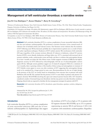
LVT (39,40). Rather than playing independent roles, the
combination of these risk factors interact with each other to
promote LVT. Figure 1 displays this relationship.
- 3. Annals of Translational Medicine, Vol 9, No 6 March 2021 Page 3 of 16
© Annals of Translational Medicine. All rights reserved. Ann Transl Med 2021;9(6):520 | http://dx.doi.org/10.21037/atm-20-7839
Table 1 Clinical studies in diagnosis and prevalence of LVT
Author, year
(reference)
Type of study
[number of patients]
Conclusion
Ezekowitz,
1982 (9)
Prospective [53] Compared to surgical or autopsy confirmation, sensitivity of indium-111 platelet scintigraphy for LVT
71%, echocardiography 77%. Specificity of scintigraphy 100%, echocardiography 93%
Gottdiener,
1983 (3)
Retrospective [123] Prevalence of LVT was 36% in nonischemic cardiomyopathy, systemic emboli in 11%
Bhatnagar,
1991 (8)
Prospective [88] After STEMI, LVT incidence was 5.5% in those receiving thrombolytics and 18% in control group,
likely due to better wall motion score indexes in the treatment group
Pizzetti,
1996 (7)
Retrospective [418] After thrombolysis treatment, 16% prevalence of LVT, 39% in those with anterior MI
Thanigaraj,
1999 (10)
Retrospective [409] Use of ECA increase diagnostic yield of LVT in 79% of patients with nondiagnostic non-contrast
images
Gottdiener,
2003 (4)
Secondary analysis
of RCT [1,343]
Prevalence of LVT was 2.1% in nonischemic cardiomyopathy treated with triple therapy
Srichai,
2006 (5)
Retrospective [361] 29% prevalence of LVT. CMR showed the highest sensitivity and specificity (88% and 99%,
respectively) compared with TTE (23% and 96%) and TEE (40% and 96%) for LVT detection
Rehan,
2006 (11)
Prospective [92] After STEMI treated with PCI and glycoprotein IIb/IIIa inhibitors, incidence of LVT was 4.3%
Kurisu,
2011 (12)
Retrospective [95] Prevalence of LVT was 5.3% in the setting of Takotsubo cardiomyopathy
Weinsaft,
2008 (13)
Retrospective [784] Delayed enhancement-CMR detected thrombus in 7% and cine-CMR in 4.7% of patients with
ischemic cardiomyopathy
Weinsaft,
2009 (14)
Retrospective [121] Contrast echo nearly doubled sensitivity (61% vs. 33%) and yielded improved accuracy (92% vs.
82%) versus non-contrast echo. Contrast echo and cine-CMR correlated well on the diagnosis of
thrombus
Delewi,
2012 (15)
Prospective [200] CMR had the highest sensitivity of 88% and specificity of 99%, followed by TEE with 40% and 96%
respectively, and TTE with 23% and 96%, respectively
Bittencourt,
2012 (16)
Retrospective [31] In a contrast-enhanced coronary computed tomography angiography dataset, a threshold of 65
Hounsfield units provided a sensitivity and specificity of 94% and 97% for detection of LVT
Mir, 2014 (17) Retrospective [85] Prevalence of LVT of 43.1% in STEMI and 5% in NSTEMI
Gianstefani,
2014 (18)
Retrospective
[1,059]
After STEMI treated with PCI, prevalence of LVT =4%. Apical akinesis noted in all LVT regardless of
the territory infarcted. No difference in mortality in patients treated with warfarin for 3–6 months
Wada,
2014 (19)
Retrospective [392] Sensitivity and specificity of non-contrast echocardiography for detection of LVT were 88% and
96%, respectively, compared with 100% each with contrast echocardiography
Robinson,
2016 (1)
Meta-analysis
[10,076]
After STEMI treated with PCI, summary rate of LVT =2.7%, 9.1% in those with anterior MI
Zeng,
2016 (20)
Retrospective [24] Iodine densities were significantly lower in the LVT than the LV cavity, whereas blood densities in the
two areas did not differ significantly
Mao,
2018 (21)
Retrospective
[1,698]
After STEMI treated with PCI, prevalence of LVT =1.6%
Rowin,
2017 (22)
Retrospective
[1,940]
In patients with HCM, incidence of apical aneurysm of 4.8% and LVT was present in 19.3% of them,
0.9% of the entire cohort
Table 1 (continued)
- 4. Cruz Rodriguez et al. Management of LVT
© Annals of Translational Medicine. All rights reserved. Ann Transl Med 2021;9(6):520 | http://dx.doi.org/10.21037/atm-20-7839
Page 4 of 16
Most reported series of LVT focus on ischemic
cardiomyopathy, and there is a paucity of studies in patients
with non-ischemic etiologies. This is compounded by
small sample size of the available studies resulting in poor
precision (wide confidence intervals) for incidence and
prevalence determinations. Previous studies reported
prevalence of LVT up to 36% in the setting of dilated
cardiomyopathy (3,9), with an incidence of 11% for embolic
events. In the setting of dilated cardiomyopathy, presence
of coronary artery disease (that was not considered to be
the cause of the cardiac dysfunction) was not associated
to the presence of thrombus nor systemic embolism (39).
A secondary analysis from the WATCH trial (n=1,343),
multicentric prospective randomized trial for the use of
Table 1 (continued)
Author, year
(reference)
Type of study
[number of patients]
Conclusion
Maron,
2008 (23)
Retrospective
[1,299]
Among patients with HCM, incidence of apical aneurysm of 1.7% and LVT was present in 9.1% of
them, 0.2% of the entire cohort
Kitkungvan,
2018 (24)
Retrospective [121] Prevalence of LVT was 7.4% in patients with chemotherapy-related LV dysfunction
Weber,
2018 (25)
Case series [11] In patients requiring VA-ECMO, prevalence of LVT was 3.1%
Hamada,
2019 (26)
Case series [5] All presented patients with HCM had apical aneurysm, only 2 with outflow obstruction
Ding,
2020 (27)
International
Registry [1,676]
Prevalence of LVT was 3.3% in the International Takotsubo Registry
CMR, cardiac magnetic resonance; ECA, echocardiographic contrast agent; HCM, hypertrophic cardiomyopathy; LVT, left ventricular
thrombus; MI, myocardial infarction; NSTEMI, non-ST elevation myocardial infarction; RCT, randomized clinical trial; STEMI, ST elevation
myocardial infarction; TEE, transesophageal echocardiogram; TTE, transthoracic echocardiogram; VA-ECMO, veno-arterial extracorporeal
membrane oxygenation.
Figure 1 Thrombogenic mechanisms in LV thrombosis. LV, left ventricle; MI, myocardial infarction; TIMI, thrombolysis in MI.
Blood stasis
Hypercoagulability Endomyocardial injury
Proinflammatory state post Ml
Thrombocytosis, leukocytosis
lncreased inflammatory markers
Transmural infarction
Poor reperfusion (TIMI 0,1)
Late presentation
ST segment elevation MI
Larger infarct size
Reduced ventricular contractility
Wall motion abnormalities, particularly apical
Large infarct size
Severe diastolic dysfunction
lncrease LV diameter
Left
Ventricular
Thrombosis
- 5. Annals of Translational Medicine, Vol 9, No 6 March 2021 Page 5 of 16
© Annals of Translational Medicine. All rights reserved. Ann Transl Med 2021;9(6):520 | http://dx.doi.org/10.21037/atm-20-7839
warfarin and antiplatelets in patients with chronic dilated
cardiomyopathy in sinus rhythm reported a prevalence of
LVT of 2.1% (4). Factors associated significantly to LVT
were younger age, lower ejection fraction (EF), higher
regional wall motion score, higher early diastolic filling
velocity, shorter deceleration time, greater LV diastolic
dimension and left atrium area.
In a cohort of patients (n=121) with chemotherapy-
related severe LV dysfunction (24), defined as LVEF <30%,
the prevalence of LVT was 7.4%. Factors associated with
presence of LVT were restrictive filling pattern (OR: 18.13,
95% CI: 4.17–78.89) and LVEF <20% (OR: 36.30, 95% CI:
7.35–179.25). Albeit significant, this report posed very wide
confidence intervals due to the small number of thrombi
detected. Cancer is known to induce a prothrombotic and
hypercoagulable state (41,42), and malignancies that have
been shown to be particularly prothrombotic such as breast
cancer (41), lymphoma (43) and leukemias (44) should
increase the level of suspicion in these patients.
There are limited reports (45) and small case series
of LVT in patients with stress-induced cardiomyopathy
(Takotsubo). The reported incidence of LVT in this
patient population is 3–5.3% (12). The largest cohort of
patients with this condition, the International Takotsubo
Registry, recorded a 3.3% prevalence of LVT and embolism
in the acute phase of the disease (27). Stress-induced
cardiomyopathy usually presents with a larger LV apical
aneurysm than anterior MI, but the lower incidence of LV
apical thrombosis could be related to its transient nature
and lack of endocardial damage compared to MI. On
the other hand, if LVT develops in this setting, the rapid
improvement in apical contraction seen in most cases could
theoretically increase the risk of embolic events.
Reports of LVT in hypertrophic cardiomyopathy
(HCM) are scarce. In a case series (n=5) of patients with
LVT and HCM (26), all had apical aneurysm, two had
LV outflow obstruction, one atrial fibrillation (AF) and
all had resolution of the LVT with anticoagulation [three
with direct oral anticoagulants (DOACs) and two with
warfarin]. In a large cohort of patients with HCM, reports
from 2008 and then 2017 (22,23) suggested incidence of
apical aneurysm of 1.7–4.8%, with LVT present in 9.1–
19.3% of them (0.2–0.9% of the entire cohort). There is
contradictory data about the presence of mid-ventricular
obstruction as the leading factor in the progression of LV
apical aneurysm formation (46-48) and LVT formation.
LVT has also been occasionally described in
amyloidosis (49), hypereosinophilic syndrome (50), and
Chagas’ disease (51).
There is also limited data of LVT in patients requiring
veno-arterial extracorporeal membrane oxygenation (VA-
ECMO). A case series (n=11), all of them due to ischemic
cardiomyopathy complicated with cardiogenic shock,
suggested a prevalence of 3.1% of the entire VA-ECMO
experience of that center (25). Despite use of inotropic
support to promote contractility and anticoagulation in the
therapeutic range at the time of diagnosis, the mortality in
this report of 100%, highlights the seriousness of LVT in
this patient population.
Diagnosis
Routine transthoracic echocardiography (TTE) after
primary PCI-treated MI is recommended to assess
biventricular function, valvulopathies, exclude post-
infarction mechanical complications and evaluate for
LVT (52).
Standard TTE is commonly used for screening, but
it has low sensitivity for LVT detection, requiring the
addition of ECA and/or use of CMR imaging (2). Despite
its widespread use and even with the most advanced
echocardiographic equipment, TTE can be technically
challenging in patients with small intercostal spaces, large
body size, chest deformities or lung disease (poor acoustic
windows), leading to potential failure to detect subtle LVT
findings. Intrinsic limitations of TTE reduce its diagnostic
accuracy, particularly in small non-protruding mural
thrombi, due to true apex foreshortening, limited near-field
resolution or consequent artifacts (near-field clutter) and
difficulty discriminating myocardium-thrombi interface.
Table 1 presents a chronological summary of studies
discussing the diagnostic modalities used to detect LVT.
Although transesophageal echocardiogram (TEE) has well
established superiority for detection of atrial thrombi, its
role for LVT is more limited. As many as 46% of patients
have nonconclusive studies (10,53) for LVT with this semi-
invasive procedure. Techniques to improve the diagnostic
image quality and improve blood-endocardial surface
include harmonic imaging (54) and ECA (54,55). The use of
ECA has been reported to be cost effective (55) and increase
the echocardiographic sensitivity for LVT detection
from 33% to 100%, and the specificity from 82% to
92% (14,19,29).
A single center study (5) in patients with ischemic heart
disease (n=361) evaluated the diagnostic accuracy of TTE,
TEE and CMR with contrast enhancement for evaluation
- 6. Cruz Rodriguez et al. Management of LVT
© Annals of Translational Medicine. All rights reserved. Ann Transl Med 2021;9(6):520 | http://dx.doi.org/10.21037/atm-20-7839
Page 6 of 16
of LVT which was confirmed by surgical or post-mortem
confirmation. The decision to use ECA was at the discretion
of the echocardiographer in this survey. Thrombus was
found to be apically located in 71% of the cases, associated
with aneurysm in 77% of the cases. It has been suggested
that the accuracy of TTE is as high as CMR when the
request for the search of a thrombus is prespecified (34).
Echocardiographic features can predict embolic potential
and guide management of LVT as described in subsequent
sections (56).
The superiority of CMR (5,57,58) with gadolinium
compared to other imaging modalities for detection of
LVT is derived to the immediate strong enhancement of
LV cavity, allowing detection of abnormal intraventricular
structures, as well as the delayed-enhancement technique
that allows visualization of LVT (black), commonly adjacent
to scarred myocardium (bright or hyper-enhanced). On
the other hand, CMR has a higher cost and is not widely
available, even in developed countries. CMR had the
highest diagnostic accuracy with sensitivity of 88% and
specificity of 99%, followed by TEE with 40% and 96%
respectively, and TTE with 23% and 96%, respectively (15).
CMR studies (13) have shown that patients with ischemic
cardiomyopathy have a fivefold higher prevalence of LVT
that patients with non-ischemic cardiomyopathy, even
with similar LVEF. LVT was more likely to be found in
areas with increased myocardial scarring, a parameter that
can only be evaluated with delayed-enhancement CMR.
Because of this reason, cine-only CMR could miss small
intracavitary and mural thrombus.
There are few studies describing the use of cardiac
computerized tomography (CT) for the detection of
LVT. Historically, it was considered that CT scanning
provides similar accuracy as TTE in detecting LVT, but
does so at the expense of increased radiation exposure
and requirement for iodine contrast use (59). Recent
case reports have suggested that metabolically inert areas
seen on positron emission tomography/CT (PET/CT),
with corresponding homogeneous hypodensity in the LV
cavity could suggest organized LVT (60), particularly in
patients with limited echocardiographic windows (61).
LVT are frequently crescent-shaped filling defects
with broad based attachments, although pedunculated
appearances have been described (62). Chronic thrombi
may develop spotty calcifications, although this finding
can also be seen in myxomas (63). CT characteristics
of LVT include lower attenuation with a threshold of
65 Hounsfield units, providing a sensitivity and specificity
of 94% and 97%, respectively (16). It has been suggested
that spectral CT dual-substance separation imaging and
derived images of iodine- and blood-based densities are a
feasible semiquantitative method to investigate LVT (20).
Although indium-111 platelet scintigraphy has been
reported to be comparable to TTE for the diagnosis of
LVT (40), it is an older technology with virtually no role in
current clinical practice.
Irrespective of the imaging modality utilized, if LV
function is found to be depressed, particularly with akinetic
or dyskinetic segments, special attention should be placed
on evaluating for LVT.
Embolic risk and mortality
The reported risk of embolic events from a LVT post-MI
ranges from 6.1% to 86% and seems to be greatest in the
first 3 months after MI (5,58,64-66). In one report, the
incidence of thromboembolism decreased from 22.3% in
pre-PCI studies to 5.5% in post-PCI reports (28), likely
due to increased myocardial salvage. Multiple studies have
demonstrated that thrombus characteristics associated
with systemic embolism include protrusion into the LV
cavity (67), mobility independent of myocardium (68-70),
patient age >68, thrombus area, length of the thrombi in
the lumen, and LVT recurrence (71). On the other hand,
thrombus described as laminated and calcified (bright and
echo dense structures) are immobile, more likely to be
chronic and less likely to embolize, although the risk is not
zero and embolization for these laminar thrombi has been
reported (72).
There is evidence to suggest that LVT should be
considered a marker of increased long-term thrombotic risk
that persists even after treatment and documented LVT
resolution by imaging (67,73). In a study with pathologic
evaluation of the thrombus (3), among patients with
documented recent embolic event, 67% had organized
thrombus and 33% recent thrombus. Furthermore, up to
40% of embolic events can occur when thrombi are neither
protuberant nor mobile (5,71). Evaluation of acute ischemic
strokes in patients with LVT (67) showed the median time
from LVT diagnosis to cerebrovascular event was 20.5 days.
Once again, 5.2% of patients had a stroke even with initial
LVT resolution. The majority (76.5%) of strokes were
characterized as cardioembolic, followed by 14.7% as small
vessel disease and 8.8% large artery atherosclerosis.
LVT is also associated to high rate of cardiovascular
events. A contemporary cohort (74) showed that close to
- 7. Annals of Translational Medicine, Vol 9, No 6 March 2021 Page 7 of 16
© Annals of Translational Medicine. All rights reserved. Ann Transl Med 2021;9(6):520 | http://dx.doi.org/10.21037/atm-20-7839
50% of patients had a major cardiovascular event and 20%
of patients died during follow up, a figure for mortality
that is much higher than reported in STEMI patients
without LVT.
Cardioversion
A small retrospective study (n=21) of patients with known
LV apical thrombus requiring cardioversion (AF in 38%
and ventricular tachycardia in 62%) reported no clinically
apparent embolic events after cardioversion, despite only
81% of patients been anticoagulated beforehand (75).
Nonetheless, most of the LV thrombi were described as
laminated (71%) and none of the patients had mobile
thrombi, which theoretically pose the highest risk. A small
case series (n=8) of patients undergoing VT ablation (76)
with documented LVT (all mural and laminated) showed
only one ischemic stroke on post-procedure day 9, in
a patient with confirmed left atrial appendage (LAA)
thrombus by TEE. All the patients underwent multiple
intraprocedural cardioversion, suggestive the relative safety
of cardioversion in the setting of laminated LVT.
Prevention
More effective revascularization strategies may reduce LVT
formation in patients experiencing a MI, but LVT remains
as a common complication of STEMI in the modern era.
Along with resulting ischemic cardiomyopathy, LVT might
be one of the predominant features affecting management
and prognosis post PCI (32). Interestingly, thrombolysis
in MI (TIMI) flow pre-PCI has been associated with
LVT, but not TIMI flow post-PCI (18). Table 2 presents a
chronological summary of studies involving prophylaxis and
treatment of LVT.
In the thrombolytic era, a randomized trial (82) of
dalteparin 150 IU/kg every 12 hours for 9 days, significantly
reduced LVT formation in patients with anterior MI,
although with increased risk of hemorrhagic complications
(both major and minor). The short in-hospital data in this
report limits the capacity to extrapolate its effectiveness as
an outpatient regimen. Similar findings were reported with
12,500 U of subcutaneous heparin (78) until discharge, but
contradictory results were reported as well in a small (n=30)
RCT (77). A subsequent randomized trial (79) compared
subcutaneous heparin high dose (12,500 U twice a day) with
low doses (5,000 units twice a day) for 10 days after anterior
MI, showing that the high dose was more effective without
incremental bleeding risk.
With current standard of care of PCI and dual antiplatelet
therapy (DAPT) after MI, small pilot randomized trial
(n=60) in patients with anterior MI with Q waves and EF
≤40% showed that preventive full therapeutic dose of
enoxaparin (1 mg/kg twice a day, maximum dose 100 mg)
for 30 days (85) had similar bleeding and thromboembolic
events compared to anticoagulation with warfarin. More
patients had probable mural thrombus at 3.5 months
with enoxaparin, albeit the difference was not statistically
significant.
A retrospective cohort (84) of 460 patients with apical
akinesis or dyskinesis after STEMI on TTE showed
that patients who received DAPT plus warfarin therapy
experienced a net increase in adverse clinical events (all-
cause mortality, strokes, reinfarction and major bleeding) at
180 days. A single center retrospective study (88), suggested
that ticagrelor based DAPT after anterior STEMI (n=641)
was associated with lower incidence of LVT when compared
to clopidogrel (OR: 0.5, 95% CI: 0.29–0.86).
Current guidelines (52,93) suggest considering short
courses (3 months) of anticoagulation in patients with large
anterior wall MI, or anterior-apical akinesis (85) as a class
IIb, level of evidence (LOE) C recommendation.
Treatment
Vitamin K antagonist (VKA)
European and United State guidelines recommend as a class
IIa, LOE C, treatment of LVT with VKAs for a minimum
of 3 to 6 months, with duration individualized to bleeding
risk (52,94) with a target international normalized ratio
(INR) of 2.5 (range, 2–3). Repeat imaging is suggested at
the end of the treatment period to evaluate resolution of
LVT as a class IIa, LOE C recommendation (52).
Lattuca et al., observed a significant decrease in risk of
major cardiovascular events with anticoagulation for more
than 3 months (48% decrease) and LVEF ≥35% (54%
decrease).
The prothrombotic effect of VKA during the start of
therapy warrants their coadministration with parenteral
anticoagulation initially (81). A small prospective study
(n=26) showed that enoxaparin 100 IU/kg twice a day,
followed by fluindione treatment decreased thrombus
size and resulted in resolution of thrombi in 73% of the
patients, suggesting that this therapy may be as effective as
unfractionated heparin (83) at 3 weeks.
- 8. Cruz Rodriguez et al. Management of LVT
© Annals of Translational Medicine. All rights reserved. Ann Transl Med 2021;9(6):520 | http://dx.doi.org/10.21037/atm-20-7839
Page 8 of 16
Table 2 Clinical studies in prevention and treatment of LVT
Author, year
(reference)
Type of study
[number of patients]
Conclusion
Arvan,
1987 (77)
RCT [30] No difference in LVT incidence after acute anterior MI with the use of heparin infusion, partial
thromboplastin time >60 seconds
Tavazzi,
1989 (78)
RCT [711] After acute anterior MI, 12,500 U of subcutaneous heparin reduced LVT incidence and mortality
Turpie,
1989 (79)
RCT [183] In patients with acute anterior MI subcutaneous heparin at a dose of 12,500 U BID was more
effective preventing LVT than 5,000 units BID
Kouvaras,
1990 (80)
Prospective [60] High-dose aspirin was noninferior to warfarin for LVT resolution after 3 months
Kontny,
1993 (81)
Prospective [229] High-dose heparin prevented LVT irrespective of warfarin therapy after acute anterior MI. Warfarin
therapy without heparin was associated with higher rates of LVT
Kontny,
1997 (82)
RCT [517] Dalteparin 150 IU/kg BID significantly reduces LVT after acute anterior MI (RR: 0.63, 95% CI:
0.43–0.92, P=0.02) but is associated with increased hemorrhagic risk.
Meurin,
2005 (83)
Prospective [19] Enoxaparin BID followed by fluindione was as effective as unfractionated heparin at 3 weeks
Le May,
2015 (84)
Retrospective [460] In patients with apical akinesis or dyskinesis, prophylactic use of warfarin increases net adverse
events (all-cause mortality, strokes, reinfarction and major bleeding)
White,
2015 (85)
RCT [60] Enoxaparin for 30 days post MI shortened hospitalization and lowered cost of care compared to
warfarin, with no statistical difference among the groups of LVT at 3 months
Smetana,
2017 (86)
Case series [10] Patients with LVT treated with rivaroxaban and apixaban showed complete thrombus resolution in
8 patients, with only one bleeding event
Robinson,
2018 (87)
Retrospective [98] DOAC-treated patients (mostly apixaban) had similar SSE-free survival
Maniwa,
2018 (65)
Retrospective
[2,301]
In patients with LVT treated with VKA, the time in therapeutic range >50% was associated with
lower embolic events without increasing bleeding events
Altıntaş,
2019 (88)
Retrospective [641] After STEMI, ticagrelor use had lower incidence of LVT than clopidogrel, OR: 0.53, 95% CI: 0.28–
0.96, P=0.039
Daher,
2020 (89)
Retrospective [59] Similar efficacy between DOAC and VKA agents in patients with LVT (70.6% vs. 71.5%)
Lattuca,
2020 (74)
Retrospective [159] Anticoagulation therapy >3 months was independently associated with less MACE (HR: 0.42; 95%
CI: 0.20–0.88; P=0.021). Reduced risk of mortality was observed among patients with total LVT
regression (15.2% vs. 25.0%; HR: 0.48; P=0.039), with higher bleeding rates
Robinson,
2020 (73)
Retrospective [514] Anticoagulation with DOAC vs. warfarin had higher rates of SSE (adjusted HR: 2.64, 95% CI:
1.28–5.43, P=0.01)
Guddeti,
2020 (90)
Retrospective [99] Resolution of LVT, rates of stroke and bleeding were not statistically different between VKA and
DOAC
Iqbal,
2020 (91)
Retrospective [84] No statistically significant differences between VKA and DOAC in rates of LVT resolution (76% vs.
65%), SSE (2% vs. 0%), or clinically significant bleeding (10% vs. 0%)
Ali, 2020 (92) Retrospective [110] Treatment with DOACs was associated with lower 1-year risk of stroke (12% vs. 6%, P=0.33),
although no difference found in ischemic stroke or thrombus resolution)
BID, every 12 hours; DOAC, direct oral anticoagulant; LVT, left ventricular thrombus; MACE, major adverse cardiovascular event; MI,
myocardial infarction; NSTEMI, non-ST elevation myocardial infarction; RCT, randomized clinical trial; SSE, stroke or systemic embolism;
STEMI, ST elevation myocardial infarction; VKA, vitamin K antagonist.
- 9. Annals of Translational Medicine, Vol 9, No 6 March 2021 Page 9 of 16
© Annals of Translational Medicine. All rights reserved. Ann Transl Med 2021;9(6):520 | http://dx.doi.org/10.21037/atm-20-7839
Resolution of LVT has been reported with VKA (32),
usually in addition to DAPT. In a small trial (n=60),
patients with LVT were randomized to aspirin 650 mg
daily, warfarin or placebo, finding that high-dose aspirin
was noninferior to warfarin for LVT resolution after
3 months (80). Maniwa et al. showed that after 5.4 years
of anticoagulation (median duration 34 months) patients
with inadequate anticoagulation (time in therapeutic range
<50%) were more likely to develop systemic embolism,
without difference in major bleeding events. Of interest, in
this report the target INR range in this cohort was 1.6–2.6,
according to the Japanese guideline of therapy for AF for
elderly population (95).
If LVT recurs after resolution, longer-term anticoagulation
could be considered (28). The recurrence rate after
6 months of anticoagulation has been reported as high
as 18.5% (96). Moreover, after treatment with VKA,
reduced mortality was observed in patients with total LVT
regression whereas an increased major bleeding risk was
observed among patients with persistent LVT (74).
Direct oral anticoagulants
DOACs are attractive alternatives for warfarin because
of their potential efficacy and safety even though there
is no randomized controlled trial that proves the effect
of DOACs in LVT (97). The growing enthusiasm about
DOACs is based on their ease of administration, lack of
requirement for INR monitoring or dietary restrictions and
overall improvement in quality of life (98).
The original indication for developing DOACs was
anticoagulation for AF, a condition with pathophysiologic
and therapeutic differences compared to LVT. In AF,
anticoagulation used is meant to prevent thrombus
formation in the LAA in addition to promote dissolution of
existing thrombi. The primary thrombogenic mechanism in
AF is stasis, associated with low LAA emptying velocities. In
LVT, the use of anticoagulation is focused on dissolution of
existing thrombi. In this setting, thrombogenic mechanisms
include stasis, hypercoagulability and endocardial
changes (33) as depicted in Figure 1. These differences in
pathophysiology could explain the differences in response to
anticoagulation, and choice of the optimal agent. In patients
with existing LAA thrombi, treatment with rivaroxaban
resolved 41.5% of thrombus (99), although this report
lacked warfarin control group.
Multiple case reports, case series (86,100,101) and small
single center retrospective studies (n<140) have reported
similar efficacy between DOAC and VKA (87,89,90,
102-104) in LVT, although all of them had very few embolic
events with short follow up. Anecdotally, in patients with
persistent LVT after DOAC therapy, resolution of the
LVT was seen in all patients after switching to VKA, with a
higher INR goal of 3–4 (87). Furthermore, regarding safety,
bleeding events and strokes comparable between the two
groups (90). A case series of 10 patients with LVT treated
with rivaroxaban and apixaban showed complete thrombus
resolution in 8 patients, with only one bleeding event (86).
On the other hand, a larger multicenter retrospective
cohort study (n=514), showed that DOAC treatment
(43.9% of total cohort, distributed as 76.2% apixaban,
24.9% rivaroxaban, 4.9% dabigatran) had an increased risk
of stroke or systemic embolism at a median of 351 days of
follow up (73). This finding was robust to sensitivity analysis
including limiting to those with effective anticoagulation
for only 3 months and restricting to 1 year of follow up.
Authors did not evaluate bleeding complications in the
groups, a theoretical advantage for DOACs. A more recent
single center study (n=110) of patients with LVT showed a
lower 1-year risk of any stroke with DOACs as compared
to warfarin (12% vs. 6%, P=0.33), although no statistical
difference was found in ischemic stroke or rate of thrombus
resolution between warfarin and DOACs (92).
In another contemporary cohort (n=159), median
duration of anticoagulation therapy was 508 days, 35%
of patients were on anticoagulation and DAPT (74).
After multivariate adjustment, factors associated with
total LVT regression were non-ischemic cardiomyopathy
(HR: 2.74; 95% CI: 1.43 to 5.26; P<0.01) and a smaller
baseline thrombus area (HR: 0.66; 95% CI: 0.45 to 0.96;
P=0.03). Recurrent LVT formation or increased size
was associated with poor treatment adherence, chronic
renal failure or prothrombotic conditions such as active
cancer, inflammatory or hematological diseases. Although
theoretically, LVT regression could be partially explained
by thrombus embolization, this study did not find a
significant difference in embolic complications between
patients with or without LVT regression. Similar cohorts
have reported that DOACs are likely to be as effective and
safe as VKA (90), including no difference in resolution of
stroke, other embolism, bleeding, rehospitalization and all-
cause mortality.
As an alternative to VKA therapy for the prevention of
recurrent stroke, the 2014 American Heart Association/
- 10. Cruz Rodriguez et al. Management of LVT
© Annals of Translational Medicine. All rights reserved. Ann Transl Med 2021;9(6):520 | http://dx.doi.org/10.21037/atm-20-7839
Page 10 of 16
American Stroke Association guidelines (105) introduced
a new recommendation to consider treatment for
3 months with a low-molecular weight heparin, dabigatran,
rivaroxaban, or apixaban in patients with ischemic stroke
or transient ischemic attack in the setting of acute MI
associated with LVT formation or anterior or apical wall-
motion abnormalities with a LVEF <40% who are intolerant
to VKA therapy because of non-hemorrhagic adverse events
(class IIb; LOE C).
Surgical thrombectomy
Surgical removal of the LVT is an option for patients with
high embolic risk undergoing other open-heart surgery or at
the time of transition from peripheral to central VA-ECMO
configuration (106). The high morbidity and mortality of
this approach outweigh the benefits of performing surgery
solely for the indication of LVT (51).
Triple therapy
As many patients with LVT have an indication for DAPT,
concern arises about increase bleeding risk with the addition
of anticoagulation. Most studies comparing outcomes
between triple therapy and DAPT typically included
patients with different indications for anticoagulation,
most commonly AF. Multiple studies have shown increase
in bleeding events with prolonged triple antithrombotic
therapy (107-109), although short them (<1 month) did not
find increase in bleeding complications (110).
In patients with STEMI-related LVT, a single center
study (n=616) with a mean duration of triple therapy of
6.4 months reported a 2.5% risk of cardioembolic event and
18.5% risk of bleeding event (111).
Studies in the use of DOACs after MI, such as the
Prevention of Bleeding in Patients with Atrial Fibrillation
Undergoing PCI (PIONEER-AF PCI) (112), Dual
Antithrombotic Therapy with Dabigatran after PCI in
Atrial Fibrillation (REDUAL-PCI) (113) or Antithrombotic
Therapy after Acute Coronary Syndrome or PCI in Atrial
Fibrillation (AUGUSTUS) (114), that evaluated different
combinations of DOAC and antiplatelet agents after PCI
all in the setting of AF, suggest safety of the triple therapy.
On the other hand, as explained above, the different
pathophysiology of AF-related thrombosis contrasted to
LVT could limit the extrapolation of these type of studies
into LVT treatment.
Future directions
There is an important unmet need for adequately powered
RCTs to address the many questions in the management
of LVT. The evidence for recommendations for optimal
anticoagulation regimens, combination with antiplatelets in
the setting of MI, length of therapy, treatment in those with
persistent LVT (chronic, “endotheliazed” thrombus), triple
therapy in the setting of LVT and use of DOACs is still
limited and in many cases anecdotal.
The information from ongoing clinical trials should
provide information that will be pivotal for the management
of LVT. In one trial (NCT01556659) patients will be
randomized to VKA plus DAPT or DAPT alone in
patients with PCI-treated STEMI and confirmed LVT.
The EARLYmyo-LVT (NCT03786757) will evaluate the
therapeutic efficacy and safety of low dose rivaroxaban
(2.5 mg BID for 24 weeks) in the prevention of post-
STEMI LVT. The EARLY-MYO-LVT (NCT03764241)
will be a prospective, multi-center and randomized trial
designed to investigate the efficacy and safety of rivaroxaban
(15 mg daily) versus warfarin (goal INR: 2–2.5) in the
treatment of post-STEMI LVT, on a background of DAPT
(aspirin and clopidogrel) for 3–6 months (115).
Summary
LVT is more prevalent in patients with anterior STEMI
(involving the apex) and reduced EF. LVT is associated with
higher risk of cardiovascular events and death, and evidence
suggests anticoagulation therapy for at least 3 months
reduces this risk. LVT should be considered a marker
of increased long-term thrombotic risk that may persist
even after thrombus resolution. Ongoing clinical trials are
expected to elucidate the best management strategies for
patients with LVT.
Acknowledgments
Authors acknowledge Dr. Anoop Ayyappan for his comments
on the section of cardiac CT for diagnosis of LVT.
Funding: None.
Footnote
Provenance and Peer Review: This article was commissioned
by the editorial office, Annals of Translational Medicine for
- 11. Annals of Translational Medicine, Vol 9, No 6 March 2021 Page 11 of 16
© Annals of Translational Medicine. All rights reserved. Ann Transl Med 2021;9(6):520 | http://dx.doi.org/10.21037/atm-20-7839
the series “Heart Failure Update and Advances in 2021”.
The article has undergone external peer review.
Reporting Checklist: The authors have completed the
Narrative Review reporting checklist. Available at http://
dx.doi.org/10.21037/atm-20-7839
Conflicts of Interest: The authors have completed the
ICMJE uniform disclosure form (available at http://dx.doi.
org/10.21037/atm-20-7839). The series “Heart Failure
Update and Advances in 2021” was commissioned by the
editorial office without any funding or sponsorship. JBCR
served as the unpaid Guest Editor of the series. The authors
have no other conflicts of interest to declare.
Ethical Statement: The authors are accountable for all
aspects of the work in ensuring that questions related
to the accuracy or integrity of any part of the work are
appropriately investigated and resolved.
Open Access Statement: This is an Open Access article
distributed in accordance with the Creative Commons
Attribution-NonCommercial-NoDerivs 4.0 International
License (CC BY-NC-ND 4.0), which permits the non-
commercial replication and distribution of the article with
the strict proviso that no changes or edits are made and the
original work is properly cited (including links to both the
formal publication through the relevant DOI and the license).
See: https://creativecommons.org/licenses/by-nc-nd/4.0/.
References
1. Robinson AA, Jain A, Gentry M, et al. Left ventricular
thrombi after STEMI in the primary PCI era: a systematic
review and meta-analysis. Int J Cardiol 2016;221:554-9.
2. McCarthy CP, Vaduganathan M, McCarthy KJ, et al. Left
ventricular thrombus after acute myocardial infarction:
screening, prevention, and treatment. JAMA Cardiolo
2018;3:642-9.
3. Gottdiener JS, Gay JA, VanVoorhees L, et al. Frequency
and embolic potential of left ventricular thrombus in
dilated cardiomyopathy: assessment by 2-dimensional
echocardiography. Am J Cardiol 1983;52:1281-5.
4. Gottdiener JS, Massie B, Ammons SB, et al. Prevalence of
left ventricular thrombus in dilated cardiomyopathy: the
WATCH trial. J Am J Cardiol 2003;41:202.
5. Srichai MB, Junor C, Rodriguez LL, et al. Clinical,
imaging, and pathological characteristics of left ventricular
thrombus: a comparison of contrast-enhanced magnetic
resonance imaging, transthoracic echocardiography,
and transesophageal echocardiography with surgical or
pathological validation. Am Heart J 2006;152:75-84.
6. Keeley EC, Hillis LD. Left ventricular mural thrombus
after acute myocardial infarction. Clin Cardiol
1996;19:83-6.
7. Pizzetti G, Belotti G, Margonato A, et al. Thrombolytic
therapy reduces the incidence of left ventricular thrombus
formation in acute anterior myocardial infarction.
Relationship to vessel patency and infarct size. Eur Heart J
1996;17:421-8.
8. Bhatnagar SK, Al-Yusuf AR. Effects of intravenous
recombinant tissue-type plasminogen activator therapy on
the incidence and associations of left ventricular thrombus
in patients with a first acute Q wave anterior myocardial
infarction. Am Heart J 1991;122:1251-6.
9. Ezekowitz MD, Wilson DA, Smith EO, et al. Comparison
of indium-111 platelet scintigraphy and two-dimensional
echocardiography in the diagnosis of left ventricular
thrombi. N Engl J Med 1982;306:1509-13.
10. Thanigaraj S, Schechtman KB, Perez JE. Improved
echocardiographic delineation of left ventricular
thrombus with the use of intravenous second-generation
contrast image enhancement. J Am Soc Echocardiogr
1999;12:1022-6.
11. Rehan A, Kanwar M, Rosman H. Incidence of post
myocardial infarction left ventricular thrombus formation
in the era of primary percutaneous intervention
and glycoprotein IIb/IIIa inhibitors. A prospective
observational study. Cardiovasc Ultrasound 2006;4:20.
12. Kurisu S, Inoue I, Kawagoe T, et al. Incidence and
treatment of left ventricular apical thrombosis in Tako-
tsubo cardiomyopathy. Int J Cardiol 2011;146:e58-60.
13. Weinsaft JW, Kim HW, Shah DJ, et al. Detection of
left ventricular thrombus by delayed‐enhancement
cardiovascular magnetic resonance prevalence and markers
in patients with systolic dysfunction. J Am Coll Cardiol
2008;52:148-57.
14. Weinsaft JW, Kim RJ, Ross M, et al. Contrast-enhanced
anatomic imaging as compared to contrast-enhanced tissue
characterization for detection of left ventricular thrombus.
JACC Cardiovasc Imaging 2009;2:969-79.
15. Delewi R, Nijveldt R, Hirsch A, et al. Left ventricular
thrombus formation after acute myocardial infarction as
assessed by cardiovascular magnetic resonance imaging.
Eur J Radiol 2012;81:3900-4.
16. Bittencourt MS, Achenbach S, Marwan M, et al. Left
- 12. Cruz Rodriguez et al. Management of LVT
© Annals of Translational Medicine. All rights reserved. Ann Transl Med 2021;9(6):520 | http://dx.doi.org/10.21037/atm-20-7839
Page 12 of 16
ventricular thrombus attenuation characterization in
cardiac computed tomography angiography. J Cardiovasc
Comput Tomogr 2012;6:121-6.
17. Mir JU, Raheel Jahangir J, Asfandyar Q, et al. Left
ventricular thrombus in patients with acute anterior wall
myocardial infarction. J Ayub Med Coll Abbottabad
2014;26:491-5.
18. Gianstefani S, Douri A, Delithanasis I, et al. Incidence
and predictors of early left ventricular thrombus after ST-
elevation myocardial infarction in the contemporary era of
primary percutaneous coronary intervention. Am J Cardiol
2014;113:1111-6.
19. Wada H, Yasu T, Sakakura K, et al. Contrast
echocardiography for the diagnosis of left ventricular
thrombus in anterior myocardial infarction. Heart Vessels
2014;29:308-12.
20. Zeng H, Zhang MC, He YQ, et al. Application of
spectral computed tomography dual-substance separation
technology for diagnosing left ventricular thrombus. J Int
Med Res 2016;44:54-66.
21. Mao TF, Bajwa A, Muskula P, et al. Incidence of left
ventricular thrombus in patients with acute ST-segment
elevation myocardial infarction treated with percutaneous
coronary intervention. Am J Cardiol 2018;121:27-31.
22. Rowin EJ, Maron BJ, Haas TS, et al. Hypertrophic
cardiomyopathy with left ventricular apical aneurysm. J
Am Coll Cardiol 2017;69:761-73.
23. Maron MS, Finley JJ, Bos JM, et al. Prevalence, clinical
significance, and natural history of left ventricular apical
aneurysms in hypertrophic cardiomyopathy. Circulation
2008;118:1541-9.
24. Kitkungvan D, Yusuf SW, Moudgil R, et al.
Echocardiographic measures associated with the presence
of left ventricular thrombus in patients with chemotherapy-
related cardiac dysfunction. Echocardiography
2018;35:1512-8.
25. Weber C, Deppe AC, Sabashnikov A, et al. Left ventricular
thrombus formation in patients undergoing femoral veno-
arterial extracorporeal membrane oxygenation. Perfusion
2018;33:283-8.
26. Hamada M. Left ventricular thrombus in hypertrophic
cardiomyopathy. Intern Med 2019;58:465-7.
27. Ding KJ, Cammann VL, Szawan KA, et al. Intraventricular
thrombus formation and embolism in Takotsubo
syndrome: insights from the International Takotsubo
Registry. Arterioscler Thromb Vasc Biol 2020;40:279-87.
28. Habash F, Vallurupalli S. Challenges in management
of left ventricular thrombus. Ther Adv Cardiovasc Dis
2017;11:203-13.
29. Menees DS, Peterson ED, Wang Y, et al. Door-to-balloon
time and mortality among patients undergoing primary
PCI. N Engl J Med 2013;369:901-9.
30. Sabaté M, Räber L, Heg D, et al. Comparison of newer-
generation drug-eluting with bare-metal stents in patients
with acute ST-segment elevation myocardial infarction:
a pooled analysis of the EXAMINATION (clinical
Evaluation of the Xience-V stent in Acute Myocardial
INfArcTION) and COMFORTABLE-AMI (Comparison
of Biolimus Eluted From an Erodible Stent Coating With
Bare Metal Stents in Acute ST-Elevation Myocardial
Infarction) trials. JACC Cardiovasc Interv 2014;7:55-63.
31. French JK, Hellkamp AS, Armstrong PW, et al.
Mechanical complications after percutaneous coronary
intervention in ST-elevation myocardial infarction (from
APEX-AMI). Am J Cardiol 2010;105:59-63.
32. Delewi R, Zijlstra F, Piek JJ. Left ventricular thrombus
formation after acute myocardial infarction. Heart
2012;98:1743-9.
33. Meurin P, Carreira VB, Dumaine R, et al. Incidence,
diagnostic methods, and evolution of left ventricular
thrombus in patients with anterior myocardial infarction
and low left ventricular ejection fraction: a prospective
multicenter study. Am Heart J 2015;170:256-62.
34. Khaled S, Hachicha Z, Elkhateeb O. Left ventricular
thrombus in myocardial infarction after successful primary
percutaneous coronary intervention: prevalence and
predictors—a middle eastern single-centre experience.
CJC Open 2020;2:104-10.
35. Celik S, Baykan M, Erdol C, et al. Doppler‐derived mitral
deceleration time as an early predictor of left ventricular
thrombus after first anterior acute myocardial infarction.
Am Heart J 2000;140:772-6.
36. Yilmaz R, Celik S, Baykan M, et al. Assessment of mitral
annular velocities by Doppler tissue imaging in predicting
left ventricular thrombus formation after first anterior
acute myocardial infarction. J Am Soc Echocardiogr
2005;18:632-7.
37. Ascione L, Antonini-Canterin F, Macor F, et al.
Relation between early mitral regurgitation and left
ventricular thrombus formation after acute myocardial
infarction: results of the GISSI-3 echo substudy. Heart
2002;88:131-6.
38. Sharma ND, McCullough PA, Philbin EF, et al. Left
ventricular thrombus and subsequent thromboembolism
in patients with severe systolic dysfunction. Chest
2000;117:314-20.
- 13. Annals of Translational Medicine, Vol 9, No 6 March 2021 Page 13 of 16
© Annals of Translational Medicine. All rights reserved. Ann Transl Med 2021;9(6):520 | http://dx.doi.org/10.21037/atm-20-7839
39. Acar Z, Ziyrek M, Korkmaz L. Mean platelet volume at
admission is a determinant of left ventricular thrombus
formation after primary percutaneous coronary
intervention for first anterior wall myocardial infarction.
Acta Cardiol 2014;69:603-9.
40. Shacham Y, Leshem-Rubinow E, Ben Assa E, et al.
Comparison of C-reactive protein and fibrinogen levels
in patients having anterior wall ST-Segment elevation
myocardial infarction with versus without left ventricular
thrombus (from a primary percutaneous coronary
intervention cohort). Am J Cardiol 2013;112:57-60.
41. Schmitt M, Kuhn W, Harbeck N, et al. Thrombophilic
state in breast cancer. Semin Thromb Hemost
1999;25:157-66.
42. Lip GY, Chin BS, Blann AD. Cancer and the
prothrombotic state. Lancet Oncol 2002;3:27-34.
43. Wada H, Sase T, Yamaguchi M. Hypercoagulant states in
malignant lymphoma. Exp Oncol 2005;27:179-85.
44. Del Principe MI, Del Principe D, Venditti A. Thrombosis
in adult patients with acute leukemia. Curr Opin Oncol
2017;29:448-54.
45. Kurisu S, Inoue I, Kawagoe T, et al. Left ventricular
apical thrombus formation in a patient with suspected
tako-tsubo-like left ventricular dysfunction. Circ J
2003;67:556-8.
46. Holloway CJ, Betts TR, Neubauer S, et al. Hypertrophic
cardiomyopathy complicated by large apical aneurysm and
thrombus, presenting as ventricular tachycardia. J Am Coll
Cardiol 2010;56:1961.
47. Fighali S, Krajcer Z, Edelman S, et al. Progression of
hypertrophic cardiomyopathy into a hypokinetic left
ventricle: higher incidence in patients with midventricular
obstruction. J Am Coll Cardiol 1987;9:288-94.
48. Hamada M, Shigematsu Y, Ohshima K, et al. Diagnostic
usefulness of carotid pulse tracing in patients with
hypertrophic obstructive cardiomyopathy due to
midventricular obstruction: a comparison with idiopathic
hypertrophic subaortic stenosis. Chest 2003;124:1275-83.
49. Feng D, Syed IS, Martinez M, et al. Intracardiac
thrombosis and anticoagulation therapy in cardiac
amyloidosis. Circulation 2009;119:2490-7.
50. Kleinfeldt T, Nienaber CA, Kische S, et al. Cardiac
manifestation of the hypereosinophilic syndrome: new
insights. Clin Res Cardiol 2010;99:419-27.
51. Massussi M, Scotti A, Lip GYH, et al. Left ventricular
thrombosis: new perspectives on an old problem. Eur
Heart J Cardiovasc Pharmacother 2020. [Epub ahead of
print]. doi: 10.1093/ehjcvp/pvaa066.
52. Ibanez B, James S, Agewall S, et al. 2017 ESC guidelines
for the management of acute myocardial infarction in
patients presenting with ST-segment elevation: the task
force for the management of acute myocardial infarction
in patients presenting with ST-segment elevation of the
European Society of Cardiology (ESC). Eur Heart J
2018;39:119-77.
53. Shaw LJ. Impact of contrast echocardiography on
diagnostic algorithms: pharmacoeconomic implications.
Clin Cardiol 1997;20:I39-48.
54. Swinburn J, Lahiri A, Senior R. Tissue harmonic imaging:
a new method for predicting left ventricular thrombus? J
Am Soc Echocardiogr 2000;13:680-1.
55. Roifman I, Connelly KA, Wright GA. Echocardiography
vs. cardiac magnetic resonance imaging for the diagnosis
of left ventricular thrombus: a systematic review. Can J
Cardiol 2015;31:785-91.
56. Vaidya VR, Prueksaritanond S, Bois JP, et al. Impact of
acute left ventricular apical thrombus on cardioversion for
atrial fibrillation. Echocardiography 2017;34:1708-11.
57. Barkhausen J, Hunold P, Eggebrecht H, et al. Detection
and characterization of intracardiac thrombi on MR
imaging. AJR Am J Roentgenol 2002;179:1539-44.
58. Mollet NR, Dymarkowski S, Volders W, et al. Visualization
of ventricular thrombi with contrast-enhanced magnetic
resonance imaging in patients with ischemic heart disease.
Circulation 2002;106:2873-6.
59. Tomoda H, Hoshiai M, Furuya H, et al. Evaluation of left
ventricular thrombus with computed tomography. Am J
Cardiol 1981;48:573-7.
60. Rinuncini M, Zuin M, Scaranello F, et al. Differentiation
of cardiac thrombus from cardiac tumor combining cardiac
MRI and 18F-FDG-PET/CT Imaging. Int J Cardiol
2016;212:94-6.
61. Ouchi K, Nakamura F, Ikutomi M, et al. Usefulness of
contrast computed tomography to detect left ventricular
apical thrombus associated with takotsubo cardiomyopathy.
Heart Vessels 2016;31:822-7.
62. Scheffel H, Baumueller S, Stolzmann P, et al. Atrial
myxomas and thrombi: comparison of imaging features on
CT. AJR Am J Roentgenol 2009;192:639-45.
63. Kassop D, Donovan MS, Cheezum MK, et al. Cardiac
masses on cardiac CT: a review. Curr Cardiovasc Imaging
Rep 2014;7:9281.
64. Vaitkus PT, Barnathan ES. Embolic potential, prevention
and management of mural thrombus complicating anterior
myocardial infarction: a meta-analysis. J Am Coll Cardiol
1993;22:1004-9.
- 14. Cruz Rodriguez et al. Management of LVT
© Annals of Translational Medicine. All rights reserved. Ann Transl Med 2021;9(6):520 | http://dx.doi.org/10.21037/atm-20-7839
Page 14 of 16
65. Maniwa N, Fujino M, Nakai M, et al. Anticoagulation
combined with antiplatelet therapy in patients with
left ventricular thrombus after first acute myocardial
infarction. Eur Heart J 2018;39:201-8.
66. Merkler AE, Alakbarli J, Gialdini G, et al. Short-term
risk of ischemic stroke after detection of left ventricular
thrombus on cardiac magnetic resonance imaging. J Stroke
Cerebrovasc Dis 2019;28:1027-31.
67. Leow AS, Sia CH, Tan BY, et al. Characterisation of acute
ischemic stroke in patients with left ventricular thrombi
after myocardial infarction. J Thromb Thrombolysis
2019;48:158-66.
68. Visser CA, Kan G, Meltzer RS, et al. Embolic potential
of left ventricular thrombus after myocardial infarction: a
two-dimensional echocardiographic study of 119 patients.
J Am Coll Cardiol 1985;5:1276-80.
69. Stratton JR, Resnick AD. Increased embolic risk in patients
with left ventricular thrombi. Circulation 1987;75:1004-11.
70. Haugland JM, Asinger RW, Mikell FL, et al. Embolic
potential of left ventricular thrombi detected by two-
dimensional echocardiography. Circulation 1984;70:588-98.
71. Johannessen KA, Nordrehaug JE, von der Lippe G,
et al. Risk factors for embolisation in patients with left
ventricular thrombi and acute myocardial infarction. Br
Heart J 1988;60:104-10.
72. Cullen JG, Korcuska K, Musser G, et al. Calcified left
ventricular thrombus causing repeated retinal arterial
emboli: clinical, echocardiographic, and pathologic
features. Chest 1981;79:708-10.
73. Robinson AA, Trankle CR, Eubanks G, et al. Off-label use
of direct oral anticoagulants compared with warfarin for
left ventricular thrombi. JAMA Cardiol 2020;5:685-92.
74. Lattuca B, Bouziri N, Kerneis M, et al. Antithrombotic
therapy for patients with left ventricular mural thrombus. J
Am Coll Cardiol 2020;75:1676-85.
75. Bangalore S, Petre L, Herweg B, et al. Cardioversion
in patients with left ventricular thrombus is not
associated with increased thromboembolic risk. J Am Soc
Echocardiogr 2006;19:438-40.
76. Rao HB, Yu R, Chitnis N, et al. Ventricular tachycardia
ablation in the presence of left ventricular thrombus: safety
and efficacy. J Cardiovasc Electrophysiol 2016;27:453-9.
77. Arvan S, Boscha K. Prophylactic anticoagulation for left
ventricular thrombi after acute myocardial infarction: a
prospective randomized trial. Am Heart J 1987;113:688-93.
78. Tavazzi L, Rovelli F, Serneri GN, et al. Randomised
controlled trial of subcutaneous calcium-heparin in acute
myocardial infarction. Lancet 1989;2:182-6.
79. Turpie AG, Robinson JG, Doyle DJ, et al. Comparison of
high-dose with low-dose subcutaneous heparin to prevent
left ventricular mural thrombosis in patients with acute
transmural anterior myocardial infarction. N Engl J Med
1989;320:352-7.
80. Kouvaras G, Chronopoulos G, Soufras G, et al. The
effects of long-term antithrombotic treatment on left
ventricular thrombi in patients after an acute myocardial
infarction. Am Heart J 1990;119:73-8.
81. Kontny F, Dale J, Abildgaard U, et al. Adverse effect of
warfarin in acute myocardial infarction: increased left
ventricular thrombus formation in patients not treated
with high-dose heparin. Eur Heart J 1993;14:1040-3.
82. Kontny F, Dale J, Abildgaard U, et al. Randomized trial of
low molecular weight heparin (dalteparin) in prevention of
left ventricular thrombus formation and arterial embolism
after acute anterior myocardial infarction: the Fragmin in
Acute Myocardial Infarction (FRAMI) Study. J Am Coll
Cardiol 1997;30:962-9.
83. Meurin P, Tabet JY, Renaud N, et al. Treatment of left
ventricular thrombi with a low molecular weight heparin.
Int J Cardiol 2005;98:319-23.
84. Le May MR, Acharya S, Wells GA, et al. Prophylactic
warfarin therapy after primary percutaneous coronary
intervention for anterior ST-segment elevation myocardial
infarction. JACC Cardiovasc Interv 2015;8:155-62.
85. White DC, Grines CL, Grines LL, et al. Comparison of
the usefulness of enoxaparin versus warfarin for prevention
of left ventricular mural thrombus after anterior wall acute
myocardial infarction. Am J Cardiol 2015;115:1200-3.
86. Smetana KS, Dunne J, Parrott K, et al. Oral factor Xa
inhibitors for the treatment of left ventricular thrombus: a
case series. J Thromb Thrombolysis 2017;44:519-24.
87. Robinson A, Ruth B, Dent J. Direct oral anticoagulants
compared to warfarin for left ventricular thrombi: a single
center experience. J Am Coll Cardiol 2018;71:A981.
88. Altıntaş B, Altındağ R, Bilge Ö, et al. The effect of
ticagrelor based dual antiplatelet therapy on development
of late left ventricular thrombus after acute anterior
ST elevation myocardial infarction. Int J Cardiol
2019;287:19-26.
89. Daher J, Da Costa A, Hilaire C, et al. Management of
left ventricular thrombi with direct oral anticoagulants:
retrospective comparative study with vitamin K antagonists.
Clin Drug Investig 2020;40:343-53.
90. Guddeti RR, Anwar M, Walters RW, et al. Treatment of
- 15. Annals of Translational Medicine, Vol 9, No 6 March 2021 Page 15 of 16
© Annals of Translational Medicine. All rights reserved. Ann Transl Med 2021;9(6):520 | http://dx.doi.org/10.21037/atm-20-7839
left ventricular thrombus with direct oral anticoagulants:
a retrospective observational study. Am J Med
2020;133:1488-91.
91. Iqbal H, Straw S, Craven TP, et al. Direct oral
anticoagulants compared to vitamin K antagonist for the
management of left ventricular thrombus. ESC Heart Fail
2020;7:2032-41.
92. Ali Z, Isom N, Dalia T, et al. Direct oral anticoagulant use
in left ventricular thrombus. Thromb J 2020;18:29.
93. Guyatt GH, Akl EA, Crowther M. Executive summary:
antithrombotic therapy and prevention of thrombosis, 9th
ed: American College of Chest Physicians evidence-based
clinical practice guidelines. Chest 2012;141:7S-47S.
94. O'Gara PT, Kushner FG, Ascheim DD, et al. 2013 ACCF/
AHA guideline for the management of ST-elevation
myocardial infarction: a report of the American College
of Cardiology Foundation/American Heart Association
Task Force on Practice Guidelines. J Am Coll Cardiol
2013;61:e78-140.
95. JCS Joint Working Group. Guidelines for
pharmacotherapy of atrial fibrillation (JCS 2013). Circ J
2014;78:1997-2021.
96. Ebrahimi M, Fazlinezhad A, Alvandi-Azari M. Long-
term clinical outcomes of the left ventricular thrombus in
patients with ST elevation anterior myocardial infarction.
ARYA Atheroscler 2015;11:1-4.
97. Turgay Yildirim Ö, Akşit E, Aydin F, et al. Efficacy of
direct oral anticoagulants on left ventricular thrombus.
Blood Coagul Fibrinolysis 2019;30:96-103.
98. Keita I, Aubin-Auger I, Lalanne C, et al. Assessment of
quality of life, satisfaction with anticoagulation therapy,
and adherence to treatment in patients receiving long-
course vitamin K antagonists or direct oral anticoagulants
for venous thromboembolism. Patient Prefer Adherence
2017;11:1625-34.
99. Lip GY, Hammerstingl C, Marin F, et al. Left atrial
thrombus resolution in atrial fibrillation or flutter: results
of a prospective study with rivaroxaban (X-TRA) and a
retrospective observational registry providing baseline data
(CLOT-AF). Am Heart J 2016;178:126-34.
100. Leow AS, Sia CH, Tan BY, et al. A meta-summary of case
reports of non-vitamin K antagonist oral anticoagulant
use in patients with left ventricular thrombus. J Thromb
Thrombolysis 2018;46:68-73.
101. Yassin AS, Abubakar H, Mishra T, et al. Rivaroxaban for
left ventricular thrombus. Am J Ther 2019;26:e511-5.
102. Cochran JM, Jia X, Hamzeh I, et al. Direct oral
anticoagulant use for left ventricular thrombus: a single
center experience. Circulation 2018;138:A16411.
103. McCarthy CP, Murphy S, Venkateswaran RV, et al.
Left ventricular thrombus: contemporary etiologies,
treatment strategies, and outcomes. J Am Coll Cardiol
2019;73:2007-9.
104. Fleddermann AM, Hayes CH, Magalski A, et al. Efficacy
of direct acting oral anticoagulants in treatment of left
ventricular thrombus. Am J Cardiol 2019;124:367-72.
105. Furie KL, Kasner SE, Adams RJ, et al. Guidelines for the
prevention of stroke in patients with stroke or transient
ischemic attack: a guideline for healthcare professionals
from the American Heart Association/American Stroke
Association. Stroke 2011;42:227-76.
106. Bolcal C, Kadan M, Kubat E, et al. Surgical treatment of a
left ventricular apical thrombus via robotic surgery. J Card
Surg 2019;34:216-8.
107. Zhao HJ, Zheng ZT, Wang ZH, et al. “Triple therapy”
rather than “triple threat”: a meta-analysis of the two
antithrombotic regimens after stent implantation in
patients receiving long-term oral anticoagulant treatment.
Chest 2011;139:260-70.
108. Teklay G, Shiferaw N, Legesse B, et al. Drug-drug
interactions and risk of bleeding among inpatients on
warfarin therapy: a prospective observational study.
Thromb J 2014;12:20.
109. Rogacka R, Chieffo A, Michev I, et al. Dual antiplatelet
therapy after percutaneous coronary intervention with
stent implantations in patients taking chronic oral
anticoagulation JACC Cardiovasc Interv 2008;1:56-61.
110. Porter A, Konstantino Y, Iakobishvili Z, et al. Short-term
triple therapy with aspirin, warfarin, and a thienopyridine
among patients undergoing percutaneous coronary
intervention. Catheter Cardiovasc Interv 2006;68:56-61.
111. Belliveau D, Quraishi A, Stewart R. Cardioembolic and
bleeding outcomes of patients treated for left ventricular
thrombus following primary percutaneous intervention
for ST-elevation myocardial infarction. Can J Cardiol
2019;35:S108-9.
112. Gibson CM, Mehran R, Bode C, et al. Prevention of
bleeding in patients with atrial fibrillation undergoing
PCI. N Engl J Med 2016;375:2423-34.
113. Cannon CP, Bhatt DL, Oldgren J, et al. Dual
antithrombotic therapy with dabigatran after PCI in atrial
fibrillation. N Engl J Med 2017;377:1513-24.
114. Lopes RD, Heizer G, Aronson R, et al. Antithrombotic
therapy after acute coronary syndrome or PCI in atrial
- 16. Cruz Rodriguez et al. Management of LVT
© Annals of Translational Medicine. All rights reserved. Ann Transl Med 2021;9(6):520 | http://dx.doi.org/10.21037/atm-20-7839
Page 16 of 16
fibrillation. N Engl J Med 2019;380:1509-24.
115. He J, Ge H, Dong JX, et al. Rationale and design of a
prospective multi-center randomized trial of EARLY
treatment by rivaroxaban versus warfarin in ST-segment
elevation MYOcardial infarction with Left Ventricular
Thrombus (EARLY-MYO-LVT trial). Ann Transl Med
2020;8:392.
Cite this article as: Cruz Rodriguez JB, Okajima K,
Greenberg BH. Management of left ventricular thrombus: a
narrative review. Ann Transl Med 2021;9(6):520. doi: 10.21037/
atm-20-7839