1. This document contains an Introduction and Conclusion sections of the original “Synopsis of the
thesis” translated to English, 5 papers in English, which were a part of thesis work, and a full original
text of the thesis in Russian, complemented with a translated list of figure captions.
Saint-Petersburg State University
ARTEM FEOFILOV
„Single-color laser ionization of some oxygen-containing benzene
derivatives in gas phase“
Specialization 01.04.05 – „Optics“
Saint-Petersburg, 2001
The work has been fulfilled at the Department of Photonics of the Institute for Physics, Saint
Petersburg State University
Scientific advisor: Prof. Dr. Akopyan M.E.
Official opponents: Prof. Dr. Kholmogorov V.E., Dr. Elizarov A.Yu.
Leading organization: S.Vavilov State Optical Institute
2. Introduction
Motivation of study
Though the first studies of laser ionization of molecular (atomic) vapors have been carried out only
30 years ago, laser photoionization spectroscopy became one of the most rapidly developing methods
of active spectroscopy. Its methods are widely used for molecular spectroscopy, studying the quantum
state distributions of fragments formed at photodissociation of molecules, and understanding
excitation energy re-distribution in molecules. High density and monochromaticity of laser radiation
enables one to “prepare” molecules and ions in special quantum states and use them in the studies of
atomic-molecular processes and to develop analytical methods of ultimate sensitivity and selectivity.
Despite plethora of methodological applications of laser photoionization spectroscopy, the number of
studies of multi-photon ionization of the molecules itself is moderate. This is explained by a
complexity of a phenomenon, which can include a sequence of single-step processes: a) absorption of
light by a molecule/ion; b) inter-molecular energy transfer, which might involve several electronic-
vibrational states of a molecule/ion; c) dissociation of molecules/ions. The mechanism of multiphoton
ionization depends both on the properties of the molecule under consideration and on the laser
radiation flux density. With these prerequisites, establishing correlations between laser ionization of
the molecules and their electronic structure becomes a non-trivial task. In addition, the complexity of
laser ionization of the molecules requires using a synergy of several experimental methods to obtain
several characteristics of the phenomenon and identify the steps of energy acquiring and re-
distribution. On the other hand, the majority of works known by the moment of statement of the
problem utilized only the method of mass-spectrometry, which cannot unambiguously establish
ionization mechanism.
Objective of work
The objective of the work was to study the laser ionization processes of oxygen-containing benzene
derivatives of various electronic structure using a synergy of experimental methods, namely, a total
current spectroscopy, mass-spectrometry, photoelectron spectroscopy, threshold electron
spectroscopy, and pulsed-field ionization threshold spectroscopy (ZEKE PFI). Correspondingly, the
work required building the laser photoionization spectrometer, which would combine the
spectrometers of these types in one body, and developing the methodology of measurements.
3. Main results, defended in thesis
Mass-spectra, photoelectron spectra, total current spectra, and ZEKE PFI spectra of a number
of oxygen-containing benzene derivatives;
Mechanisms of energy re-distribution in stepwise ionization processes for the molecules
considered;
Channels of fragment ion formation;
Laser photoionization spectrometer for carrying out comprehensive experiments of gas phase
photoionization of molecules.
Objects of study
The choice of the objects is explained by their electronic structure, which is characterized by higher
molecular orbitals (MO) of n- and –type. Varying the substituents, one can change the order of MO
of different type in molecule and its ion that allows studying a correlation of electronic structure with
laser ionization mechanisms of the molecule.
Intellectual merits
The intellectual merits of the work include the following: a) for the first time, stepwise ionization
processes of 1,2-dihydroxy, 1,4-dihydroxy, 1,2-dimethoxybenzene, 2,4-dihydroxybenzaldehyde, and
3,4-dimethoxypropiophenone have been examined in the framework of one study using a variety of
experimental methods; b) for all molecules under consideration, mass-spectra and photoelectron
spectra have been obtained at laser radiation flux varying from ~1E5 W/cm2
to 3E7 W/cm2
; c) it has
been shown that a common mechanism for all molecules considered is “ionization-dissociation”;
d) for 3,4-dimethoxypropiophenone, a contribution of “dissociation-ionization” mechanism has been
found; e) for the majority of peaks in mass-spectra, the most probable formation channels have been
proposed; f) it has been shown that an intramolecular hydrogen bond leads to typical signatures in
mass-spectra of laser ionization.
Practical utility
A practical utility of the work is related to a new experimental setup: a laser photoionization
spectrometer of high functionality, which can be used not only for experiments with laser ionization
of gases and vapors, but also for a whole range of problems in the fields of molecular spectroscopy
and chemical physics.
4. Evaluation of the work
The main results of the work have been published in 6 peer-reviewed publications and have been
reported at 4 international and national meetings and conferences: ICONO’98 (Moscow, 1998),
“Optics-99” (St. Petersburg), “Laser diagnostics in science and technology” (St. Petersburg, 2000),
ICP2001 (Moscow, 2001), and at 3 grant report meetings.
The work has been supported by Russian Fund for Fundamental Research (N 98-03-32716a) and by
several grants of Center for Fundamental Research of Ministry of Education.
Conclusion
The stepwise photoionization processes have been studied for a number of oxygen-containing benzene
derivatives. The objective of the work was to explain the mechanisms of laser radiation energy re-
distribution and fragmentation of the molecules under consideration. The work required building a
laser photoionization spectrometer. The main results of the work are summarized below:
a new experimental setup has been built, which consists of the laser photoionization
spectrometer capable of registering total current, mass-spectra, photoelectron spectra, and
threshold electron spectra. An approach of synergetic usage of this combination of methods
for studying photoionization processes has been developed;
for the first time, stepwise ionization processes of 1,2-dihydroxybenzene (hydroquinone), 1,4-
dihydroxybenzene (catechol), 1,2-dimethoxybenzene (veratrol), 2,4-dihydroxybenzaldehyde,
and 3,4-dimethoxypropiophenone vapors have been studied. For each of the aforementioned
molecules, mass-spectra and photoelectron spectra have been recorded in a broad range of
laser radiation flux densities (1E5−3E7 W/cm2
), and total current and threshold electron
current spectral dependencies have been obtained in spectral range of 315−217 nm;
it has been found that at the lower limit of laser flux densities used in the experiments mass-
spectra contain only molecular ion while the fragmentation increases with flux density
increase, up to formation of light hydrocarbon ions (12−15 a.m.u.), which requires an
absorption of 3−4 quanta;
it has been shown that for all benzene derivatives under consideration a common mechanism
of fragment ion formation caused by laser irradiance is “ionization-dissociation”. For 3,4-
dimethoxypropiophenone, a contribution of “dissociation-ionization” mechanism has been
found: an excitation to S2(1
A’) (*) leads to a carbonile group -bond break with subsequent
two-photon ionization of the fragments;
5. it has been found that differences of fragmentation paths for 1,2-dihydroxybenzene and 1,2-
dimethoxybenzene are linked with the peculiarities of electronic structure of the latter, which
are: higher density of vibrational states, lack of internal hydrogen bond, and difference of
ground and ion equilibrium configurations;
it has been established that presence of an internal hydrogen bond in 1,2-dihydroxybenzene
and 2,4-dihydroxibenzaldehyde leads to a fragment ion corresponding to depletion of H-
bonded fragment from parent ion M+
and to peculiarities in ratio of fragment peak intensities
in mass-spectra that is related to a dissociation of this fragment ion;
for 1,4-dihydroxybenzene and 1,2-dimethoxybenzene, M+
metastable decay processes have
been found with rate coefficients of ~3E6 s−1
and ~2E7 s−1
, respectively;
the channels of fragment ion formation have been suggested for each benzene derivative under
consideration.
Figure captions
Fig. 1.1. A diagram of single-color stepwise ionization and dissociation processes
Fig. 1.2. Ionization through S1-state
Fig. 2.1. Laser photoionization spectrometer. 1,9: amplifiers; 2,8: microchannel plate detectors; 3,7:
grids; 4: electron optic system elements; 5: photomultipliers; 6: luminophore screen; 10: convex lens;
11: mirror.
Fig. 2.2. Spectrum of tunable dye laser LJI-506
Fig. 2.3. Spectral dependence of 85
Rb+
ion current at stepwise ionization
Fig. 2.4. Photoelectron spectrum of stepwise ionization of Rb through 6P3/2 level.
Fig. 2.5. Mass-spectrum of laser ionization of 1,2-dimethoxybenzene at wavelength of 278.5 nm and
flux of 1.6E7 W/cm2
.
Fig. 2.6. Total current and threshold electron current spectral dependencies for stepwise ionization of
1,2-dihydroxybenzene vapors
Fig. 2.7. Time diagram of the ZEKE PFI spectrometer’s operation (top to bottom): laser pulse,
Rydberg states population, ionizing field pulse, photoelectorns/ZEKE-electrons/formation of
electrons in the ionization area, time of flight/detected signal.
Fig. 2.8. ZEKE PFI spectrum of naphthalene vapors
Fig. 2.9. Electron optics module. 1: microchannel plates; 2: ZEKE PFI spectrometer electrodes; 3:
ionization area; 4: time-of-flight analyzer electrodes.
6. Fig. 2.10a. Saddle-shaped potential surface in the ionization area (variant I)
Fig. 2.10b. Potential surface with small gradients (variant II)
Fig. 2.11a. Transmission function of electron optics system (variant I).
Fig. 2.11b. Transmission function of electron optics system (variant II).
Fig. 2.12a. 0.2 eV electron trajectories for variant I.
Fig. 2.12b. 0.2 eV electron trajectories for variant II.
Fig. 2.13. Volume charge effects shown in the case of time of flight photoelectron spectrum of 1,2-
dihydroxybenzene vapors (298 nm, 4E7 W/cm2
, 2E−4 Pa). 1: fast electrons; 2: photoelectrons,
“caught and released” by a potential well.
Fig. 2.14. Using volume charge effects for determining the contact potential difference (CPD):
a) potential along Z axis of the spectrometer before compensation for CPD; b) accelerating voltage;
c) compensating the CPD in the area 4−5; d) compensating the CPD in the area 1−2. 1: detector, 2:
grid, 3: drift space, 4: ionization chamber electrode, 5: ionization area.
Table 3.1. Objects of study
Table 3.2. Electron structure and properties of objects #1 and #2
Fig. 3.1. High occupied molecular orbitals of 1,4-dihydroxybenzene and 1,2-dihydroxybenzene
Fig. 3.2a. 1,2-dihydroxybenzene spectra. 1: total current spectrum; 2: threshold electron spectrum
Fig. 3.2b. 1,4-dihydroxybenzene spectra: 1: absorption spectrum, 2: total current spectrum; 3: ZEKE
PFI spectrum
Fig. 3.3. Vibrational state density in 1,4-dihydroxybenzene at 300 K.
Fig. 3.4a. Time of flight photoelectron spectrum of 1,2-dihydroxybenzene (triangles: 4E6 W/cm2
,
solid line: 4E7 W/cm2
)
Fig. 3.4b. Time of flight photoelectron spectrum of 1,4-dihydroxybenzene (triangles: 1E6 W/cm2
,
solid line: 1E7 W/cm2
)
Fig. 3.5a. Distribution of 1,2-dihydroxybenzene ion excited states (at excitation wavelength of
281.0 nm (top) and 303.5 nm (bottom))
Fig. 3.5b. Distribution of 1,4-dihydroxybenzene ion excited states (at excitation wavelength of
266.0 nm (top) and 283.0 nm (bottom))
Fig. 3.6a. Mass-spectra of 1,2-dihydroxybenzene at wavelength of 281 nm and fluxes of 3E6, 5E6,
9E6, 2E7 W/cm2
(top to bottom)
Fig. 3.6b. Mass-spectra of 1,4-dihydroxybenzene at wavelength of 266 nm and fluxes of 6E5, 1.8E6,
4E6, 1.4E7 W/cm2
(top to bottom)
7. Fig. 3.7. Mass-spectra obtained for stepwise ionization of 1,4-dihydroxybenzene (top, 280.5 nm,
1.4E7 W/cm2
) and 1,2-dihydroxybenzene (bottom, 298.1 nm, 2E7 W/cm2
)
Fig. 3.8. High occupied molecular orbitals of 1,2-dimethoxybenzene in flat (top) and non-flat (bottom)
conformations
Table 3.3. Electron structure of object #3 (1,2-dimethoxybenzene)
Fig. 3.9. 1,2-dimethoxybenzene spectra: 1: absorption spectrum, 2: total current spectrum; 3: threshold
electron spectrum
Fig. 3.10. Time of flight photoelectron spectrum of 1,2-dimethoxybenzene at 266.1 nm (triangles:
3E6 W/cm2
, solid line: 1E7 W/cm2
)
Fig. 3.11. Distribution of 1,2-dimethoxybenzene ion excited states (at excitation wavelength of
289.6 nm (top) and 266 nm (bottom))
Fig. 3.12. Fragmentation of 1,2-dimethoxybenzene at 266 nm and its dependence on laser radiation
flux density
Table 3.4. Electron structure and properties of objects #4 and #5
Fig. 3.13. High occupied molecular orbitals of 2,4-dihydroxybenzaldehyde
Fig. 3.14. High occupied molecular orbitals of 3,4-dimethoxypropiophenone
Fig. 3.15a. 2,4-dihydroxybenzaldehyde spectra: 1: absorption spectrum, 2: total current spectrum
Fig. 3.15b. 3,4-dimethoxypropiophenone spectra: 1: absorption spectrum, 2: total current spectrum
Fig. 3.16a. Mass-spectra obtained at stepwise ionization of 2,4-dihydroxybenzaldehyde through S2
(top) and S3 (bottom) states at flux of 7E6 W/cm2
Fig. 3.16b. Mass-spectra obtained at stepwise ionization of 3,4-dimethoxypropiophenone through S2
(top) and S3 (bottom) states at flux of 8E6 W/cm2
Fig. 3.17a. Time of flight photoelectron spectrum of 2,4-dihydroxybenzaldehyde at 266 nm (triangles:
2E6 W/cm2
, solid line: 1.2E7 W/cm2
)
Fig. 3.17b. Time of flight photoelectron spectrum of 3,4-dimethoxypropiophenone at 266 nm
(triangles: 2E6 W/cm2
, solid line: 2E7 W/cm2
)
Fig. 3.18. M+
and 165+
concentration change in time for 3,4-dimethoxypropiophenone fragmentation
Fig. 3.19. Fragmentation of 2,4-dihydroxybenzaldehyde at 311 nm and 6E6 W/cm2
Fig. 3.20. Fragmentation of 3,4-dimethoxypropiophenone at 306 nm and 8E6 W/cm2
8. ZEKE-PFI spectroscopy of naphthalene vapours via S2 electronic state
Akopyan ME., Ivanov TS, Kleimenov VI., Feofilov AG.
Institute ofPhysics, St-Petersburg State University, Old Peterhoff Ulianovskaja 1,
198904. St-Petersburg, Russia.
Phone: 812-4284561. Fax: 812-4287240. E-mail: valmolsphyslguspbsu
ABSTRACT
A new group of electrons in the naphthalene ZEKE PFI spectrum has been observed
in the energy range 70000 - 72000 cm' by the one-color two-step laser excitation
technique. It can be assigned to the field ionization of the high n Rydberg states
converging to the first electron-excited ionic state of naphthalene. The lifetimes of the
superexcited Rydberg states with the energy excess (above the first ionization energy)
up to 1 eV are about 1 ts. The improved value of the second adiabatic ionization
potential is 71823±30 cm' (8.904±0.004 eV).
Keji1'ord: photoionization rnethodc, ZEKE-PFI spectroscopy, naphthalene.
INTRODUCTION
In this work we have performed the zero-kinetic-energy pulsed-field ionization
(ZEKE PFI) studies of naphthalene vapors with the help of a new laser spectrometer.
The method of ZEKE-PFI photoelectron spectroscopy has become a widely used tool
for investigation of the molecular ion energy spectra. Naphthalene, as a prototype
aromatic molecule with a conjugated it-system is a traditional object of spectroscopic
studies18. Energy spectra of neutral molecule of naphthalene and its cation were
successfully investigated by absorption and luminescence spectroscopy and by laser
photoionization spectroscopy as we119'4. So we have chosen the naphthalene to test
our spectrometer. The obtained ZEKE-PFT spectrum has revealed a new group of
electrons. We assign it to the field ionization of the high n Rydberg states converging
to the first electron-excited state of cation. These states are populated resonantly via
52 electron state of naphthalene. The new group enabled us to improve the value of
the second adiabatic ionization potential - 71823±30 cm' (8.904±0.004 eV).
EXPERIMENT
The experimental setup for ZEKE-PFT spectroscopy is based on a new laser
spectrometer. This spectrometer enables one to carry out the following methods of
photoionization spectroscopy: total current spectroscopy; mass-spectroscopy;
photoelectron spectroscopy; ZEKE- and ZEKE-PFI spectroscopy. A tunable dye
laser, pumped by the 2-nd harmonic of Nd-YAG, is used to populate the high n
Rydberg states. The beam of the frequency-doubled radiation is focused into the
ionization chamber by the short-focus lens F=14O mm). The ionization chamber is
filled with naphthalene vapors at the pressure of about iO Torr and at room
temperature. The laser wavelengths were calibrated by Hg lamp with the accuracy of
0.05 nm. Wavelengths were scanned with the 0.02 nm step. The laser pulse duration is
12 ns, the energy in pulse is 1-15 1J
Photoelectrons are removed from the viewing field of detector by the dc field of 0. 1
V/cm. The Rydberg states with n>1 30 are ionized by the delayed (0-1 ps) electric
SPIE Vol. 3732 • 0277-786X/99/$1O.OO
131
9. 132
field pulse ('—4 V/cm). Thus formed electrons (ZEKE PFI electrons) are detected by
chevron pair ofmicrochannel plates (MCP).
RESULTS
The one-color two-step ZEKE-PFI spectrum is shown in Fig. 1 .It consists of the two
groups of electrons - nearly 300 nm and nearly 279 nm, respectively. The first one
corresponds to the two-step excitation of the Rydberg states converging to ground
ionic state ofnaphthalene D0. This group (Fig.2) has been studied thoroughly in other
laboratories (see the two-color excitation ofjet cooled naphthalene'3). It is assigned to
the following sequence of processes:
hv /i F
So(v"O) —* S1(v') —* SR(VR) —+ Do(v) + e,
where So is a ground electron state; S - first excited state; S - Rydberg states
converging to the different vibrational terms of the ground D0 state of cation; v", v',
VR and v - series of vibrational quantum numbers; E -ionizingelectric field.
Thus formed electrons have almost zero kinetic energy because the ionized Rvdberg
states lie 4-6 cm beiow the threshold (at field intensity -1 V/cm).
Analysis of ZEKE-PF! spectra via S1 state made it possible to identify the vibrations
of D0 state of cation13"4. We used this information to analyze the structure of the first
group of electrons.
1011.
I
2,0
r
I i.5j.
r ! I H 3g
1°r Ik . 1g
r , i
I fljj I,,/.. ft. !V J , 0,5 V '.1 4 0°
2b xlO v_'v AI
0 - 0.0 b
275 280 205 290 295 300 305 310 300 302 304 306 308 310
WiveIenglh. Pn Wat'thngth, nm
ZEKE-PFI spectrum ofnaphthalene °"i" OUP
(Fi.1 (Fig.2j
The structure can depend both on the one-photon absorption spectrum structure in the
region of S0 —* S1 transition and on the satisfying the conditions of the double optical
resonance:
2vI1+E(M)-E"(M) (1)
where v is the wave number, I - i-th adiabatic ionization energy, E(MT) and E"(M) -
vibrational energies of the ionic and initial electron states, correspondingly. Energy is
measured in cm1. The equation (1) isn't exact due to neglecting the rotational
excitation of molecules and ions, laser radiation bandwidth and dispersion of "zero"
electron energies (4-6 cm').
10. Spectrum of naphthalene vapors luminescence excitation was obtained3 at the
conditions, similar to ours. Its comparison with the one-color ZEKE-PFT spectrum
reveals no correlation. Therefore, we assign the obtained structure to the set of
electron-vibrational states of cation. The long-wave peak (65674±20cm')
corresponds to the first adiabatic ionization potential of naphthalene (65687±7cm')13.
The next peak is at 200±30 cm' distance. We assign it to the excitation of 4a
vibration of cation (176 cm')'4. Wide peak with maximum at 302.5 nm is at
450±30cm' distance from 0° peak. This peak is complex. Its high intensity is defined
by the superposition ofthe following vibrations: 3b111 (423 cm'), 4b3g (425 cm') and
8big(455 cm'). The long-wave shoulder can correspond to 3b2g (365 cm') and/or 8b2
(365 cm') vibrations. Short-wave increase of the intensity of electron background is
provided by the noticeable role of hot transitions and by the short-wave increase of
the vibrational states density of cation. Hot transitions are responsible for red
shadowing of all peaks, because most of the low-frequency ion vibration energies are
less than that ofthe initial, So state6"4.
One can see that it is possible to obtain the useful information from one-color
experiments, which are far easier to carry out.
The second group of ZEKE-PFI 10
electrons (near 279 nm) has been [ 4/ D.
observed for the first time. This group is 1
depicted in Fig.:3 . We can assign it to the
/pulsed-field ionization of the Rydberg 4 j
states converging to the first electron-
'
excited state of cation D1. The second ' 2
adiabatic ionization energy was
determined by photoelectron 2Th 276 278 219 281 4
spectroscopy. The value of this energy . ,,
varies within 8.7-9.0 eV limits''7. The Unknown group
two-step ionization (So — . . — D1)
becomes possible at the wavelengths less than '285 nm. Both S1 and S2 electron-
vibrational levels can serve as an intermediate one in this process. The most accurate
value of the difference between S2(O°) and So(O°) terms is 35815±5 cm' (4.44 eV),
derived from the dependence of radiative lifetimes of electron-vibrational states of
napthalene upon excitation energy. This value is approximately in the center of the
gap between D,(O°) and S0(O°). The one-color two-step excitation is possible due to
this intermediate level. Two-photon energy on the short-wavelength limit of laser
radiation band exceeds the second ionization threshold by 2OO cm' . Accordingly,
the second group is formed involving ground (or low) vibrational levels of S2 state.
When the equilibrium geometries of S2. SR and D, states are similar, the Frank-
Condon factors of the transitions are high'4. In fact, in energy region higher, than
S2(O°), molecular states of naphthalene are mixed due to electron-vibrational
interaction of S1 and S2 states2'3'8. The oscillator force of So — S2 transition is 50
times greater than that of So —p 5i transition8"8 Frank-Condon factors for latter
transitions with energies higher than 3800 cm' are very small. This leads to the
optical coupling of So and S2 components of molecular state. The intensity ratio of
two our groups is defined by the oscillators' forces ratio.
133
11. 134
The results of semi-empirical calculations tell us that the equilibrium geometries of
SI, D0 and D1 are similar (average deviation is 0.8%, maximal deviation - 2.7%)14. In
this case the transitions with big changes of vibrational energy will be characterized
by small Frank-Condon factors and the preference will be given to transitions with the
conservation of vibrational energy (Av1 = O)'. The fact. that in most of multi-photon
(photoelectron) spectra transitions with small Av1 have maximal intensity12, just adds
to our arguments.
Molecular states of naphthalene, lying above the 5.2(00) level, can have admixture of
low-lying triplet states3'4. However. the same. as for Si state, reasons. lead us to the
thought, that these states add a little to our signal.
The short-wave group is almost structureless, though there are two distinct peaks at
the distance 174±20 cm'. It corresponds (concerning the uncertainty in determining
the wavelengths) to the. 4a vibration of D0 state of naphthalene cation'4. This
vibration is seen in the first group. There are no data for out-of-plane vibrations in D1,
but whereas potential surfaces ofD0 and D1 states are quite close, we assign the main
peak to the generation ofcation in ground state D1(O°) and the second one -in Di(4a1)
state. In this case the second adiabatic ionization potential value is 71823±30 cm
(8.904±0.004 eV). This value improves the less accurate values, obtained by
photoelectron spectroscopy'517.
CONCLUSION
The new laser spectrometer was successfhlly tested on a naphthalene vapors. We have
observed a new group of electrons near 279 nm in one-color two-step ZEKE-PFI
spectrum. This group appears due to the following chain ofelementary processes:
hv liv E
So(v"=O) —+ S2(v') —+ SR(vR) —* Di(v)+ e
The superexcited Rydberg states converging to D1 have long lifetimes. (The decay of
these states via all decay channels including autoionization is characterized by —4 .0 ts
period).
We assume that one of the two distinct peaks in a new group corresponds to the
second adiabatic ionization potential, the improved value of which is 7 1823±30 cm'
(8.904±0.004 eV) and the second one - to the generation of cation in Di(4a) state,
correspondingly (the energy of 4a1 vibration is 174±20 cm4).
REFERENCES
1. Beck S. M., Powers D. E., Hopkins J. B., Smalley R. E., J ('hem. Phys. 73(5), pp.
2019-2027 (1980).
2. Stockburger M., Gattermann H., Klusmann W., J ('hem. Phys. 63(10), pp. 4519-
4528 (1975).
13. 136
17. Moomaw W. R., Kleier D. A., MarkgrafJ. H., Thoman J. W., Ridyard J. N. A., .1
Phyc. Ghem. 22(17), pp. 4892-4898 (1988).
18. George G. A., Morris G. C., .1. Mo!. Spectros. 26(1), pp. 67-7 1 (1968).
19. Compton RN., Miller J.C., Laser Applications in Physical Chemistry.
Ed. Evans D.K. N. Y.: Marcel Dekker, 1989. pp. 221-257.
14. High EnergyChemistry,Vol.34, No. 5, 2000,pp. 315-319. Translatedfrom KhimiyaVysokikhEnergii, Vol.34, No. 5, 2000,pp. 365-370.
OriginalRussianTextCopyright9 2000by Akopyan, V.Kleimenov,M. Kleimenov,Feofilov.
LASER
CHEMISTRY
Stepwise Photoionization of 1,2-Dimethoxybenzene Vapor
M. E. Akopyan, V. I. Kleimenov, M. V. Kleimenov, and A. G. Feofilov
Research Institute of Physics, St. Petersburg State University, ul. Ul'yanovskaya l a, St. Petersburg, 198904 Russia
ReceivedFebruary21, 2000
Abstract--Processes ofstepwiseionizationof 1,2-dimethoxybenzenevaporby radiationin therange295-275 nm
were studiedby the techniquesoftotal current spectroscopy,mass spectrometry,electronicabsorption spectros-
copy,and thresholdelectron spectroscopy.A two-stepionizationprocess via the firstelectronicallyexcited state
of the molecule yielding the molecular ion was found to prevailat an average laser radiation intensity less than
106W/cm2.Molecular ions possess a considerable (up to 1eV) vibrational excitation energy.As the radiation
intensityincreases,the progressivelystronger and deeper degradationtakes place via dissociation of molecular
and, probably,fragment ions due to absorption of at least one additional photon.
Investigation of the processes of multiphoton ion-
ization of gases and vapors is a promising line of
research in laser chemistry. Such studies are necessary
not only for designing theoretical models for interac-
tion of laser radiation with molecules but also for devel-
oping analytical methods of superlative sensitivity and
selectivity and applying laser photoionization spectros-
copy to investigation of intramolecular energy redistri-
bution processes [1].
We have launched studies on the processes of laser-
induced ionization of oxygenated benzene derivatives
in the vapor phase. In the previous paper [2], we
reported the results of ionization of vaporized hydro-
quinone (1,2-dihydroxybenzene) via the transient S1
state. The electronic structure of 1,2-dimethoxyben-
zene o-(CH30)2C6H4is close to that of hydroquinone,
although the presence of methyl groups as substituents
results in a higher density of vibrational states in that
electronic state which is intermediate in ionization and
can thus alter the photophysical characteristics of its
molecule and the energy accumulation mechanism.
Breen et al. [3] used two-step ionization of
1,2-dimethoxybenzene to resolve the structure of the
SoS t transition 0--0band due to excitation of torsional
vibrations of CH30 groups. They obtained only the
spectral dependence of molecular-ion current in the
two-step ionization of a supersonic beam in a narrow
spectral range near the 0--0transition of the first absorp-
tion band.
E~ER~E~
The laser photoionization spectrometer and the
experimental procedure used in this work are described
elsewhere [2, 4]. 1,2-Dimethoxybenzene vapor at a
pressure of about 10-4 Pa and a temperature of 300 K
was ionized by a focused radiation beam from a dye
laser having a short-wavelength tuning limit of 275 nm
or by the 266-nm fourth-harmonic radiation from a
solid state laser. The laser energy per pulse was varied
from 2 to 50 laJ. The experimental setup allowed spec-
tral dependences for total ionization current and thresh-
old electron (with the kinetic energy close to zero) cur-
rent, mass spectra, and photoelectron spectra to be
obtained.
The absorption spectrum of 1,2-dimethoxybenzene
vapor in a bath gas (3000-6000 Pa Xe) was obtained on
a laboratory spectrometer based on a commercial com-
plex KSVU with a King oven-type heated cell having
quartz windows. The resolution for recording spectra
was 1 nm, and a temperature was about 350 K.
Quantum-chemical calculations were performed by
the ZINDO/S CI method using the ground state geom-
etry determined by the analysis of HeI photoelectron
spectra [6].
The enthalpies of formation of charged and neutral
species used in thermochemical calculations were bor-
rowed from Takhistov [6] unless otherwise stated, and
the enthalpy of formation of 1,2-dimethoxybenzene
was taken as 215.9 kJ/mol [7].
RESULTS AND DISCUSSION
Figure 1 shows the absorption spectrum and spectral
dependence of the total ionization current and the
threshold electron current normalized to the light flux.
The contribution of resonance processes to the total
current does not exceed 1% over the entire spectral
region. The quantum-chemical calculations allowed the
absorption in the examined spectral region to be
assigned to the SI(IAI)~--So(IA1) ~* transition. The
absorption spectrum was obtained at a higher tempera-
ture as compared to the ionization spectra. A larger con-
tribution of hot transitions from thermally populated So
levels is responsible for enhanced relative intensity of
the long-wavelength part of absorption spectrum.
The difference between ionization and absorption
spectra is also observed on the short wavelength side of
0018-1439/00/3405-0315$25.009 2000MAIK"Nauka/Interperiodica"
15. 316 AKOPYAN et al.
Fig.1.(I) Absorptionspectrumandspectraldependenceof(2)thetotalionizationcurrentand(3)thethresholdelectroncurrentof
1,2-dimethoxybenzenevapor.
the 0-0 transition band. The absorption maximum at
278.7 nm is shifted with respect to the 0--0 band of the
SI~---S o transition (279,81 nm) toward shorter wave-
lengths, and the ionization spectra show a small red
shift of the maximum. The difference is not surprising
since the spectral dependences of ionization currents
are determined not only by the absorption spectrum but
also by a variation in distribution of molecules over
vibrational levels in the preionization state S~and in the
probability of ionization transitions from these levels,
depending on laser radiation frequency.
The adiabatic ionization energy (I0 of
1,2-dimethoxybenzene is 7.61 eV [7]; so, the absorption
of two photons at an energy higher than 3.8 eV will be
sufficientfor generatingthe molecularion.As we can see
in Fig. 1, the observed ionization threshold corresponds
to a higher energy (-4.2 eV) and is determined by the
probability for absorption of first photon in the St~----So
transition by thermally excited molecules.
The absence of electrons with a kinetic energy (e)
greater that 2E- I~(where E is the photon energy) from
photoelectron spectra over the entire range of light
intensity (Fig. 2) suggests that molecular ions M+ are
indeed produced only via two-photon ionization of the
molecule M:
M+2hv ,M ++e(e).
Therefore, the ladder climbing mechanism of genera-
tion of M+ upon absorption of three or more photons
can be ruled out.
In Fig. 2, the photoelectron spectra obtained at an
identical wavelength but different laser radiation inten-
sities are compared. Molecular ions are predominantly
produced at a low power density of exciting radiation,
whereas the contributions of molecular and fragment
ions become comparable at a high density (Fig. 3).
Within the limits of precision, the spectra are indistin-
guishable. This allows us to conclude that, as in the
case of laser ionization of hydroquinone, the major
pathway of formation of fragment ions is photodissoci-
ation of molecular and, probably, heavy fragment ions.
Note that M+ions generated by laser ionization have
a broad vibrational energy distribution Ev = 2E- I1 - e
in the ground electronic state. At a wavelength ~, =
280.9 nm (E = 4.41 eV), there is the maximum proba-
bility of formation of M+with Ev = 0.61 eV, and at ~,=
266.1 nm (E = 4.66 eV), the most abundant ions have
E v = 1.04 eV. The half-widths of the distributions also
somewhat increase (from 0.4 to 0.5 eV) with increasing
the photon energy (from ~, of 280.9 to 266.1 nm,
respectively). The distributions are narrower than those
in the HeI photoelectron spectra [5]. This indicates that
the equilibrium configuration in the S~ state is interme-
HIGHENERGYCHEMISTRY Vol.34 No. 5 2000
16. STEPWISE PHOTOIONIZATION OF 1,2-DIMETHOXYBENZENE VAPOR 317
Fig.2.Photoelectronspectraof 1,2-dimethoxybenzenevaporionizedby280.9-nmradiationata laserenergyof(dashedline)3and
(solidline)13[aJ/pulse.
diate between equilibrium configurations of the ground
states of M and M+.
Figure 3 shows representative mass spectra of ions
generated by 280.9-nm laser radiation in the 0--0 band
of the Sl - Sotransition. The pattern of change in mass
spectra remains qualitatively the same for other photon
energies. As in the case of hydroquinone studied previ-
ously [2], the severity of fragmentation of M+ is
strongly enhanced by increasing the power density of
laser radiation.
At the lower fluence limit, the most abundant frag-
ment ion is the one having a mass number of 123 cor-
responding to detachment of CH3 group from M+. The
appearance energy of this ion is equal to 9.03 eV, as
determined by the one-photon photoionization mass
spectrometry [7], which corresponds to a two-photon
ionization threshold wavelength of 274.6 nm. However,
the ion (M-CH3)+ is also observed in mass spectra at
~,> 274.6 nm, and the extrapolation of the 123+/M+
ionic current intensity ratio to zero fluence by five sets
of measurements at different wavelengths gives the
value 0.02 + 0.02. Therefore, we believe that the contri-
bution of the two-quantum ionization process yielding
the ions at 123+is insignificant under the given experi-
mental conditions and the observed peaks at mass num-
ber 123 in the mass spectra are due to photodissociation
of M+:
M++ hv - C7H70~ +CH 3 (1.42 eV). (1)
Given in the parentheses is the threshold energy for
photodissociation of unexcited M+, as calculated from
thermochemical data. This threshold value suggests
that it is sufficient to absorb one photon by the molecu-
lar ion to open up channel (1). The process of detach-
ment of radicals R, such as H, CH3, and C2H5, was also
observed upon laser ionization of RO-monosubstituted
benzenes [8-11]. In agreement with our results, its was
assumed in all cases that ionization follows the two-
photon ionization-M+photodissociation mechanism.
With low light fluxes, ion peaks having a lower
intensity appear at mass numbers 95, 80, 65, and 41
(Fig. 3). The formation processes of these ions, at least
at the lower limit of laser radiation intensity, involve
one-photon dissociation of M+. The ion peak at 95+ is
broadened on the side of higher mass (Fig. 3), which
indicates that the formation takes place not only in the
ionization space but also in the acceleration area upon
traveling of the predecessor ion to the drift space. In
contrast, the peak at 123+is broadened on the low mass
side, which is explained by the metastable degradation
123+ - 95+ in the region between the drift space and
the detector. Observation of the metastable degradation
allows the sequence of processes M+ ,. 123+ - 95+
induced by photon absorption to be unambiguously
detected. The mass number 95 corresponds to two pos-
sible empirical formulas of ions, C6H7O+ and CsH30 2 .
Generation processes of these ions may be as follows:
HIGH ENERGYCHEMISTRY Vol. 34 No. 5 2000
17. 318 AKOPYAN et al.
Intensity,arb. units
1.0
0.8
0.6
0.4
0.2
J
" L~---- J~ I[__ ? _ ~
I I I I I I
20 40 60 120
./
80 100
Mass number
,I I
140 160
Fig.3.Massspectraof 1,2-dimethoxybenzeneionizedinthevaporphaseby280.9-nmlaserradiationatanenergyof3(lowerspec-
trum)and 13td/pulse(upperspectrum).
M§ + hv - C6H7O+ + ca 3+ CO, (2)
9 C5H302 + CH3+ C2H4. (3)
Process (3) rather than (2) includes a much more
complex skeletal rearrangement of M§ In addition, the
collision-induced dissociation mass spectra of ions at
95§ generated from 1,2-dimethoxybenzene by electron
impact reportedly resemble those of protonated phenol
C6HsOHH§ [12]. Therefore, process (2) should be pre-
ferred. The threshold energy is estimated from the pro-
ton affinity (829 kJ/mol [13]) and the formation
enthalpy (-96 kJ/mol [14]) of phenol at 2.59 eV; so,
reaction (2) can indeed proceed as a one-photon pro-
cess in the spectral region of our interest.
The mass number 80 also corresponds to two possi-
ble empirical formulas, C6H~ and C5H40§ It is more
likely that the hydrocarbon ion forms, having the struc-
ture of 1,3-cyclohexadiene cation. Neutral fragments
cannot be unequivocally detected; nonetheless, the
least endothermic process is
M++ hv 9 +cyclo-C6H 8 + H2CO + CO
(4)
(1.73 eV).
The threshold energy was calculated using the ion-
ization energy of 9.29 eV [15] and the formation
enthalpy of 108 kJ/mol [14] for 1,3-cyclohexadiene.
Note that the formation of the C5H40§ ion having the
cyclopentadienone cation structure cannot be ruled out
from the thermodynamic point of view. The spontane-
ous or photon absorption-induced dissociation of CaH~
ions can yield light hydrocarbons ions prevailing in
mass spectra at high laser radiation intensities.
The ion at 65§ is certainly the hydrocarbon ion
CsH~ having the most stable cyclic structure. In the
electron ionization mass spectrum, its formation is
associated with the degradation of M§ by successive
elimination of CH3, CO, and H2CO [16]. Our data are
consistent with this mechanism.
M § , cyclo-CsH~+CH 3
+ H2CO + CO (4.86 eV).
(5)
Although channel (5) is closed for unexcited M§ in
terms of energy consideration, we have shown above
that the excitation energy of ions generated via two-
photon ionization of M§ is sufficient to open this chan-
HIGHENERGYCHEMISTRY Vol.34 No. 5 2000
18. STEPWISE PHOTOIONIZATION OF 1,2-DIMETHOXYBENZENE VAPOR 319
nel over the entire spectral range examined. The pro-
posed mechanism of formation of ions 65§ agrees with
the observation of low-intensity tailing toward higher
masses for most of the obtained mass spectra, which
may be explained by the metastable degradation
95§ ,. 65§ + H2CO with a rate constant of the order
of 107 s-1.
The ion at 41§can also have two empirical formulas,
C3H~ or OC+CH. The formation of a hydrocarbon ion
seems to be the most probable. The most stable ion is
CH2=CHCH~ [6] and, of the neutral fragments having
the empirical formula C5H~O2, the most stable species
is the radical "C(O)CH=CHCH2C(O)H with the
enthalpy of formation of-83.7 kJ/mol [17] The thresh-
old energy for photodissociation of M§ yielding the
aforementioned fragments is equal to 3.52 eV, i.e., it
can be initiated by absorption of one photon. However,
the energetics admits that the C3H~ ion can form by
one-photon dissociation of M+ in combination with
other neutral fragments.
At a laser radiation energy exceeding -10 #/pulse,
peaks due to hydrocarbon ions C2H4, C2H~, C4H2,
C4H3, C4H4, etc. have an intensity comparable to that
of the mass spectral peaks considered above (Fig. 3).
The formation process of these ions can involve the dis-
sociation of both M§ and heavier fragment ions trig-
gered by absorption of one or more photons. Their vari-
ety and the lack of information do not allow us to spec-
ulate on the particular nature of such processes.
Despite the identical mechanism of energy accumu-
lation (ionization-dissociation), the laser ionization
mass spectra of hydroquinone [2] and 1,4-dimethoxy-
benzene strongly differ over the entire studied range of
laser energy fiuence.
ACKNOWLEDGMENTS
This work was supported by the Russian Foundation
for Basic Research, project no. 98-03-32716a, and the
Competition Center of Basic Natural Sciences, Minis-
try of General Education and Vocational Training.
REFERENCES
1. Letokhov, V.S., Lazernaya fotoionizatsionnaya spek-
troskopiya (Laser Photoionization Spectroscopy), Mos-
cow: Nauka, 1987.
2. Akopyan, M.E., Kleimenov, V.I., and Feofilov, A.G.,
Khim. Vys. Energ., 2000, vol. 34, no. 2, p. 149 [High
Energy Chem. (Engl. transl.) 2000, vol. 34, no. 2,
p. 107].
3. Breen, P.J., Bernstein, E.R., Secor, H.V., and See-
man, J.I., J. Am. Chem. Soc., 1989, vol. 111, no. 6,
p. 1959.
4. Akopyan, M.E., Ivanov, V.S., Kleimenov, V.I., and Feo-
filov,A.G., Opt. Spektrosk., 1999, vol. 86, no. 6, p. 978.
5. Kleimenov, M.V., Sukhov, D.A., Kleimenov, V.I., and
Andreev,V.A., Khim. Drev., 1992, no. 6, p. 49.
6. Takhistov, V.V., Organicheskaya mass-spektrometriya
(Organic Mass Spectrometry), Leningrad: Nauka, 1990.
7. Ponomarev, D.A., Takhistov, V.V., Misharev, A.D., and
Orlov, V.M., Zh. Obshch. Khim., 1994, vol. 64, no. 6,
p. 1006.
8. Polevoi, A.V., Matyuk, V.M., and Potapov, V.K., Khim.
Vys. Energ., 1995, vol. 29, no. 3, p. 165 [High Energy
Chem. (Engl. transl.) 1995, vol. 29, no. 3, p 147].
9. Lemaire, J., Dimicoli, I., and Botter, R., Chem. Phys.,
1987, vol. 115, no. 1, p. 129.
10. Chang Ta-Chau, and Johnston, M.V, J. Phys. Chem.,
1987, vol. 91, no. 4, p. 884.
11. Stiller, S.W. and Johnston, M.V., J. Phys. Chem., 1985,
vol. 89, no. 13, p. 2717.
12. Florencio, H., Heerma, W., and Vijfhuizen, P.C., Org.
Mass Spectrom., 1978, vol. 13, no. 12, p. 735.
13. Meot-Ner, M., J. Phys. Chem., 1980, vol. 84, no. 21,
p. 2716.
14. Stull, D., Westrum, E., and Sinke, G., The Chemical
Thermodynamics of Organic Compounds, New York:
Wiley, 1969. Translated under the title Khimicheskaya
termodinamika organicheskikh soedinenii, Moscow:
Mir, 1971.
15. Levin, R.D. and Lias, S.G., Ionization Potential and
Appearance Potential Measurements, Washington, DC:
U.S. Natl. Bur. Stand., 1982, p. 193.
16. Terent'ev, P.B., Mass-spektrometriya v organicheskoi
khimii (Mass Spectrometryin Organic Chemistry), Mos-
cow: Vysshaya Shkola, 1979, p. 109.
17. Xian Zhong, and Bozzelli, J.W., J. Phys. Chem., 1998,
vol. 102, no. 20, p. 3537.
HIGH ENERGYCHEMISTRY Vol. 34 No. 5 2000
19. HighEnergyChemistry,Vol.34,No.2,2000,pp. 107-111.TranslatedfromKhimiyaVysokikhEnergii,Vol.34,No.2, 2000,pp. 140-144.
OriginalRussianTextCopyright9 2000byAkopyan,Kleimenov,Feofilov.
LASER
CHEMISTRY
Stepwise Ionization of Hydroquinone Vapor
by Monochromatic Radiation
M. E. Akopyan, V. I. Kleimenov, and A. G. Feofilov
Research Institute of Physics, St. Petersburg State University, ul. Ul'yanovskaya l a, St. Petersburg, 198904 Russia
Received August 12, 1999
Abstract Processes of stepwise ionization ofhydroquinonevapor by radiation in the range 315-275 nm were
studied using photoionization spectroscopy techniques. The two-step ionization process yielding molecular
ions was found to prevail at a laser power density up to -107 W/cm2.As the radiation intensity increases, the
progressively stronger and deeperdegradation takes place via dissociation of the molecularand, probably, frag-
mentionsdue to absorption of at least one additional photon. The slow process of the formation ofC5H60+ions
at an effective rate constant of the order of 106 s-1 was observed.
Investigation of the processes of multiphoton ion-
ization of gases and vapors is a promising line of
research in laser chemistry. Such studies are necessary
not only for deriving the models of interaction of laser
radiation with molecules but also for the development
of analytical methods of extreme sensitivity and selec-
tivity and the application of laser photoionization spec-
troscopy in studying intramolecular energy redistribu-
tion processes [1].
The mechanisms of processes of multiphoton ion-
ization of gases and vapors are quite various and
depend on both characteristics of laser radiation and
spectral-kinetic properties of molecules that are sub-
jected to ionization. Therefore, in order to reveal the
detailed mechanism of energy accumulation by a mol-
ecule and the processes responsible for the formation of
different ions, it is frequently necessary to use a few
experimental methods that allow information on differ-
ent characteristics of this phenomenon to be obtained
[2].
In this paper, we report the results of the study of
multiphoton ionization of vaporized hydroquinone by
tunable laser radiation in the region 315-275 nm using
the techniques of total ion current spectroscopy, mass
spectrometry, photoelectron spectroscopy, and thresh-
old electron spectroscopy. The two-step ionization of
hydroquinone was previously used to resolve the struc-
ture of the absorption spectrum in the region of SoS l
transition, which was due to the existence of isomers
[3-6]. In the cited works, only the spectral dependence
of the molecular-ion current upon two-step ionization
of a supersonic beam at the first transition band was
reported.
EXPERIMENTAL
Experiments were performed on the modified laser
photoionization spectrometer described briefly in the
earlier paper [2]. Hydroquinone vapor at a pressure of
about 10-4Pa was ionized by the second-harmonic radi-
ation from an LZhI-506 dye laser pumped with the sec-
ond harmonic (532 nm) of an LTI-PCh solid state laser.
The Rhodamine 6G and Rhodamine C dyes were used,
thus providing wavelength (~,) tuning in the range 315-
275 nm. The wavelength scale was calibrated accurate
to 0.05 nm with a PRK-4 mercury lamp. The minimum
~, scanning step was 0.01 nm, the UV pulse duration
was 12 ns, the laser energy was 1-60 ~, and the beam
waist ~, of focused radiation was ~0.1 mm. In order to
create the necessary concentration of molecules, a hyd-
roquinone sample, the leak system, and the vacuum
chamber were maintained at a temperature of about
310-320 K.
The ion mass and electron energy analyses were
based on the time-of-flight principle at the drift length
of 0.45 m. Space-resolved trains of particles arrived to
a chevron pair of multichannelplates, from which a sig-
nal passed to a Pico-200-100 analog-to-digital con-
verter (United Kingdom) having a time resolution of
10 ns. In order to detect threshold electrons with a
kinetic energy close to zero, we employed their capture
by space charge in the ionization region. The space
charge was removed by a pulsed electric field with a
field strength of 4-5 V/cm 160-170 ns after a laser
pulse, and the total current of released electrons was
measured with an ATsP14 CAMAC analog-to-digital
converter. Data collection, acquisition, and processing
was performed system by an IBM PC 486 computer.
The field strength in the ionization region was
200 V/cm for the mass spectrometric analysis, whereas
the field gradient in the ionization region in the electron
energy analysis did not exceed 0.02 V/cm and the vapor
pressure and the intensity of laser radiation was
selected in such a manner so that to make the influence
of the space charge negligible.
0018-1439/00/3402-0107525.00 9 2000 MAIK "Nauka/lnterperiodica"
20. RESULTS AND DISCUSSION
Intensity, arb. units
5r
Figure 1 shows the spectral dependence of the total
current and the threshold-electron current for hydro-
quinone after normalization with respect to the light
flux. The arrow marks the position of the 0-0 band of
S~., So transition for the most stable trans-isomer of
hydroquinone as given in Humphrey and Pratt [7]
(33 500.054 cm-1). The diffuse structure of the spectra
is in satisfactory agreementwith the one expected from
the reported high-resolutionabsorption spectrum of the
vapor [8] and the spectrum of (1 + 1) multiphoton ion-
ization of hydroquinone in a supersonic beam [3, 6].
Thus, the ionization in the spectral region of interest is
associated with the absorption of the first photon in the
Sl - Sotransition. The contribution of resonance pro-
cesses to the total current at 2L= 281.3 nm does not
exceed 1%. We fail to measure the spectra of threshold
electrons upon ionization of higher Rydberg states by
an electric field pulse. The negative result of these
experiments suggests that the laser excitation of hydro-
quinone vapor does not lead to the population of the
higher Rydberg states having a lifetime longer than
170 ns (the minimal time delay between a laser radia-
tion pulse and a Rydberg state-ionizing electric field
pulse).
Figure 2 depicts representative mass spectra. The
abundance pattern of the mass spectra depends both on
photon energy and, especially strongly, on laser energy.
At an energy that does not exceed ~10 ktJper pulse, the
molecular ion M§ prevails in the mass spectrum. Since
the adiabatic ionizationenergy of the more stable trans-
isomer of hydroquinoneis 7.932 eV [5], the absorption
of two photons at the wavelength no greater than
312.6 nm would be sufficient for generating molecular
ions. As the energy per pulse increases, the abundance
of ion M§ increases reaching a plateau, which may be
associated with both saturation effect and photodissoci-
ation ofM§(see below).At ~,= 298.1 nm and an energy
per pulse of 3.5 I.tJ, the contribution of fragment ions
with masses of 81, 55, 53, and 39 (in order of increasing
0--0
L
I
108 AKOPYAN et al.
275 280 285 290 295 300 305 310 315
Wavelength, nm
Fig. 1. Spectral dependence of the total current (upper curve) and the threshold electron current (lower curve) upon laser ionization
of hydroquinone vapor.
HIGH ENERGY CHEMISTRY Vol. 34 No. 2 2000
21. STEPWISEIONIZATION OF HYDROQUINONEVAPORBY MONOCHROMATIC RADIATION 109
.=
~2
1
20 40
I I I I
60 80 100 120 140
Mass
Fig.2.Massspectraofhydroquinoneionizedinthevaporphasebylaserradiationat (1,2)281.3and(3,4)298.1nmandanenergy
of (I, 3)4 and(2,4)40~/pulse.
ion current) to the total current is about 5%, and the cur-
rent of molecular ions does not exceed 15% of the total
current at the energy per pulse of 65 laJ. As the laser
radiation intensity increases, the contribution of lighter
ions increases. The peak at the mass to charge ratio of
82 (spectrum 2 in Fig. 2) displays in the region of high
masses a long tail due to degradation of M§ yielding
this fragment during the flight from the ionization
region to the detector (metastable decomposition). The
effective rate constant of the process was evaluated
from the tail length as (2-3) x 106 s-1.
In Fig. 3, the photoelectron spectra obtained at the
same wavelength (281.3 nm) but differing (approxi-
mately by an order of magnitude) laser energies are col-
lated. The spectra have been corrected for the analyzer
transmittance function. The solid-line spectrum refers
to the radiation intensity at which M+ions dominate in
the mass spectrum, and the second spectrum has been
measured under conditions when the contributions of
the molecular and the fragment ion to the total current
were comparable. The spectra coincide within the
experimental error (-10%). This similarity suggests
that the major pathway of formation of fragment ions
(with a branching ratio of at least 0.9) is photodissoci-
ation of molecular and, probably, heavy fragment ions.
Alternative mechanisms, the ionization of neutral frag-
ments produced by dissociation of the molecule from a
transient state or the autoionization of the molecule
after absorption of at least three photons, suggest the
dependence of photoelectron spectra on radiation
intensity.
The analysis of the obtained photoelectron spectra is
out of the scope of the present work. Let us only high-
light the points significant for further discussion. Com-
parison of the spectra with the HeI photoelectron spec-
trum of hydroquinone reported in Palmer et al. [9]
shows that, under our conditions, the molecular ions are
formed only in the ground electronic state X2Bg.How-
ever, a part of molecular ions has the vibrational excita-
tion energy close to the maximum attainable value at a
given photon energy, which ranges from 0.19 at ~, =
295.0 nm to 1.08 eV at 275.2 nm. This circumstance
should be taken into account in the analysis of possible
formation pathways for fragment ions. Note that
molecular ions with the limiting vibrational excitation
are formed in the resonance processes of vibrationally
induced autoionization or ionization of higher Rydberg
states by dissipated electric fields. The HeI photoelec-
tron spectrum also enables us to identify electronic
transitions in the ion M§that result in its fragmentation,
these are the energy-overlapped transitions
2Au',---X2Bg (~*~ type) and 2Bu.~---X2Bg ((~*Tttype).
Let us try to identify the formation processes for
some fragment ions on the basis of the model of pre-
ferred degradation of ions by less endothermic chan-
HIGHENERGYCHEMISTRY Vol.34 No. 2 2000
22. 110 AKOPYAN et al.
Intensity,arb. units
8
AAAA
AAAA
AA
2
0 A 9
I I I I I I
0 0.2 0.4 0.6 0.8 1.0
Energy,eV
Fig.3. Photoelectronspectrumofhydroquinonevaporion-
izedby281.3-nmradiationatalaserenergyof(solidline)4
and(triangles)40Ill/pulse.
nels, which is widely used in mass spectrometry [10].
Steric hindrances will be taken into account by exclud-
ing the O atom from migration to atoms that occur far
from it. The heats of reactions were determined with
the use of the formation enthalpy of the hydroquinone
molecular ion, 27.9 kJ/mol, obtained from the values of
the aforementioned ionization energy and the heat of
formation as given in [11]. The heats of formation for
fragments were borrowed from the book [10].
It is the molecular ion alone which can be the pre-
cursor of the fragment ions appearing at low laser radi-
ation intensities. Based on the above estimates, we pro-
pose that these ions may be formed as follows:
C6H602 +hv 9 c-C5H6O+ +CO (262 nm), (1)
9 c-C5H50 ++HCO (523 nm), (2)
, H2C=CHC-O + + CH2=CHCO (353 nm), (3)
- H2CCO++ c-C4H40 (377 nm), (4)
-- c-Call~ + H2CCHO + CO (247 nm). (5)
Given in the parentheses are the threshold wavelength
values for photodissociation of unexcited molecular
ions. The energy estimations show that the internal
energy of the ion M§ generated by absorbing two pho-
tons by a molecule is insufficient for degradation,
which agrees with the finding that only molecular ions
are produced at low light fluxes. The absorption of one
photon by M§ in the spectral region relevant to this
work opens degradation channels (2)-(4). Allowance
for the vibrational excitation energy in M§ (see above)
results in lowering of the k threshold of process (1)
upon absorption of one photon below 295 nm. From the
mass spectra recorded with a step size of 0.5 nm in this
spectral region, we determined the threshold wave-
length as 294 + 1 nm. The CO and HCO elimination
processes were observed earlier upon laser ionization
of phenol [12] and cresols [13], respectively.
The cyclic ion C3H~ represents the most stable iso-
mer, and process (5) seems to be the least endothermic.
Thus, the formation of the c-C3H~ ion requires the
absorption of two photons by the molecular ion. This
requirement is consistent with the stronger fluence
dependence for the current of ions with a mass number
of 39 than for the current of ions produced in processes
(1)-(4). The absorption of at least four photons seems
to be required for the generation of ions with mlz 63.
The formation of the most stable ethynylcyclopropeny-
lium ion [10] in the process
C6H602 + hv - HC-C-c-C3H 2 + HCO + H20 (6)
requires at least 6.15 eV. With absorption of two pho-
tons, reaction (6) can proceed via a consecutive mech-
anism involving the formation of the c-C5H50§ ion
(process (2)) which dissociates upon absorption of the
second photon. The successive degradation route is
supported by the observed invariability of the current
intensity ratio of the ions at m/z 63 and 81 with varying
the light flux over a wide (more than by an order of
magnitude) range, 0.50 + 0.02 at ~, = 298.1 nm and
0.45 • 0.05 at ~,= 305 nm.
At laser energies exceeding 50 or 15 gJ/pulse in the
long- or the short-wavelength region, respectively, the
processes of formation of light ions having a mass of
§ + + + +
27 (C2H3), 28 (C2H4, CO ), 29 (HCO), and 26 (C2H2)
prevail. Their formation requires the absorption of at
least four photons; however, the available data are
insufficient for deciding which of the plausible pro-
cesses are favorable.
ACKNOWLEDGMENTS
This work was supported by the Russian Foundation
for Basic Research, project no. 98-03-32716a, and the
Competition Center of Basic Natural Sciences, Minis-
try of General and Vocational Education.
REFERENCES
1. Letokhov, V.S., Lazernaya fotoionizatsionnaya spek-
troskopiya (Laser Photoionization Spectroscopy), Mos-
cow: Nauka, 1987.
2. Kleimenov, V.I., Feofilov, A.G., Akopyan, M.E., Ale-
ksandrov, M.S., Ivanov, V.S., and Medynskii, G.S.,
Khim. Vys. Energ., 1998, vol. 32, no. 4, p. 291 [High
Energy Chem. (Engl. transl.), 1998, vol. 32, no. 4,
p. 2571.
3. Tembreull,R., Dunn,T.M.,and Lubman, D.M., Spectro-
chim.Acta, PartA, 1986,vol.42, no. 8, p. 899.
4. Dunn,T.M., Chem.Phys. Lett., 1985,vol. 121,nos.4-5,
p. 453.
5. Oikawa,A., Abe, H., Mikami, N., and Ito, M., Chem.
Phys. Lett., 1985,vol. 116,no. 1,p. 50.
HIGHENERGYCHEMISTRY Vol.34 No. 2 2000
23. STEPWlSE IONIZATION OF HYDROQUINONE VAPOR BY MONOCHROMATIC RADIATION 111
6. Tzeng, W.B., Narayanan, K., Hsieh, C.Y., and
Tung, C.C., Spectrochim. Acta, Part A, 1997, vol. 53,
no. 14, p. 2595.
7. Humphrey, S.J. and Pratt, D.W., J. Chem. Phys., 1993,
vol. 99, no. 7, p. 5078.
8. Beck, C.A.,J. Chem. Phys., 1950, vol. 18, no. 9, p. 1135.
9. Palmer, M.H., Moyes, W., Speirs, M., and Rid-
yard, J.N.A., J. Mol. Struct., 1979, vol. 52, no. 2, p. 293.
10. Takhistov, V.V., Organicheskaya mass-spektrometriya
(Organic Mass Spectrometry), Leningrad: Nauka, 1990.
11. Zhigiang Wang Day, EN. and Pachter, R., Chem. Phys.
Lett., 1995, vol. 237, nos. 1--2,p. 45.
12. Matyuk, V.M., Polevoi, A.V., Potapov, V.K., and
Prokhoda, A.L., Khim. Vys.Energ., 1982, vol. 16, no. 2,
p. 99.
13. Ta-Chau Chang and Johnston, M.V., J. Phys. Chem.,
1987, vol. 9l, no. 4, p. 884.
14. Hopkinson, A.C. and Lien, N.H., Z Am. Chem. Soc.,
1986, vol. 108, no. II, p. 2843.
HIGH ENERGYCHEMISTRY Vol. 34 No. 2 2000
25. 266
HIGH ENERGY CHEMISTRY Vol. 35 No. 4 2001
AKOPYAN et al.
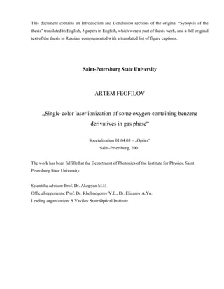
fuse character of the spectral dependences shown in
Fig. 1 as compared with hydroquinone.
In the measurements of ZEKE-PFI spectra, elec-
trons with zero kinetic energy are detected; i.e., the
condition of optical double resonance between the ini-
tial, intermediate, and ionic states is fulfilled. This sig-
nificantly reduces the set of transitions contributing to
the detected signal. Therefore, ZEKE-PFI spectra dis-
play the structure which can be attributed to both the
cation energy spectrum [8] and the spectrum of vibra-
tional levels in the intermediate state. The vertical bars
in Fig. 1 mark the position of intense S1 S0 transi-
tion bands in the REMPI spectrum of supersonic cate-
chol molecular beam [5]. It is seen that the structure is
associated with the spectrum of the intermediate S1
state.
The effective lifetimes of Rydberg states was deter-
mined by measuring a decrease in the intensity of
ZEKE-PFI electron signals with increasing the delay of
a Rydberg state-ionizing field pulse after a laser pulse.
For all λ ≤ 283 nm, the obtained values fall in the range
150 ± 15 ns.
Figure 2 depicts the laser ionization photoelectron
spectra of catechol obtained at λ = 283 and 266 nm. The
abscissa is graduated in the excitation energy of M+
ions generated by two-photon ionization, Ee = 2E – I1 –
ε, where E is the photon energy and ε is the photoelec-
tron energy. For the spectrum at λ = 283 nm corre-
sponding to the region of hot transitions, a correction
for thermal energy in the S0 state is introduced. It is seen
that the most abundant molecular ions are M+ with an
excitation energy of about 0.3 eV at λ = 283 nm or
0.9 eV at λ = 266 nm. Moreover, a portion of M+ ions
has the maximum excitation energy attainable at a fixed
photon energy—from 0.6 eV at λ = 293 nm to 1.1 eV
at λ = 266 nm.
Within the measurement error, the spectra are not
changed by varying the radiation intensity over the
entire accessible range, including the region of strong
fragmentation. This finding in combination with the
detection of M+ ions in mass spectra suggest that, as in
the case of previously studied hydroquinone and vera-
trole, the principal pathway of formation of fragment
ions is the photodissociation of molecular and fragment
ions. Alternative schemes, the ionization of neutral
fragments produced via dissociation of the molecule
from an intermediate state or the autoionization of
the molecule after absorption at least three quanta, sug-
gest that the spectra should depend on the radiation
intensity.
The absorption of a 283-nm photon primarily leads
to the population of the S1(00) state. If ionization results
from the absorption of the second photon by the mole-
cule in this state, close vibration energy distributions of
M+ produced by single-photon ionization transitions
from the S1 and S0 states should be expected because of
a small difference in the equilibrium interatomic dis-
tances, except for the torsional coordinates (see above)
of the S1 and S0 states [7]. The most probable transition
to the ground state of the cation D0 in the HeI photo-
electron spectrum [3] is shifted by approximately
0.4 eV relative to the 0–0 ionization transition. The
spectrum shown in Fig. 2 is qualitatively consistent
with the one predicted in terms of the above consider-
ations.
The photoelectron spectrum at λ = 266 nm is char-
acterized by a low probability of transitions to the M+
states in the region of the equilibrium configuration and
by a shift of the most probable transitions toward higher
Ee(M+) energies. When a 266-nm photon is absorbed,
the vibronic S1 states with the total energy of vibra-
tional excitation of 0.25–0.5 eV including the contribu-
tion of hot transitions are populated. At the qualitative
level, the change in the shape of the photoelectron spec-
trum can be explained as follows. As the energy of
vibrational excitation increases, the Franck–Condon
region of ionization transitions is extended for the start-
ing ionization state. In addition, rapid intramolecular
redistribution of vibrational energy over isoenergetic
states leads to the situation that some excitation energy
is localized in Franck–Condon inactive modes and is
transferred to Ee(M+) without change (the ∆vi = 0 rule
[13]).
Assuming that the ionization were preceded by the
intersystem crossing to a triplet T state or internal con-
version to the S0 state, the vibrational excitation energy
in the state prior to ionization would be increased by the
difference in the electronic energy between the S1 and
T or between S1 and S0 states. Then, the 283-nm spec-
trum would resemble the spectrum obtained at λ =
266 nm. Therefore, we believe that the molecular ions
under the conditions of our experiments are produced
by two-step ionization via the S1 state.
0–0
1
2
3
3
2
1
0
4
3
2
1
0
274 276 278 280 282 284 286 288 292290
λ, nm
I, arb. units
Fig. 1. (1) Absorption spectrum and (2) total ionization cur-
rent and (3) and ZEKE-PFI electron current spectra of cate-
chol vapor.
26. HIGH ENERGY CHEMISTRY Vol. 35 No. 4 2001
STEPWISE PHOTOIONIZATION OF 1,2-DIHYDROXYBENZENE VAPOR 267
Let us consider a possible contribution of ionization
transitions to the first electronically excited state of the
cation D1 with the vertical ionization energy of 9.25 eV
[3]. As the transition into the D0 state, they correspond
to the removal of π electrons and the intensity distribu-
tions in the first two bands of the HeI photoelectron
spectrum are close [3]. This resemblance allows the
adiabatic ionization energy yielding the D1 state to be
estimated at 8.8 eV. However, the probability of the 0−0
D1 S1 transitions is very low and becomes compa-
rable to that of the D0 S1 transitions in the overlap-
ping Ee region when the threshold is exceeded by 0.3–
0.4 eV. Therefore, a small contribution of the transi-
tions to the D1 state may be expected only at λ =
266 nm.
At low laser intensities, only M+ is generated. An
increase in the intensity progressively enhances frag-
mentation (Fig. 3). The character of fragmentation
upon laser-induced ionization of catechol is close to
that observed for hydroquinone [1]. The most signifi-
cant difference is the appearance of a mass spectral
peak at the mass to charge ratio of 92 (92+) correspond-
ing H2O elimination from M+:
(1)
which is the most abundant fragment ion at a radiation
flux density of ~3 × 10⁶ W/cm2. The formation of this
ion is due to the intramolecular H-bonding of the cate-
chol, and can be used for analytical purposes to distin-
guish between the isomers. There are also other differ-
ences in the peak intensity distribution in the mass
C6H6O2
+
hν C6H4O
+
H2O,+ +
spectra of catechol and hydroquinone. For example, the
ions at 38+ and 39+ of the group of ions are the
most abundant for catechol and hydroquinone, respec-
tively, which can also be explained by the easier migra-
tion of the H atom to neutral fragments because of
H-bonding. The shape of peaks at 92+ and 82+ suggests
their formation in the processes of metastable degrada-
tion of M+ with the effective rate constants of ~106 s–1.
The following formation channel is proposed for the
ion at 82+:
(2)
The elimination of CO from M+ was postulated in the
analysis of mass spectra of two-color laser-ionization
[14] and electron-ionization [15] mass spectra of phe-
nol. The fragment ion was shown [15] to have the
1,3-cyclopentadiene structure. Thus, we should expect
the appearance of an ion with the structure of 1,3-cyclo-
pentadienol. We determined the enthalpy of formation
of this ion from the formation enthalpy of the neutral
(−51 kJ/mol [16]) and I1 (8.3 ± 0.3 eV). The latter value
was obtained from the ionization energy of 1,3-cyclo-
pentadiene and typical values of a change in the ioniza-
tion energy of cyclic compounds with the double bond
upon replacement of H by OH. The calculated heat of
reaction (2) corresponds to the threshold λ = 886 nm;
therefore, this channel is opened by the absorption of a
single photon over the entire spectral range studied.
This conclusion is consistent with the finding that the
ion at 82+ appears with the group of ions displayed in
the mass spectrum at low radiation intensities. The
C3Hn
+
C6H6O2
+
hν C5H6O
+
CO.+ +
266 nm
283 nm
6
4
2
0
6
4
2
0
–0.3 –0.1 0.1 0.3 0.5 0.7 0.9 1.1 1.3
I, arb. units
Ee(M+), eV
Fig. 2. Photoelectron spectra of catechol.
27. 268
HIGH ENERGY CHEMISTRY Vol. 35 No. 4 2001
AKOPYAN et al.
alternative formation channel of the 82+ ion via C2H4
elimination from M+ involves more complex rearrange-
ment, and by our estimates, is more endothermic.At the
same time, we did not observe a similar process upon
laser-induced ionization of 1,2-(CH3O)2C6H4 [2],
which may be explained by the absence from this mol-
ecule of the hydroxylic H to migrate to the ring.
The ions at 81+, 63+, and 55+ are assumed to form by
the same processes as in the case of hydroquinone ion-
ization:
and the ion at 53+ which has not been considered previ-
ously [1] is presumably formed by
+hν cyclo- +CO+HCO(306nm). (6)
+ hν cyclo-C5H5O+ + HCO (585 nm),(3)
HC≡C-cyclo-
+ HCO + H2O
(210 nm),(4)
H2C=CHCO+
+ CH2=CHCO
(381 nm),(5)
C6H6O2
+
C3H2
+
C6H6O2
+
C4H5
+
Given in the parentheses are the threshold λ values for
the photodissociation of unexcited M+. The energy of
one photon is sufficient for triggering channels (3), (5),
and (6). As in the case of hydroquinone, to initiate pro-
cess (4), it is necessary that at least two photons be
absorbed in the spectral region relevant to this work.
The absorption of another photon by M+* with an exci-
tation energy exceeding the threshold values for several
dissociation channels is low under our experimental
conditions. Therefore, we assume that the second pho-
ton is absorbed by the fragment ions at 92+ or 81+ hav-
ing higher absorption cross sections, rather than by the
M+* ions:
(7)
(8)
Pathway (7) is absent for hydroquinone, and the ion at
63+ has a higher abundance in the catechol mass spec-
trum.
M
+
hν C6H4O
+
H2O;+ +
C6H4O
+
hν C3H3O
+
HCO,+ +
M
+
hν C5H5O
+
HCO;+ +
C5H5O
+
hν C3H3O
+
H2O.+ +
×10
10
5
0
0 20 40 60 80 100 120
M, amu
I, arb. units
Fig. 3. Mass spectra of catechol ionized by 283-nm laser radiation at an intensity of 3 × 10⁶ (lower spectrum) and 1.5 × 10⁷ W/cm2
(3 and 15 µJ/pulse, respectively).
28. HIGH ENERGY CHEMISTRY Vol. 35 No. 4 2001
STEPWISE PHOTOIONIZATION OF 1,2-DIHYDROXYBENZENE VAPOR 269
The absorption of two photons is also necessary for
the formation of the ions at 38+. The elimination of the
neutral fragments CO and CH2=CHOH is an energeti-
cally favored process having a threshold energy of
6.49 eV. To calculate it, we used the formation enthalpy
of equal to 1376 kJ/mol [17]. Based on the above
considerations, the process should be assumed to occur
via the formation of the intermediate 82+ ion:
(9)
The process involving H2O, CO, and C2H2 elimination
is more endothermic (threshold energy 7.65 eV), and it
also occurs in two steps:
(10)
The formation of the ion at 92+, which is absent from
the mass spectrum of hydroquinone, by ionization of
catechol followed by its dissociation via pathway (10)
explains the higher abundance of the 38+ ion in the cat-
echol mass spectrum.
Light hydrocarbon ions contributing considerably to
the mass spectrum at the intensities near the upper limit
of our experimental range are most probably produced
by photodissociation of heavier fragment ions; more
definite information on their origin does not seem
obtainable at present.
ACKNOWLEDGMENTS
This work was supported by the Russian Foundation
for Basic Research, project no. 98-03-32716a, and the
Competition Center of Basic Natural Sciences, Minis-
try of Education of the Russian Federation.
REFERENCES
1. Akopyan, M.E., Kleimenov, V.I., and Feofilov, A.G.,
Khim. Vys. Energ., 2000, vol. 34, no. 2, p. 140 [High
Energy Chem. (Engl. transl.) 2000, vol. 34, no. 2,
p. 107].
2. Akopyan, M.E., Kleimenov, V.I., and Feofilov, A.G.,
Khim. Vys. Energ., 2000, vol. 34, no. 5, p. 371 [High
Energy Chem. (Engl. transl.) 2000, vol. 34, no. 5,
p. 315].
3. Palmer, M.H., Moyes, W., Speirs, M., and Rid-
yard, J.N.A., J. Mol. Struct., 1979, vol. 52, no. 2, p. 293.
4. Bürgi, T. and Leutwyler, S., J. Chem. Phys., 1994,
vol. 101, no. 10, p. 8418.
5. Gerhards, M., Peri, W., Schumm, S., Jacjby, C., and
Kleinermanns, K., J. Chem. Phys., 1996, vol. 104,
no. 23, p. 9362.
6. Dunn, T.M., Tembreull, R., and Lubman, D.M., Chem.
Phys. Lett., 1985, vol. 121, nos. 4–5, p. 453.
7. Gerhards, M., Schumm, S., Unterberg, C., and Kleiner-
manns, K., Chem. Phys. Lett., 1998, vol. 294, no. 1,
p. 65.
8. Akopyan, M.E., Ivanov, V.S., Kleimenov, V.I., and Feo-
filov, A.G., Opt. Spektrosk., 1999, vol. 86, no. 6, p. 978.
9. Takhistov, V.V., Organicheskaya mass-spektrometriya
(Organic Mass Spectrometry), Leningrad: Nauka, 1990.
10. Sabbah, R. and Buluku, E.N.L.E., Can. J. Chem., 1991,
vol. 69, no. 3, p. 481.
11. Tzeng, W.B., Narayanan, K., Hsieh, C.Y., and
Tung, C.C., Spectrochim. Acta A, 1997, vol. 53, no. 14,
p. 2595.
12. Humphrey, S.J. and Pratt, D.W., J. Chem. Phys., 1993,
vol. 99, no. 7, p. 5078.
13. Compton, R.N. and Miller, G.C., Laser Applications in
Physical Chemistry, Evans, D.K., Ed., New York: Dek-
ker, 1989, p. 221.
14. Matyuk, V.M., Polevoi, A.V., Potapov, V.K., and
Prokhoda, A.L., Khim. Vys. Energ., 1982, vol. 16, no. 2,
p. 99.
15. Terent’ev, P.B., Mass-spektrometriya v organicheskoi
khimii (Mass Spectrometry in Organic Chemistry), Mos-
cow: Vysshaya Shkola, 1979, p. 105.
16. Zhong Xian and Bozzelli, J.W., J. Phys. Chem. A, 1998,
vol. 102, no. 20, p. 3537.
17. Smith, D., Adams, M.G., and Ferguson, E.E., Int.
J. Mass Spectrom. Ion Processes, 1984, vol. 61, no. 1,
p. 15.
C3H2
+
M
+
hν C5H6O
+
CO;+ +
C5H6O
+
hν C3H2
+
CH2=CHOH.+ +
M
+
hν C6H4O
+
H2O;+ +
C6H4O
+
hν C3H2
+
C2H2 CO.+ + +
30. OPTICS AND SPECTROSCOPY Vol. 92 No. 1 2002
STEPWISE IONIZATION OF THE 2,4-DIOXYBENZALDEHYDE 37
In order to analyze the mass spectrum of ions and
the energy spectrum of electrons, we used the time-of-
flight method with the drift-region length being equal to
0.45 m. The spatially separated particle packets fall
onto a chevron pair of microchannel plates, the signal
from which is fed to a Pico-200-100 analog-to-digital
converter that has a time resolution of 10 ns. The data
acquisition, accumulation, and processing were per-
formed using an IBM PC 486.
The field in the ionization region was 200 V/cm dur-
ing the mass analysis, whereas the field gradient in the
same region was no higher than 0.02 V/cm when the
electron-energy spectrum was analyzed; the vapor pres-
sure and the laser-radiation intensity were chosen so
that the effect of the space charge was negligible. The
resolution in the energy analysis of photoelectrons was
determined from the spectra of stepwise ionization of
Rb using both components of the 62P3/2, 1/2 state. The
full width of the peaks at half-maximum was 0.04 eV at
the energy of 0.56 eV, and increased to 0.15 eV at an
energy of 1.70 eV.
The absorption spectrum of the organic-compound
vapors in a buffer gas (3000–6000 Pa of Xe) was mea-
sured for wavelengths down to 200 nm using a labora-
tory spectrometer based on a KSVU commercial instal-
lation equipped with a heatable cell (of the King type)
with quartz windows. The spectra were measured at a
temperature of about 350 K with a resolution of 1 nm.
The quantum-chemical calculations of excited
states were performed on the basis of the ZINDO/S CI
method using the ground-state geometry optimized
according to the MINDO3 method.
RESULTS AND DISCUSSION
In Fig. 1, we show the absorption spectra and the
spectral dependences of the total ionization current for
the 2,4-dioxybenzaldehyde (DOB) and 3,4-dimethox-
ypropiophenone (DMP) vapors; the latter dependences
were normalized to the squared luminous flux. Two
absorption bands peaked at 311 and 269 nm for DOB
and at 305 and 259 nm for DMP fall within the spectral
range of our experiments. These bands are structure-
less. The peaks of the first absorption bands are appre-
ciably shifted in reference to the long-wavelength cut-
offs, which indicates that the equilibrium configura-
tions of molecules are changed markedly as a result of
excitation. The same is also true for the second DMP
band. The corresponding band in the DOB spectrum
features a much steeper falloff from the peak to longer
wavelengths; this means that the equilibrium configura-
tions of the ground and excited states are close to each
other for DOB.
Quantum-chemical calculations made it possible to
attribute the bands in the DOB spectrum to the
S2(1A') S0(1A') and S3(1A') S0(1A') transitions of
the ππ* type. According to the calculations, the lowest
singlet excited electron state S1(1A'') of the nπ* type is
located below the S2 state by 1 eV; however, the
S2(ππ*) S0 state does not manifest itself in the
absorption spectra we measured, because, according to
the available data for benzaldehyde, the absorption
coefficient for transition to the nπ* state is smaller than
that for the ππ* transition by a factor of 60–70. In addi-
tion, the calculations indicate that no less than four trip-
let states terms are located in the energy region below
S2(ππ*), with the lowest of these terms being repre-
sented by a level of the ππ* type. The inference about
the ππ* type of the T1 state in the oxy- and methoxy-
benzaldehyde derivatives had been made [18] on the
basis of measurements of the phosphorescence in fro-
zen solutions.At the same time, taking into account dif-
ferences in the exactness of calculations of the singlet
and triplet states, we may assert that the T1(ππ*) and
T2(nπ*) states of oxybenzaldehydes are nearly degener-
ate (see, for example, [19]).
Arrows in Fig. 1 indicate the threshold wavelengths
that correspond to the values of 0.5Ii (i = 1, 2), where Ii
are the adiabatic energies of ionization accompanied
with the generation of molecular ions in the ith elec-
tronic state. These energies were estimated to within
±0.1 eV using the HeI photoelectron spectra [16]. For
DOB, the halved value (≈4.3 eV) of the first adiabatic
ionization energy exceeds the energy of the peak of the
first absorption band; thus, the ionization threshold is
controlled by the adiabatic ionization energy. However,
a sharp increase in the current in the vicinity of 282 nm
coincides with the onset of the second absorption band.
For DMP, the long-wavelength absorption cutoff coin-
cides with 0.5I1; as a result, a peak corresponding to the
S2 S0 transition appears in the total-current spectral
dependence. The shift of this peak in reference to the
peak in the absorption spectrum may be explained by
an increase in the cross sections for ionization of
vibronic states as λ decreases; these states become
occupied when the first photon is absorbed. A subse-
quent increase in the current in the vicinity of 285 nm
sets in at a somewhat smaller wavelength than the onset
of the second absorption band in DMP, which is caused
by an increase in the ionization cross section due to the
generation of M+ ions in their first excited electronic
state. The aforementioned special features of the total-
current spectral dependences indicate that the laser-
induced ionization is initiated by photon absorption in
the S2 S0 and S3 S0 transitions.
Under comparable experimental conditions (similar
pressures and luminous fluxes), the currents observed
in DOB and DMP subjected to laser-induced ionization
are much lower than those recorded previously for the
dioxy- and dimethoxy-benzene derivatives [1, 2]. The
same relation between the currents in the case of
266-nm laser ionization of benzaldehyde and 4-oxy-
benzaldehyde compared to that of phenol has been
reported [7]. A decrease in the efficiency of stepwise
ionization is caused by the redistribution of energy in
the intermediate state with formation of the products
31. 38
OPTICS AND SPECTROSCOPY Vol. 92 No. 1 2002
AKOPYAN et al.
whose ionization is impaired under experimental con-
ditions. In particular, this redistribution may be related
to the dissociation processes. Since the highest photon
energy in our experiments is 4.66 eV and the fragment-
ionization energy exceeds this value, the fragments can
be ionized only as a result of absorption of two photons.
This is unlikely, at least in the lower part of the lumi-
nous-flux range. Indeed, by reducing the laser-radiation
intensity, we managed to attain complete (to within the
background-signal amplitude) absence of the fragmen-
tation ions in the mass spectra of DOB and DMP. Thus,
for low luminous fluxes, the scheme of processes
occurring during laser excitation of the DOB and DMP
vapors can be represented as
M(S0) + hν M(Si, i = 2, 3) M(Γ), (1)
M(Γ) + hν M+ + e, (2)
Xi (dissociation products). (3)
In this scheme, we take into account the fact that the
state Γ (initial for ionization and dissociation) may dif-
fer from the state which is optically populated owing to
intramolecula energy redistribution. Process (3) does
1
2
3
4
5
6
7
8
0
1
2
(a)
OH
H
OH
HCO
H
H
J, arb. units
270
1
275 280 285 290 295 300 305 310 315265
2
3
4
5
6
7
8
0
1
2
(b)
H
OëH3
OCH3
C=O
H
H
CH2
CH3
9
10
11
12
Fig. 1. The (1) absorption and (2) total current spectra of (a) 2,4-dioxybenzaldehyde and (b) 3,4-dimethoxypropiophenone.
32. OPTICS AND SPECTROSCOPY Vol. 92 No. 1 2002
STEPWISE IONIZATION OF THE 2,4-DIOXYBENZALDEHYDE 39
not contribute to the recorded currents but affects the
ionization efficiency.
In the upper part of the luminous-flux range, neutral
fragments Xi can be ionized as a result of two-photon
absorption; i.e.,
Xi + 2hν + e. (4)
As the radiation intensity increases, the rate of process (4)
becomes higher; however, dissociation yield k/(k + σiΦ)
(where k is the rate constant of dissociation for all avail-
able channels, σi is the cross section of the intermedi-
ate-state ionization, and Φ is the photon-flux density)
becomes lower compared to the channel of intermedi-
ate-state ionization (2). We can state that the relative
contribution of two ionization mechanisms varies with
increasing radiation intensity only after an analysis of
the kinetics for processes (1)–(4). However, the infor-
mation needed for such analysis is lacking. Notably, the
M+-ion signal is most intense in the mass spectra
obtained under the luminous flux that is available in our
experiments. Therefore, the ionization–dissociation
mechanism is effective in the entire intensity range.
As far as we know, photodissociation of the DOB
and DMP vapors has not been studied. If benzaldehyde
is excited to the S2 and S3 states, the CO group becomes
split off with a high quantum yield [20]; the photo-
chemistry of acetophenone is related to the breakage of
the C6H5C(O)–CH3 bond [21]. Dissociation occurs
from the triplet state (states) with the rate constants that
Xi
+
are no larger than 5 × 10⁷ s–1 [9, 20, 21]. The 110+ and
165+ ions are observed in the mass spectra of the laser-
induced ionization of DOB and DMP, respectively; the
emergence of these ions may be attributed to ionization
of fragments produced by dissociation from the inter-
mediate state via the channels that are similar to those
mentioned above (Figs. 2, 3). However, these ions can
also be generated owing to photodissociation of M+
groups. It is noteworthy that a contribution of dissocia-
tion from the intermediate state to the charged-particle
current may be significant only under the condition that
k ≥ σiΦ.
If this condition is satisfied, the photoelectron spec-
tra of the laser-induced ionization should depend on the
luminous flux because the intermediate-state and disso-
ciation-fragment spectra do not generally coincide. A
low level of electron signal with energy resolution
made it possible to obtain only photoelectron spectra of
a rather low quality (Figs. 4, 5). For DOB, the intensity
distribution in the photoelectron spectrum at λ =
266 nm does not change more than 10% when the irra-
diation intensity is increased by a factor of 6. Arrows in
Figs. 4 and 5 indicate the electrons’ kinetic energies ε
that correspond to the formation of unexcited M+ ions
from unexcited molecules as a result of two-photon
absorption: εm = 2hν – I1(M). Electrons with high
kinetic energies are not observed in the spectra. An
appreciable fraction of electrons have a kinetic energy
in the range of 0–0.5 eV, which corresponds to genera-
0
10
J, arb. units
M, au
(b)
30 50 70 90 110 130 150
(a)
1
2
3
2
1
0
Fig. 2. The mass spectra of stepwise ionization of 2,4-dioxybenzaldehyde for ionization via the (a) S2 and (b) S3 intermediate states
for the luminous flux amounting to ~7 × 10⁶ W/cm2.
33. 40
OPTICS AND SPECTROSCOPY Vol. 92 No. 1 2002
AKOPYAN et al.
0
0
J, arb. units
M, au
(b)
160 200
(a)
1
2
3
2
1
0
60 100 120 1404020 80 180
3
×10
×10
6
5
4
Fig. 3. The mass spectra of stepwise ionization of 3,4-dimethoxypropiophenone for ionization via the (a) S2 and (b) S3 intermediate
states for the luminous flux amounting to ~8 × 10⁶ W/cm2.
0
J, arb. units
ε, eV
2.2
1
2
3
2.01.81.61.41.21.00.80.60.40.20.0–0.2
4
5
6
7
8
Fig. 4. The photoelectron spectra of 2,4-dioxybenzaldehyde under excitation with λ = 266 nm and for the luminous fluxes amount-
ing to ~2 × 10⁶ W/cm2 (triangles) and ~1.2 × 10⁷ W/cm2 (the solid line).
34. OPTICS AND SPECTROSCOPY Vol. 92 No. 1 2002
STEPWISE IONIZATION OF THE 2,4-DIOXYBENZALDEHYDE 41
tion of the M+ ions with the excitation energies in the
range of 0.3–0.8 eV.
Electrons with ε < εm are also dominant in the pho-
toelectron spectra of DMP. We emphasize that an
increase in λ brings about a shift of εm in the photoelec-
tron spectra; this shift is linearly proportional to the
photon energy. For low luminous fluxes, an increase in
the electron current in the energy range corresponding
to formation of electron-excited M+ ions with I2 ≈
8.7 eV is observed in the spectra obtained under excita-
tion with λ = 266 and 275 nm for low luminous fluxes.
However, in contrast to DOB, a significant increase in
the fraction of electrons with energies in the range of
0.3–1.0 eV is observed in the photoelectron spectra of
MDP as the luminous flux increases (Fig. 5). Such
changes may be caused by both the two-photon ioniza-
tion of the products of dissociation from the intermedi-
ate state and the processes of intramolecular energy
redistribution in this state, which do not induce the dis-
sociation.
In any case, a discrepancy between the DOB and
DMP photoelectron spectra should be caused by spe-
cial features of nonradiative transitions after light-
induced population of the states. A DOB molecule
belongs to the class of bifunctional derivatives of ben-
zene with an intramolecular H bond; for these deriva-
tives, in contrast to other aromatic aldehydes, fluores-
cence with a large Stokes shift is observed if the
S2(ππ*) S0 transition is excited [22–26]. This fluo-
rescence is related to radiative transition from the
S2(ππ*) state after a fast intramolecular proton trans-
port. Such fluorescence was observed under excitation
of the 2-oxybenzaldehyde vapors [25, 26] whose elec-
tronic structure is close to that of DOB. In this case, the
rate constant for the proton transport is equal to ~109 s–1
and is controlled by the competition with the internal
conversion S2(ππ*) S1(nπ*) [23]. An intense phos-
phorescence (rather than fluorescence) with character-
istics typical of radiative decay of the T1(nπ*) state was
observed in propiophenone under excitation to the
S2(ππ*) and S3(ππ*) states [27]. If these special fea-
tures of nonradiative transitions are retained for the
molecules under investigation, we should expect that
the singlet states in the state Γ initial for ionization have
a larger weight in DOB than in DMP.
Although an analysis of experimental data does
make it possible to completely exclude a certain contri-
bution of the dissociation–ionization mechanism to the
results of our experiments, we attempt to identify the
processes of formation of the fragmentation ions in
terms of the ionization–dissociation mechanism. To
this end, we employ the model (used widely in mass
spectrometry) that implies that dissociation of ions via
the least endothermic channels is predominant (see, for
example, [28]). We take into account the steric inhibi-
tions by giving preference to the processes that are
unrelated to the motion of heavy ions to large distances.
When estimating the heat of reactions, we used the pub-
lished values of the enthalpies of formation [28, 29].
0
J, arb. units
ε, eV
2.2
2
2.01.81.61.41.21.00.80.60.40.20.0–0.2
4
6
10
8
1
2
12
Fig. 5. The photoelectron spectra of 3,4-dimethoxypropiophenone irradiated with photons of λ = 266 nm for luminous fluxes
amounting to (1) ~2 × 10⁶ and (2) ~2 × 10⁷ W/cm2.