1. Evaluation of soft tissue changes using ConeBeam CT scans during treatment of
esophageal cancer.
A. van den Hoek, M. Kamphuis, R. de Jong, N. van Wieringen, M. Hulshof
ACADEMIC MEDICAL CENTRE AT THE UNIVERSITY OF AMSTERDAM, Department of Radiation
Oncology, Amsterdam, The Netherlands
Keywords: inter-fraction variation, soft tissue changes, Conebeam-CT, esophagus cancer
Purpose/objective
The verification of the set-up of patients treated for esophageal cancer recently changed from
portal imaging to Conebeam CT (CBCT) in our department. The protocol, registration on the
bony anatomy and using an off line decision rule, remained the same. As the use of CBCT
and experience grew, it became apparent that the CBCT scans contained more information
about the patient. Besides information of patient set-up also soft tissue changes became
visible. The purpose of this study is to score soft tissue changes during treatment of esophagus
that have an impact on GTV coverage or may alter the dose distribution.
Materials/methods
Twenty seven patients with an esophagus carcinoma are included in this study. Patients had
either preoperative or definitive chemoradiotherapy with 23x1.8 Gy or 28x1.8 Gy
respectively. Both groups had once weekly carboplatin and paclitaxel. CBCT scans were
acquired for position verification and used for an extended No Action Level correction
protocol (eNAL): 4 scans in the first week and weekly follow-up. Inter-fraction anatomical
changes were determined for a period of 4 weeks in all patients: for the first week only the
second scan was evaluated. Soft tissue changes were assessed after performing a full
registration (6 degrees of freedom) using XVI 4.2 (Elekta, Sweden). Changes that caused the
GTV to be outside of PTV were scored. Furthermore we scored any change in anatomy
between CT en CBCT larger than 1cm, e.g. change in circumference, pleural effusion and
atelactasis.
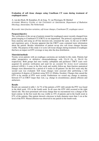
Results
Results are summed in table 1. In 7% of the patients a GTV shift outside the PTV was found
in the third week, 15% in the fourth week. In all cases the GTV shift occurred at the right
lateral side. The most prominent soft tissue change that we encountered was a change of the
heart contour. In the first week this was visible in 15% of patients and in the fourth week in
67% of the patients. One patient showed a decrease in body diameter more than 1 cm at the
level of the PTV. Pleural effusion was observed in one patient in the fourth week.
2. 0.0%
10.0%
20.0%
30.0%
40.0%
50.0%
60.0%
70.0%
GTV outside PTV Any change in
anatomy between CT
en CBCT > 1cm
Change in
circumference
Pleural effusion or
atelectasis
week 1
week 2
week 3
week 4
Conclusion/discussion
Results are showing that the PTV margin is exceeded by a shift of the GTV in a substantial
part of the group. These shifts occurred on the right lateral side. We have to underline that we
assessed changes after a full registration and other uncertainties, such as rotational errors
which can not be corrected for during treatment, were not taken into account.
Soft tissue changes occur frequently over the course of radiotherapy treatment. The effect on
the dose distribution needs to be studied. Changes in the volume of the heart and its
determinants are subject of further investigation.
Using 3D kV-imaging for position verification as well as for tracking anatomical changes
during treatment is highly recommended.