
Recommended
More Related Content
What's hot
What's hot (20)
Similar to The occurrence of entamoeba gingivalis among patients with periodontists
Similar to The occurrence of entamoeba gingivalis among patients with periodontists (20)
Recently uploaded
Recently uploaded (20)
The occurrence of entamoeba gingivalis among patients with periodontists
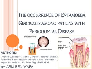
- 1. THE OCCURRENCE OF ENTAMOEBA GINGIVALIS AMONG PATIONS WITH PERIODONTAL DISEASE AUTHORS: Joanna Luszczak1*, Michal Bartosik1, Jolanta Rzymowska1, Agnieszka Sochaczewska-Dolecka2, Ewa Tomaszek3, Joanna Wysokinska-Miszczuk3, Anna Bogucka-Kocka1 BY: ARIJ BEN WAFA
- 2. INTRODUCTION Entomoeba gingivalis is a protozoan endoparasite It colonizes the gingival tissue and was the first amoeba described in humans E. gingivalis was first of all observed by Gros in 1849 in the tartar of teeth but its detail description was given by Von Prowazak in 1904. Entamoeba gingivalis is considered by some authors to be a pathogenic protozoan, and to be commensal by others.
- 3. Morphologically, this amoeba resembles Entamoeba histolytica, and diagnosis requires great attention. E. gingivalis has a large number of pseudopodia that allow it to move quickly it does not have resistant forms (cysts), but only trophozoites, which vary in size from 10 to 35 ìm
- 4. This parasite inhabits the area around the teeth and gums, including the spaces between teeth, and especially in teeth cavities, dental tartar, necrotic mucosa of cells and the gingival fringes of the gums . According to some studies, this amoeba is also an important agent in the induction of periodontitis . E. gingivalis is recognized as causing gum itch, fatigue halitosis, severe headaches, sore palate and periodontal tissue damage
- 5. STUDIES AND RESEARCHERS Many researchers have found that the infection rates of this amoeba in patients with periodontitis are higher than those of healthy control groups . According to some studies, E. gingivalis produces progressive, periodontal disease among immunocompromised patients . A Swedish study showed its association with human immunodeficiency virus type 1 (HIV-1) and reported that no other parasite was ever found in this type of immunodeficiency
- 6. DIAGNOSIS The diagnosis of Entamoeba gingivalis is still based on direct microscopy of tissue scrapings or on preculture in specialized parasitological media
- 7. MATERIALS AND METHODS Diagnostic material in the form of swabs was collected from 102 patients with periodontal disease, who were undergoing treatment in the Department of Periodontology The same was undertaken with 32 people assessed as being healthy. all 134specimens, 88 were taken from women and 46 from men. samples from the gingival pouch, subgingival areas and gums were obtained by using sterile swabs and then were examined for the presence of Entamoeba gingivalis.
- 8. Entamoeba gingivalis cells were identified by observation of morphology and the characteristic movement of their pseudopodia. Next, the correlation between the prevalence of E. gingivalis infection and the patients’ sex and age was calculated. for Entamoeba gingivalis trophozoites Such protozoa were found in 103 patients (77%), but not in 31 patients (23%) Among the samples derived from women, the protozoa were in 68 (77%), and among men, in 35 (77%). No correlation was seen between gender and the presence or absence of protozoa
- 9. RESULTS The study involved 134 persons: 88 women and 46 men. In the studied group, 102 people had some periodontal disease, while the remaining 32 patients were healthy. Such protozoa were found in 103 patients (77%), but not in 31 patients (23%) .Among the samples derived from women, the protozoa were in 68 (77%), and among men, in 35 subjects (77%). No correlation was seen between gender and the presence or absence of protozoa
- 10. The correlation between the person’s age and the presence of protozoa was strongly marked among women but it could not be observed in men Among women, the protozoa was observed in three groups of age (40-49, 60-69 and above80years) 31patients in total 100% presence of protozoa was observed Among men, the protozoa was observed in only two groups of age (60-69 and above 80 years) (only four patients)
- 11. In both groups, a correlation between the occurrence of amoebae and other diseases in the oral cavity have been noted, Regarding patients with some periodontal disease (102 individuals) 83 persons (81%) showed the presence of amoeba, This protozoa was not found in only 19persons(19%) By sex, this constitutes, for women (in total, a group of 68 patients), 55 individuals (82%); and for men (in total, a group of 35 patients), 28 persons (80%). Among those who did not have oral diseases (31 cases), the amoeba was evident in 20 (65%). By sex, for women (a population of 21), 13 persons (62%); and for men (a population of 35), 28 people (80%).
- 12. DISCUSSION E. gingivalis , potential to turn into opportunistic pathogens The activity of E. gingivalis in oral cavity disease lesion progression is associated with a capacity for erythrocytophagy and the presence of enzymes important in the development of periodontitis. infection rates of E. gingivalis in patients with periodontitis were higher than those of healthy groups. occurrence of E. gingivalis is correlated with the age of the host, so oral protozoa are rarely found in children, while the frequency of infection increases with age
- 13. PATHOGENESIS amoeba is an important cause of periodontal disease, generating gum itch, sore palate, halitosis, fatigue, severe headaches, and periodontal tissue damage
- 14. CONCLUSIONS Our study has led us to the conclusion that infections with Entamoeba gingivalis should be regarded as an factor associated with the pathological changes occurring in patients with periodontal diseases.