Production of natural biopharmaceuticals from the microalgae living in the dead sea
•
1 like•186 views

International peer-reviewed academic journals call for papers, http://www.iiste.org

Recommended
Recommended
More Related Content
What's hot
What's hot (8)
Viewers also liked
Viewers also liked (20)
Similar to Production of natural biopharmaceuticals from the microalgae living in the dead sea
Similar to Production of natural biopharmaceuticals from the microalgae living in the dead sea (20)
More from Alexander Decker
More from Alexander Decker (20)
Recently uploaded
Recently uploaded (20)
Production of natural biopharmaceuticals from the microalgae living in the dead sea
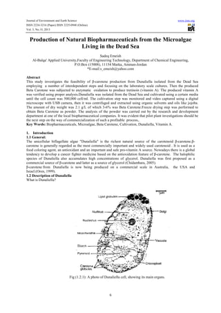
- 1. Journal of Environment and Earth Science ISSN 2224-3216 (Paper) ISSN 2225-0948 (Online) 0948 Vol. 3, No.10, 2013 www.iiste.org Production of Natural Biopharmaceuticals from the Microalgae he Living in the Dead Sea Sadeq Emeish Al-Balqa' Applied University,Faculty of Engineering Technology, Department of Chemical Engineering, Balqa' P.O.Box (15008), 11134 Marka, Amman Amman-Jordan *E-mail:s_emeish@yahoo.com Abstract This study investigates the feasibility of β β-carotene production from Dunaliella isolated from the Dead Sea employing a number of interdependent steps and focusing on the laboratory scale cultures. Then the produced Beta Carotene was subjected to enzymatic oxidation to produce tretinoin (vitamin A). The produced vitamin A a was verified using proper analysis.Dunaliella was isolated from the Dead Sea and cultivated using a certain media Dunaliella until the cell count was 500,000 cell/ml. The cultivation step was monitored and video captured using a digital cell/ml. microscope with USB camera, then it was centrifuged and extracted using organic solvents and oils like jojoba. The amount of dry weight was 2.1 g/L of which 3 3-6% was Beta Carotene.Freeze drying step was performed to Freeze obtain Beta Carotene as powder. The analysis of the powder was carried out by the research and development department at one of the local biopharmaceutical companies. It was evident that pilot plant investigations should be the next step on the way of commercialization of such a profitable process,. Key Words: Biopharmaceuticals, Microalgae, Beta Carotene, Cultivation, Dunaliella, Vitamin A. 1. Introduction 1.1 General: The unicellular biflagellate algae "Dunaliella" is the richest natural source of the carotenoid β-carotene.βcarotene is generally regarded as the most commercially important and widely used carotenoid . It is used as a d food coloring agent, an antioxidant and an important and safe pro-vitamin A source. Nowadays there is a global vitamin tendency to develop a cancer fighter medicine based on the antioxidation feature of β carotene. The halophilic β-carotene. species of Dunaliella also accumulates high concentrations of glycerol. Dunaliella was first proposed as a glycerol. commercial source of β-carotene and latter as a source of glycerol (Chidambara, 2005). carotene β-carotene from Dunaliella is now being produced on a commercial scale in Australia, t the USA and Israel.(Oren, 1999). 1.2 Description of Dunaliella What is Dunaliella? Fig (1.2.1): A photo of Dunaliella cell, showing its main organs. 6
- 2. Journal of Environment and Earth Science ISSN 2224-3216 (Paper) ISSN 2225-0948 (Online) 0948 Vol. 3, No.10, 2013 www.iiste.org It is a unicellular, bi-flagellate, naked green alga which is morphologically similar to chlamydomonas with the flagellate, main difference being the absence of a cell wall in Dunaliella. It has two flagella of equal length and a single cup shaped chloroplast. In Dunaliella Salina and Dunaliella Parva cup-shaped the chloroplast accumulates large quantities of β carotene (as droplets) so that the cells appear orange β-carotene orange-red rather than green. (Avron et al, 1999). The first description of the genus was given by Teodoresco who reported occurrence of the organism we know today as Dunaliella . At present a total of 29 species as well as a number of varieties and forms are recognized. Cell shape in this genus is very variable, being oval, spherical, cylindrical, ed. ellipsoidal, egg-pear-or spindle shaped. Cell in any given species may change shape with changing conditions, or often coming spherical under unfavorable conditions. Cell size also varies with growth conditions and light intensity(Ben-Amotz et al, 2012). Dunaliella species are commonly observed in salt lakes in all parts of the world from tropical to temperate to polar regions where they often impart an orange orange-red color to the water. When exposed to stress conditions such as high light intensity or nutrient starvation, two stereoisomers of β βcarotene, all-trans and 9-cis β-carotene, accumulated, reaching up to 14% of the cell's dry weight, with the carotene, pigment being deposited into plastid.(Emeish, 2012) astid.(Emeish, We now know that not all Dunaliella species produce massive amounts of carotene and those that can do so only under suitable conditions.The following figures illustrates some of Dunaliella types. Fig. (1.2.3): A photo of different Dunaliella types. Fig. (1.2.3): A photo of different Dunaliella types. 7
- 3. Journal of Environment and Earth Science ISSN 2224-3216 (Paper) ISSN 2225-0948 (Online) 0948 Vol. 3, No.10, 2013 www.iiste.org 1.3 β –carotene 1.3.1 Structure: Fig. (1.3.1): β-carotene structureC40H56 Its chemical composition is: 1,3,5,7,9,11,13,15,17-octadeca-nonaene-1,18-diyl)bis[2,6,6 diyl)bis[2,6,61,1'-(3,7,12,16-16-Tetramethyl-1,3,5,7,9,11,13,15,17 trimethylcyclohexane]. And its molecular weight is 536.87 g. Fig.(1.3.2)Produced Beta Carotene photo under microscope The pigment responsible for the brightly red coloration displayed by Dunaliella was recognized already very early as a carotenoid. (Abusara et al, 2011).It can be found in carrot, orange, and many other yellow, orange or red colored fruits and vegetables. It also accumulates in large quantities in the chloroplast of Dunaliella cells as droplets so that the cells appear orange orange-red rather than green. A typical composition, specialize the Dunaliella β-carotene from other sources, expressed as percentage of total tion, β-carotene is: 10% 15-cis- β-carotene, 41% 9 carotene, 9-cis- β-carotene, 42% all-trans- β-carotene, and 6% other isomers. carotene, (Avron et al, 1997) In the alga, this β-carotene seems to act a sun screen to protect the chlorophyll and the cell DNA from high irradiance, which characterizes the normal habitat of Dunaliella. 1.3.2 Biosynthesis of β-carotene: The general biosynthesis of β-carotene can be divided into four stages: carotene 1. Formation of Geranylgeranyl pyrophosphate (GGDP) from Mevalonic acid. 2. Condensation to form phytoene. 3. Desaturation of phytoene to lycopene. 4. Cyclization of lycopene to form β β-carotene. The unique caroteneogenesis in Dunaliella has been used to study the last two stages, (3) and (4), by applying rapid solvent extraction. 1.4 Literature review A hundred years have passed since the description of the genus Dunaliella, the unicellular alga which is responsible for most of the primary production of various types of compounds and phenomena in hypersaline various environments worldwide as salt mushroom formation phenomena within the Dead Sea solar ponds.(Daghistani et al, 2003). Dunal First sighted in 1838 in saltern evaporation ponds in south France by Michel Flix Dunal who reported occurrence of the organism we know today as Dunaliella Salina in the salterns of Montpellier, on the coast of France. 8
- 4. Journal of Environment and Earth Science ISSN 2224-3216 (Paper) ISSN 2225-0948 (Online) Vol. 3, No.10, 2013 www.iiste.org In the century that has elapsed since its formal description the genus Dunaliella has become a convenient model organism for the study of salt adaptation in alga. The establishment of the concept of organic compatible solutes to provide osmotic balance was largely based on the study of Dunaliella species. Moreover, the massive accumulation of β-carotene by some strains under suitable growth conditions has led to interesting biotechnological applications. In the course of 19th century, Dunal's red flagellate alga has been observed by other biologists as well in salt lakes and hypersaline sites in many countries. Different names were attached to the organism by each investigator.(Emeish et al, 2003). It was named after its discoverer by Teodoresco in 1905, this year saw the publication of two papers presenting in-depth descriptions of Dunaliella as anew genus one by Teodoresco and the second by Carla Hamburger. Teodoresco studied materials collected from a Romanian salt lake, while Hamburger worked with samples sent to her from salterns of Sardinia. Both authors presented detailed drawings of the organisms and provided extensive information on its morphology, cell structure, reproduction, behavior and ecology. A formal description of the genus Dunaliella, observed by Dunal who had first seen these organisms in salterns in France almost seventy years earlier was published in 1906. In the Dead Sea, Dunaliella cells have been reported since 1940s. The first quantitative estimates of the Dunaliella population in the lake were made in 1964 and showed very high numbers up to 4 x10⁴cells per ml of surface water. Stephens and Gillespie in 1976 reported measurements of the primary production in the south arm of Great Salt Lake performed in 1973. An in-depth taxonomic treatment of the genus was given in Massyuk's 1973 monograph. Dead Sea from 1980 onwards has yielded a clear picture of the factors that determine development of the alga in this unusual environment. A Master thesis under the title of: "Optimal Growth Conditions for the Production of β-Carotene and Glycerol from a Halophilic Alga Dunaliella Species Isolated from the Dead Sea" was presented In January, 2004 by Nader Abu Sara from Jordan University of Science and Technology .(Abusara,2004). In that research Dunaliella species were isolated from scattered ponds of the southern part of the Dead Sea and the conditions that gave the highest growth and β-carotene production were studied. Also the effects of different physical (temperature and light intensity) and chemical (different nitrogenous and sulfate compounds) factors on the growth and carotenoids production rates were reported. (Wheater, 2007). With a study published in 2004 by Liska, Dunaliella research has entered the era of modern proteomics. That was a worthy conclusion of the first century of Dunaliella research, and provides us with a preview of the kind of information that may be obtained in the coming years. 1.5 The Dead Sea 1.5.1 Ecology: The Dead Sea is a terminal lake in the Syrian-African rift valley with a rhomb–shaped. The Dead Sea water level (425 m below mean sea water is the lowest exposed surface on earth; which is 76 km long, up to 18 km wide and 400 m deep, while its bottom (825 m below mean seawater level) forms the lowest continental surface. The Dead Sea has a unique Ca-chloride composition [i.e., Ca2+ > (SO42- + HCO3-)] forming one of the world's saltiest lake with a total dissolved salt concentration of 340 g /L and a density of about 1.235 g /ml. At that time, the lake was stratified and consisted of 2 basins: a deep one on the north and a shallow one in the south, in 1976, the southern basin were detached and today the area is occupied by a series of evaporation ponds that are filled by pumping water from the northern basin.(Haugen, 1986).The rise and decline of a bloom of the algae in the Dead Sea is well observed. In the beginning of 1980 a massive amount of fresh water from the Jordan river and rain floods in flowed to the lake causes a sufficient dilution of the upper water layers, and as a consequence a dense Dunaliella bloom developed; the algal bloom remained present for a few months only.During the winter of 1991-1992 another massive amounts of rain water being transported to the Dead Sea, which caused an increase in level of almost 2 m. A dilution of the upper 5 meters to a salinity as low as 245 g / L triggered a short-lived mass development of Dunaliella and a prolonged bloom of red algae.(Emeish et al, 2003) The negative water balance of the Dead Sea since 1930 to 1999 has resulted in a decrease in its water level by about 20 m, this has been a companied by an increase in the salinity of the brine and by various change in the chemical and physical characteristics of the lake. 1.5.2 Dunaliella in the Dead Sea: The eukaryotic alga Dunaliella was first reported to be present in the Dead Sea in 1940 by Elzari-Volcani. (Oren.1999). Twenty different species of microalgae and halophilic bacteria were isolated and identified from eastern shores 9
- 5. Journal of Environment and Earth Science ISSN 2224-3216 (Paper) ISSN 2225-0948 (Online) Vol. 3, No.10, 2013 www.iiste.org of the Dead Sea. Multispectral LANDSAT images of the Dead Sea area were analyzed to obtain spatial and temporal information on the development of the algae bloom. Two images were obtained: the first, on April 15 1992 at the beginning of Dunaliella bloom, and the second, on June 22 from the same year, when the algal bloom had declined and the Dead Sea appeared red. For comparison, a LANDSAT image was also obtained on May, 5, 1991, at the time when algae were absent. The spectral distribution of the reflected solar radiation is modified by pigments present in the micro algae.The main interaction commonly measured is the absorption of sunlight by the red band of chlorophyll contained in the algal plastid. 2. Methodology: 2.1Cultivation: The microalgae was cultivated in a 5 litters vessels exposed to neon lamp from upper and lower sides at room temperature (about 25ºC) which kept constant during the different seasons. Ordinary air was supplied. Modified Johnson medium (table 2.1.1) was used as nutrient source for algae cultivation.The cultivation of Dunaliella was monitored by using a digital microscope with USB camera connected to computer to follow the algal cultivation under different light intensities . Table 2.1.1: Modified Johnson Medium (Borowitzka, 1990) To 5 L of distilled water add : NaCl 24% per unit volume MgCl2.6H2O 7.65g MgSO4.7H2O 2.6g KCl 1.02g CaCl2.2H2O 1.02g KNO3 5.1g NaHCO3 0.2g KH2PO4 0.2g Fe-solution 51ml Trace- element solution 51ml Na2EDTA 945mg FeCl3.6H2O 1220mg H3BO3 305mg (NH4)6Mo7O24.4H2O 190mg CuSO4.5H2O 30mg CoCl2.6H2O 25.5mg ZnCl2 20.5mg MnCl2.4H2O 20.5mg Adjust PH to 7.5 with HCl 2.2 Cell Counting: To be sure that the cells are ready to be harvested, cell counting step was undertaken.Counting of Dunaliella cells was performed using a Hemocytometer slide of 0.1 mm depth. No sample dilution has been done, 100 µl of the culture was taken and tested to count the cell number, and the trial was repeated 3 times. A simple filtration followed by long spontaneous drying in dark was used to approximate the algal cell density. 2.3 Harvesting: After the cultivation media reaches to a suitable cells density it was isolated from the cultivation media and transferred into smaller vessels. 2.4 Centrifugation: Then 25 ml culture media was centrifuged at 4000 rpm for 5 min. The sample was re suspended in 5ml distilled water. Continues flow centrifuge of type HERMLE z300 rotates by 4000 rpm was employed to perform the mentioned. 2.5 Extraction: β-carotene was extracted with hexane, ether, jojoba oil or light vegetable oil separately by adding 25 ml cell suspension to 15 ml solvent. When the extraction was performed, oil was heated to 70ºC; after it was cooled down to 40º C, then β -carotene was added.(See Fig.(2.5.1)) 10
- 6. Journal of Environment and Earth Science ISSN 2224-3216 (Paper) ISSN 2225-0948 (Online) Vol. 3, No.10, 2013 www.iiste.org Figure (2.5.1) Extraction of β-carotene 2.6 Freeze Drying: β-carotene crystallization was accomplished using freeze dryer equipment at 0.32 hpa pressure and -85°C temperature for four hours. 2.7 Spectrophotometer Analysis Scanning of the absorption spectrum of the hexane extracted pigments present in the supernatant was recorded at the wavelengths 350 – 700 nm using a scanning spectrophotometer. β-carotene was estimated spectro photo metrically at wave lengths 454 and 480 nm according to pharmacopeia, and then the concentration was determined . 5. The absorbance of the final solution is then measured at 440 nm against a cyclo hexane blank, and a spectrophotometer plot was obtained. 2.8 Enzymatic Oxidation It is a process by which the enzyme is used as a catalyst in the oxidation reaction, to allow a new pathway for the reaction to occur. The enzymatic oxidation reaction occurs in a different potential range depending on the material reacted and the product produced. The produced Beta Carotene was transferred into a one liter capacity bioreactor and was subjected to enzymatic oxidation under inert atmosphere and at 37 C0 , and then subjected to freeze drying for 24 hours, and the product was tretinoin (vitamin A). It was analyzed properly.The structure of vitamin A is shown in Figure (2.8.1). Fig.(2.8.1)The structure of vitamin A Astaxanthin, the structure of which structure is shown in Figure (2.8.2) can be found in certain percentages in Beta Carotene and associated with vitamin A. 11
- 7. Journal of Environment and Earth Science ISSN 2224-3216 (Paper) ISSN 2225-0948 (Online) Vol. 3, No.10, 2013 www.iiste.org Fig.(2.8.2) Astaxanthin increases muscles endurance and strengthens the body's immune system. 2.9 Infrared Spectroscopy Infrared spectroscopy (IR spectroscopy) is the spectroscopy that deals with the infrared region of the electromagnetic spectrum, that is light with a longer wavelength and lower frequency than visible light. It covers a range of techniques, mostly based on absorption spectroscopy]. IR spectrometers can accept a wide 64 range of sample types such as gases, liquids, and solids. The infrared portion of the electromagnetic spectrum is usually divided into three regions related to the visible spectrum: a. The near-infrared: the wavelength is approximately 0.8–2.5 µm. b. The mid-infrared: the wavelength is approximately 2.5–25 µm. c. The far-infrafred: the wavelength is approximately 25–1000 µm. 3.Results and Discussions . a. Results: 3.1 Cultivation: Dunaliella cells were found in samples collected from the Dead Sea. The cells were observed under digital microscope with USB camera. The Dunaliella cells were cultivated in an inorganic medium (Johnson media); ordinary air was supplied at room temperature 25±2 under high illumination. 3.2 Cell counting: The number of Dunaliella cells was found to be about 500,000cell/ml using Hemocytometer slide under microscope .Cell filtration was performed for five samples and a sequential curve was plotted with slope near to 0.0407 (approximately horizontal line resulted) and an intercept of 2.1129 indicating a cell density of 2.1129 g dry weight per litter see Fig. (4.2.1).1 m3 cultivation media was found to contain approximately 114.1 g βcarotene. Sequencial curve y = 0.0407x + 2.1129 10 g dry weight/L 8 6 4 2 0 0 1 2 3 4 Sample # 5 Fig (3.2.1): Sequential curve of filtered samples 12 6 7
- 8. Journal of Environment and Earth Science ISSN 2224-3216 (Paper) ISSN 2225-0948 (Online) Vol. 3, No.10, 2013 www.iiste.org Fig (3.3.1): β-carotene extracted with jojoba oil 3.4 Freeze Drying: A centrifuged suspended in distilled water samples were dried using freeze dryer device at a temperature of -85 °C and 0.32 hpa pressure, the run time was four hours to obtain crystals. 1g Beta Carotene powder was obtained. 3.5Spectro photometric Analysis: Scanning spectrum of the extracted pigments is shown in Fig. 4.5.1. The highest peak was appeared at 440 nm wave length and indicated an optical density value near to 2.7.The shorter peak indicates related carotenoids such as 9-cis, 13-cis, and 15-cis Astaxanthin. b. Discussions: Absorption Spectra of the Pigments 3 2.5 Absorbance O p tic a l D e n s it y 2 1.5 1 0.5 0 350 400 450 500 550 Wave Lenght (nm) 600 650 700 Fig. (3.5.1) Absorption spectra of Beta Carotene extracted with hexane. Although β-carotene can be synthesized or extracted from other natural sources such as carrots, Dunaliella is still the richest and best natural source of this carotenoid. The most suitable media for the isolated Dunaliella species lab scale cultivation was found to be Modified Johnson Medium, which gave the highest 13
- 9. Journal of Environment and Earth Science ISSN 2224-3216 (Paper) ISSN 2225-0948 (Online) Vol. 3, No.10, 2013 www.iiste.org growth and β-carotene production. Pigment analysis using spectrophotometer indicates the presence of βcarotene, in addition to other related pigments. As can be seen from Figure (4.2.1) the dry weight per liter cultivation media was 2.1 gram of which 5.8% was β-carotene. Then in 1m3 cultivation media 114.1 g highly pure β-carotene exists. To obtain 1 kg of highly purified β-carotene we need 8.8 m3 cultivation media, its price is about 5000 US$/kg. The low cell densities achieved by the algae and their small cell size however, make harvesting more difficult and costly. The best method for concentration the algal material without smashing its clusters was found to be gathering it in a separatory funnel and permit it to sediment by gravity force. The best extraction solvents were hexane, ether, jojoba oil or light vegetable oil for dietary use. It was noted that sun-dried or heat dried samples of Dunaliella were rapidly degraded in terms of keeping βcarotene not oxidized, so freeze drying process was employed and showed high efficiency in drying samples without oxidizing any part of β-carotene. Further more the use of β-carotene as a food or food additive and a nutritional supplement means that a high quality product is required .This means that great care must be taken in the extraction and formulation steps. The extracted beta carotene was subjected to enzymatic oxidation using 15,15’-beta- beta carotene dioxygenase enzyme with a purity of 99.9 %, for one hour at 37 °C with and without nitrogen atmosphere. The product was analyzed by using IR- Spectrophotometer and UV- Spectro photometry to detect the presence of beta-carotene and Vitamin A at a specific wave length. The wave number of IR spectrometer can be identified by the sharpest peak appears at the chromatogram, the wave number affected by many variables such as concentration, temperature. The appearance of more than one peak indicates the presence of cartenoids besides vitamin A. After the enzymatic oxidation, the samples were analyzed by IR –Spectrophotometer, the results obtained at wave number 1636.19cm-1 and with the presences of broad peaks at the wave number 625.82,2082.66,3447.87 cm-1. While pure Vitamin A have a wave number range ( 1512.03 -2920.02 ) cm-1. This indicated that the result obtained in the range of the standard Vitamin A. Figure (4.5.2) shows the produced natural vitamin A. Figure (3.5.2) The produced natural vitamin A 4.Conclusions and Recommendations 4.1 Conclusions 1. Dunaliella salina was cultivated using a certain media and the cell count was performed to be 500,000 cells per ml after two weeks of cultivation. 2. Beta- carotene was extracted using n-hexane, jojoba oil and ethanol as a solvent. 3. UV- visible spectro photometric analysis was carried out for beta carotene. 14
- 10. Journal of Environment and Earth Science ISSN 2224-3216 (Paper) ISSN 2225-0948 (Online) Vol. 3, No.10, 2013 www.iiste.org 4.The produced β-carotene was associated with related carotenoids such as 9-cis, 13-cis, and 15-cis Astaxanthin. 5. The enzymatic oxidation was carried out using a fermenter with and without nitrogen and with the enzyme 15,15’-beta- beta carotene dioxygenase. 6. The freeze drying step was performed at Jordan Bio- Industries Center (JOVAC). 7. The resulted vitamin A powder was analyzed using IR and UV-spectro photometric analysis at one of the local pharmaceutical companies. 8. The resulted natural vitamin A powder was within the expected limits as far as the quality and quantity analysis are concerned. 9. According to the obtained results a spin off company will be established with BASF/The Chemical Company/Germany, to produce natural vitamin A for the whole world. 4.2 Recommendations 1. Using a pilot plant of a fully controlled fermentor to carry out the enzymatic oxidation step. 2. Using a local cultivation media in the cultivation step. 3. Putting more emphasis on the purification steps. 4. Using two different enzymes to produce natural Astaxanthin from natural beta carotene. 5. A mixture of n-decane and CH2CH2Cl is be used as a solvent in the extraction process. 5. References Oren, A. 1999. Microbiology and biogeochemistry of halophilic microorganisms-an overview. In: Microbiology and biogeochemistry of hypersaline environments. Oren, A. (Edit). CRC Press. London. Pp1-10. Oren, A. 1999b. The Rise and decline of a bloom of halophilic algae and Archaea in the Dead Sea: 1992-1995. In: Microbiology and biogeochemistry of hypersaline environments. Oren, A. (Edit). CRC Press. London. pp 129- 139. Mordhay Avron& Ami Ben-Amotz, Morphology .In: Dunaliella physiology, Biochemistry, and Biotechnology. CRC press Boca Raton Ann Arbor London Tokyo Nader Faried J. AbuSara, a thesis under the title "Optimal Growth Conditions for the Production of β-carotene and Glycerol from a Halophilic Microalgae Dunaliella sp. Isolated from the Dead Sea" Jordan University of Science and Technology (J.U.S.T) January, 2004. Mordhay Avron , Amin Ben-Amotz , Dunaliella Physiology , Biochemistry , And Biotechnology , Haifa, Israel / 1997. Caroline Wheater , Beta-carotene: Webster's Timeline History, Icon Group International , 1946 - 2007 . Richard Passwater , β-carotene and Other Carotenoids (Keats Good Health Guides) , Georgiana GordonStrachan ,1988. Leiv Haugen and Terje Bjornson , Beta Carotene: Dietary Sources, Cancer and Cognitio, Icon Group International , 1986 . Emeish, S., Daghistani, H., Barjakli, Y., Halophilic Algae and Bacteria of the Dead Sea as a renewable source of valuable chemicals, Journal of Applied Science, 2003, Volume 5, No.2, P.15-26. AbuSara, N., Emeish, S.,Sallal, A., The Effect Of Certain Environmental Factors on Growth and Beta Carotene Production by Dunaliella s.p. Isolated from the Dead Sea, Jordan Journal of Biological Sciences, Volume 1, Number 1, 2011, P.29-36 Emeish, S.,Production of Natural Beta Carotene from Dunaliella Living in the Dead Sea, Jordan Journal of Earth and Environmental Sciences (JJEES), 2012. Daghistani, H., Emeish, S., Preliminary Study of the Halophilic Microorganisms of the Dead Sea, Dirasat, Volume 30, No. 2, 2003,75-86. Borowitzka, M., A., The mass culture of Dunaliella salina, algal biotechnology laboratory, school of biological and environmental sciences, Murdoch university, Murdoch, 1990, W.A.6150, Australia. Chidambara, K., N., Production of β-carotene from cultured Dunaliella sp. And evaluation of biological activities, Thesis submitted to the university of Mysore, 2005, India. Ben-Amotz, A., Mori, N., Method for producing β-carotene rich Dunaliella powder, Patent application number 20120288519, 2012. 15
- 11. This academic article was published by The International Institute for Science, Technology and Education (IISTE). The IISTE is a pioneer in the Open Access Publishing service based in the U.S. and Europe. The aim of the institute is Accelerating Global Knowledge Sharing. More information about the publisher can be found in the IISTE’s homepage: http://www.iiste.org CALL FOR JOURNAL PAPERS The IISTE is currently hosting more than 30 peer-reviewed academic journals and collaborating with academic institutions around the world. There’s no deadline for submission. Prospective authors of IISTE journals can find the submission instruction on the following page: http://www.iiste.org/journals/ The IISTE editorial team promises to the review and publish all the qualified submissions in a fast manner. All the journals articles are available online to the readers all over the world without financial, legal, or technical barriers other than those inseparable from gaining access to the internet itself. Printed version of the journals is also available upon request of readers and authors. MORE RESOURCES Book publication information: http://www.iiste.org/book/ Recent conferences: http://www.iiste.org/conference/ IISTE Knowledge Sharing Partners EBSCO, Index Copernicus, Ulrich's Periodicals Directory, JournalTOCS, PKP Open Archives Harvester, Bielefeld Academic Search Engine, Elektronische Zeitschriftenbibliothek EZB, Open J-Gate, OCLC WorldCat, Universe Digtial Library , NewJour, Google Scholar