
Recommended
More Related Content
What's hot
What's hot (20)
Similar to Oral Cholecystography (OCG): A Radiographic Study of the Gallbladder
Similar to Oral Cholecystography (OCG): A Radiographic Study of the Gallbladder (20)
Recently uploaded
Recently uploaded (20)
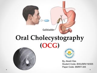
Oral Cholecystography (OCG): A Radiographic Study of the Gallbladder
- 1. By, Akash Das Student Code- BWU/BRI/19/005 Paper Code- BMRIT-305 Oral Cholecystography (OCG)
- 2. • Definition: Oral Cholecystography or Oral Cholecystogram is the radiographic study of gallbladder by the oral administration contrast media. Oral Cholecystography (OCG) • It was developed in 1924 by American surgeons E.A Graham & W. H Cole. • But now days it is largely superseded by ultrasound and MRCP. • This is the initial examination for the investigation of biliary tract. Fig- 1: Gallbladder
- 3. • Inflammation of the organ. • Other abnormalities like polyps. • Tumors • Gallstones • To visualizing cystic duct and common bile. • To demonstrate suspected pathology in the gallbladder. Fig- 3: Gallstone Formation Fig- 2: Polyp Inside Gallbladder
- 4. • Pr severe hepatorenal disease. • Acute cholecysititis • Iodine sensitivity • Pregnancy • Dehydration • An IV cholecystography within the previous week evious cholecystectomy. Fig- 4: Acute Cholecysititis Fig- 5: Pregnancy
- 5. • Patient should take low residue diet for 2 days prior to examination. • A laxative 2 days prior to the examination. • The CM is taken with water 14 hours prior to the examination. • A fat-containing meal after preliminary film, if the gallbladder is not visualize by contrast media. • Food is forbidden until the examination is completed. Fig-6: Gall Stone Formation Fig-7: Radiographic Appearance of GB
- 6. 1. Biloptin 2. Telepaque 3. Cholebrin 4. Solu-Biloptin 1. Prone 20º LAO 2. Supine 20º RPO 3. Erect 20º LAO 4. Fatty meal provided. • Prone 20º LAO 30 minutes after a fatty meal. Fig- 9: Positioning of patient for OCG Fig- 8: Biloptin Capsules
- 7. • A night before the examination 6 tablet of Teleopaque or Biloptin is given orally. • After 12-16 hours a prone oblique view with right side raised to 20º is taken for gallbladder visualization. • After the preliminary film taken, the patient lie in the supine position and appropriate spot film of the gallbladder are taken. • Ask the patient to eat fatty meal. • After 30-40 min. films are taken to assess the contractibility of the gallbladder and small filling defect. (stones or polpyps) • Cystic and common bile duct also visualized in post fatty meal films. * If the GB not visualized a "double dose" OCG may have to be performed where the patient takes in all 12 tablets of CM.
- 8. • Mild gastrointestinal disturbances • Skin reactions • Uricosuric action • Impaired renal function • Psedoalbuminuria • Abnormal thyroid function tests • Increased effect of protein-bound drugs because of shared binding with albumen Fig- 11: Gastrointestinal Disturbances Fig- 10: Skin Reactions
- 9. There are many ways by which we can protect our self from scattered radiation, like- By using • Protective apron:0.5mmPb • Lead shield:0.5mm Pb • Lead protective gloves. • Film badge or TLD monitor. And also we should always the rule of TDS & ALARA principle We can also use beam limiting devices like cone to minimize the radiation dose. Fig- 14: Beam Limiting Device (Cone) Fig- 13: Cardinal’s principle (TDS) Fig- 12: Lead apron, shield, gloves & TLD
- 10. THANK YOU