major manifestation of lung diseases
•Download as PPT, PDF•
2 likes•230 views

A Medical lecture about major manifestation of lung diseases in medicine.

Recommended
More Related Content
What's hot
What's hot (20)
Similar to major manifestation of lung diseases
Similar to major manifestation of lung diseases (20)
More from med zar
More from med zar (17)
Recently uploaded
Recently uploaded (20)
major manifestation of lung diseases
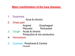
- 1. Major manifestation of the lung diseases 1. Dyspnoea. Acue & chronic. 2. Chest pain Anginal Esophageal Pleuretic Pericardial 3. Cough Acute & chronic Productive & non productive 4, Hemoptysis 5. Cyanosis Peripheral & Central Causes
- 2. Dyspnoea: is unpleasant or uncomfortable breathing, Or Subjective awareness of the sensation of breathing. The experience of dyspnea likely results from a complex interaction between chemoreceptor stimulation, mechanical abnormalities in breathing, and the perception of those abnormalities by the CNS. causes according to physiological bases: 1. Increased ventilatory rate a. Increase PaC02 e.g. COADs. b. Decrease Pa 02, cyanotic CHD, COAD c. Metabolic acidosis. d. Exercise e. Fever 2. Reduced Ventilatory capacity a. Decrease lung volume b. Increase resistance to air flow c. Pleural pain
- 4. D.D of chronic exertional dyspnoea: 1. COADs. 2. Heart diseases. 3. Interstitial lung diseases. 4. Large pleural effusion. 5. Severe anemia. 6. Chronic Pulmonary Thrombo-Embolism. 7. Psychogenic breathlessness: 8. Hysterical hyperventilation.
- 5. Psychologenic breathlessness Is very important to be confident of the diagnosis a. We should exclude other causes b. Anxiety could be associated with other organic Conditions c. In some conditions such as Primary Pulmonary Hypertension & Pulmonary thrombo-embolisation There are no signs on examination d. Anxiety related e. Absent of symptoms on exertion f. Unable to take deep breath with frequent sighing/ Erratic breathing g. No signs on examination h. Not able to do spirometry i. Short breath holding time in the absence of severe respiratory diseases n. PaCO2 is low & PaO2 is high in ABG
- 6. Hysterical hyperventilation. Severe hyperventilation associated with Carpo-pedal spasm Respiratory Alkalosis
- 7. Causes of acute dyspnoea a.Cardiac, Acute myocardial infarction, pul. Oedema, pul. Embolism, Papillary muscle dysfunction or rupture, Pericardial effusion or tamponade b. Respiratory, Acute severe asthma, acute exacerbation of COAD, Pneumothorax, Pulmonary embolism Pneumonia, ARDS, Inhaled foreign body, lobar collapse, Laryngeal oedema, etc. c. Metabolic, Diabetic ketoacidosis, Renal failure, Lactic acidosis, Aspirin overdose. E. Other causes, Diaphragmatic paralysis.
- 8. Paroxysmal nocturnal dyspnoea Left ventricular failure Nocturnal dyspnoea could be due to Asthma Sleep apnoea syndrome Interstial lung diseases GERD
- 9. Chest pain: Anginal getting worse on exertion and relieved by rest Pleuretic getting worse on coughing and inspiration Pericrdial, Oesophageal. getting worse on drinking hot fluid
- 10. Cardiac causes: 1. Ischemic heart diseaeses, Angina, MI, Coronary spasm. 2. Aortic dissection, sudden and tearing chest and back pain. 3. Pericarditis, Pleuretic chest pain. 3. Myocarditis. 4. Hypertrophic cardiomyopathy. 5. Mitral valve prolapse 6. Cardiac arrythmias. 7. Aortic stenosis,
- 11. Pulmonary causes: Causes pleuretic chest pain. Is a chest pain exacerbated by forceful breathing or coughing Causes include 1. Pleurisy 2. Pulmonary embolism 3. Pneumonia 4. Pneumothorax 5.Tracheobronchitis 6. Mediastinal emphysema 7. Pericarditis
- 12. Chest Wall causes.. All causes pleuretic chest pain Costocondritis – Tietze syndrome Rib fracture Chest wall injury Chest wall or rib malignancy Myalgia Herpes zoster infection Neuritis-radiculitis Thoracic outlet obstruction Sickle cell disease
- 13. Gastrointestinal Oesophagitis Gastro esophageal reflux disorder Esophageal Motility disorders Biliary Colic Gastointestinal perforation Assessment of patients hemodynamic stability. If the patient is stable a detailed history followed by physical examination would help to limit the number of differentials. Investigations like ECG, Chest X ray, ECHO, Cardiac enzyme levels will rule in or rule out the commonest causes. If the patient is unstable, effort must be taken to rule out the critical etiologies like aortic dissection, myocardial infarction, pneumothorax, pulmonary embolism and infarction, cardiac arrhythmia, GI perforation, pancreatitis etc.
- 14. Mechanism of Cough Cough is a protective reflex serving a normal physiologic function of clearing excessive secretions and debris from the pulmonary tract. Is the most frequent symptom of respiratory diseases. It is productive or non productive It is caused by stimulation of sensory nerves in the mucosa of the pharynx, larynx, trachea and bronchi cough
- 15. The cough reflex It has 3 components: an afferent sensory limb, a central processing center, and an efferent limb. The Afferent sensory path way is mainly through vagus Nerve Receptors are located throughout the airway from the pharynx to the terminal bronchioles, with the greatest concentration located in the larynx, carina, and the bifurcation of larger bronchi. Afferent impulses are transmitted to the cough center of the brain, which is poorly localized mainly in medulla ablongata and in the nucleus tractus solitarius of the medulla. And also other brain parts Efferent impulses leave the medulla and travel to the larynx and tracheobronchial tree via the vagus, and superior laryngeal nerve while the phrenic and spinal motor nerves of C3 to S2 supply the intercostals muscles, abdominal wall, diaphragm, and pelvic floor.
- 16. Acute cough. Less than 3 weeks Acute coughs can be divided into infectious (caused by an infection) and noninfectious causes. Infectious causes of acute cough include viral upper respiratory infections. sinus infections, pneumonia, lung abscess whooping cough. Noninfectious causes of cough include Foreign body inhalations Asthma Exposure to irritants such as smoke or perfumes, change of air temperature and environmental allergies. •Angiotensin-converting enzyme inhibitor •Psychological
- 17. Chronic cough More than 8 weeks, This include 1. Enviromental irritants: cigarette smoke, irritant substance 2. Lung diseases asthma, emphysema, and chronic bronchitis, bronchiectasis, Less common causes of lung-induced chronic cough include. Bronchogenic carcinoma, sarcoidosis, Fibrosing alveolitis. 3. Heart diseases Congestive heart failure. 4. Upper respiratory tract diseases: Chronic sinusitis, Chronic post nasal drip, and chronic pharyngitis & GERD 5. ACEI 6, psycholodical
- 18. Chronic cough continues Causes of chronic cough with normal CX-Ray 1. Chronic bronchitis 2. Asthma 3. Post nasal drip, 4. Gastro-esophageal reflux 5. Eosinophilic bronchitis ( non asthmatic) 6. ACE inhibitor
- 19. Complications of cough 1. fainting spells due to decreased blood flow to the brain when coughs are prolonged and forceful 2. Insomnia 3. cough-induced vomiting 4. Subconjectival hemorrhage 5. Spontaneous pneumothorax or Mediastinal emphysema 6. Fractures of ribs 7.Cough induced urination and defecation 8. Prolapse of uterus in female 9. Abdominal and pelvic hernia
- 20. Hemoptysis. Is the expectoration of blood or of blood-stained sputum It comes from the bronchi, larynx, trachea, or lungs Is alarming symptom and the patients nearly always seek medical advice. We should be able to differentiate it from Hematemesis, and some times from epistaxis. Hemoptysis must always be assumed to have a serious cause until this is excluded. Many episodes remain unexplained, even after full investigations and are likely to be caused by simple bronchial infections. A full history should be taken All relevant investigations should be done.
- 21. Hemoptysis Common cases less common causes a. Acute bronchits 1. bronchial adenoma b. Bronchiectasis 2. foreign body c. Bronchogenic CA. 3. lung abscess d. Pulmonary Embolism 4. truma e. MS & Pul. Oedema 5. hydatid cyst f. Pneumonia 6. aspergilloma g. TB 7. actinomycosis Other causes: Idiopathic Pul. Haemosiderosis A-V malformation Polyareritis Nodosa Aortic aneurysm Blood disorders, leukemia, hemophelia anticoagulant, chronic bronchitis Diagnosis Treatment; according to the cause in severe cases Tranexamic acid 1gm 8 hrly oraly Bronchial artery embolisation
- 22. Cyanosis is the appearance of a blue discoloration of the skin or mucous membrane due to hypoxia. The onset of cyanosis is 2.5 g/dL of deoxy-hemoglobin. The bluish color is more readily apparent in those with high hemoglobin counts than it is with those with anemia. Also the bluish color is more difficult to detect on deeply pigmented skin. Cyanosis is divided in to two main types: Central, Tongue and mucous membrane of the mouth Peripheral, the extremities are affected, fingers, extremities. Ear It develops when the arterial blood O2 saturation is about 85%. The causes are diseases of CNS, Cardiovascular, Respiratory systems. Les common causes include, Methhemoglobinemia, polycythemia, high altidude, and hypothermia. SPO2 of less than 92% is indication for O2 therapy
- 24. Summary Major manifestation of the lung diseases are common presenting symptoms of respiratory lesions.