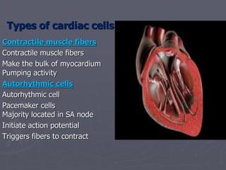
1. Types of cardiac cells
Contractile muscle fibers
Contractile muscle fibers
Make the bulk of myocardium
Pumping activity
Autorhythmic cells
Autorhythmic cell
Pacemaker cells
Majority located in SA node
Initiate action potential
Triggers fibers to contract
2. Properties of the cardiac cells
► Cardiac cell have four important properties
1. automaticity
2. excitability
3. conductivity
4. contractility
► Automaticity
► Unique function of pacemaker cells in SA
node
► Generates an action potiential
► Cell membrances of pacemaker
3. ►Excitability
► Ability to reach threshold potential
► Respond to stimulus
► Less excitable the cell
► If strong stimulus is needed
► More excitable
► Weak stimulus need for activation
► Ischemia and hypoxia increase
excitability
4. ► Conductivity
► Unique ability of heart cells
► Transmit electrical current from cell to cell
► Contractility
► ability to shorten & contract
► In response to electrical stimulus
5. The Heart:
► Heart pumps blood through the body
► Contraction and relaxation of the
cardiac muscle
► Intercalated discs
► Allow impulses to travel rapidly
► From cells to cell
► Function as one rather than individual
cells
Conduction System
6. Conduction System
► Cardiac conduction system
► Autorhythmic fibers
► Conduction system controls the heart rate
► Creates the electrical impulses
► Spreading of impulse throughout the heart
► Make the heart contract
► Pump blood out of heart
7. Nodal Firing
Rates
► Sa node = 75 b.p.m
► AV node = 50 b.p.m
► AV bundle = 30 b.p.m
► Purkinje fibers = 30 b.p.m
What would happen if the SA node could not conduct an impulse to the
AV node?
Heart block (no gap jxn’s found between atria & ventricles)
8. Components of the Conduction System
► Sinoatrial Node
Located in back wall of the right atrium
Near the entrance of vena cava
Initiates impulses 70-80 b.p.m
Without any nerve stimulation
Establishes basic rhythm of the heartbeat
Pacemaker of the heart
Impulses move through atria
Atria to contract
Impulses reach the second part of the
conduction system
9. Pacemaker Potential
► SA node do not maintain rapid membrane
potential
► Suring the period of diastole
► Exhibits slow spontaneous depolarization
pacemaker Potential
► Membrane potential begins at about − 60 mV
► Gradually depolarizes to − 40 mV which is the
threshold
10. ► Spontaneous depolarization
► In response to hyperpolarization “funny current”
► Hyperpolarization− 90 mV stimulus opens Na-channel
► These channel permeable to both Na + and K +
► K + move out & Na enter
► Spontaneous depolarization occurs during diastole
► Diastolic depolarization
11. ► Once depolarization reaches threshold –40mV
► Opening of voltage-gated Ca 2 + channels
► Inward diffusion of Ca 2 +
► Results in contraction of these myocardial cells
► Repolarization is produced voltage-gated K + channels
► Outward diffusion of K +
Sinoatrial node
Atrioventricular node
AV-bundle
Right and
left bundle
branches
Purkinje fibers
12. Components of the Conduction System
► Atrioventricular Node
located in the bottom of the right atrium near the septum
Cells in the AV node conduct impulses more slowly
Delay as impulses travel through the node
Allows time for atria to finish contraction before ventricles
begin contracting
14. Atrioventricular Bundle
► “Bundle of His”
From the AV node,
impulses travel through
to the right and left
bundle branches
These branches extend
to the right and left
sides of the septum and
bottom of the heart
15. Atrioventricular Bundle
These branch form the
Purkinje fibers
Transmit the impulses to the
myocardium (muscle tissue)
The bundle of His
Purkinje fibers transmit
quickly and cause both
ventricles to contract at the
same time
Like a “phone tree”
16. Atrioventricular Bundle
► As the ventricles contract
► Blood move through pulmonary trunk and the aorta
► Once ventricles complete contraction
► Undergo relaxation
► SA node initiates another impulse
► Start another cardiac cycle
17.
18. 1 - Sinoatrial node (SA node)
2 - Atrioventricular node (AV node)
3 – Bundle of His
4 - Right & Left Bundle Branches
which lead to Purkinje Fibers