

Recommended
More Related Content
What's hot
What's hot (20)
Similar to Down's syndrome
Similar to Down's syndrome (20)
More from Abdulmalik Abdulateef
More from Abdulmalik Abdulateef (20)
Recently uploaded
Recently uploaded (20)
Down's syndrome
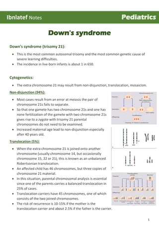
- 1. 1 Down's syndrome Down's syndrome (trisomy 21): This is the most common autosomal trisomy and the most common genetic cause of severe learning difficulties. The incidence in live-born infants is about 1 in 650. Cytogenetics: The extra chromosome 21 may result from non-disjunction, translocation, mosaicism. Non-disjunction (94%): Most cases result from an error at meiosis the pair of chromosome 21s fails to separate. So that one gamete has two chromosome 21s and one has none fertilization of the gamete with two chromosome 21s gives rise to a zygote with trisomy 21 parental chromosomes do not need to be examined. Increased maternal age lead to non-disjunction especially after 40 years old. Translocation (5%): When the extra chromosome 21 is joined onto another chromosome (usually chromosome 14, but occasionally chromosome 15, 22 or 21), this is known as an unbalanced Robertsonian translocation. An affected child has 46 chromosomes, but three copies of chromosome 21 material. In this situation, parental chromosomal analysis is essential since one of the parents carries a balanced translocation in 25% of cases. Translocation carriers have 45 chromosomes, one of which consists of the two joined chromosomes. The risk of recurrence is 10-15% if the mother is the translocation carrier and about 2.5% if the father is the carrier. Ibnlatef Notes Pediatrics
- 2. 2 If a parent carries the rare 21:21 translocation, all the offspring will have Down's syndrome. If neither parent carries a translocation (75% of cases), the risk of recurrence is <1%. Mosaicism (1%): In mosaicism some of the cells are normal and some have trisomy 21. This usually arises after the formation of the zygote, by non-disjunction at mitosis. The phenotype may be milder in mosaicism. Causes: The cause of the extra full or partial chromosome is still unknown. Maternal age is the only factor that has been linked to an increased chance of having a baby with Down syndrome resulting from nondisjunction or mosaicism. However, due to higher birth rates in younger women, 80% of children with Down syndrome are born to women under 35 years of age. There is no definitive scientific research that indicates that Down syndrome is caused by environmental factors or the parents' activities before or during pregnancy. The additional partial or full copy of the 21st chromosome which causes Down syndrome can originate from either the father or the mother. Approximately 5% of the cases have been traced to the father. Clinical features: Typical craniofacial appearance: o Round face and flat nasal bridge. o Upslanted palpebral fissures. o Brushfield spots in iris (pigmented spots).
- 3. 3 o Epicanthic folds (a fold of skin running across the inner edge of the palpebral fissure). o Small mouth and protruding tongue, Small ears. o Flat occiput and third fontanelle. Other anomalies: o Short neck. o Single palmar creases (present in 6-7% of normal population). o Incurved fifth finger. o Wide 'sandal' gap between toes. o Hypotonia. o Congenital heart defects (40%). o Duodenal atresia. o Hirschsprung's disease. Later medical problems: o Delayed motor milestones. o Moderate to severe learning difficulties. o Small stature. o Increased susceptibility to infections (due to hypotonia, especially in respiratory tract). o Hearing impairment from secretory otitis media. o Visual impairment from cataracts, squints, myopia. o Increased risk of leukemia and solid tumors. o Risk of atlantoaxial instability. o Hypothyroidism and coeliac disease. o Epilepsy. o Alzheimer's disease. Diagnosing Down's syndrome: Screening during pregnancy: o Combined test Blood test + nuchal translucency ultrasound scan (nuchal translucency is a collection of fluid at the back of the baby's neck) between 11 and 14 weeks of pregnancy. o Blood test that can be offered between 14 and 20 weeks of pregnancy, this blood test is less accurate than combined test. Diagnosis during pregnancy: o Chorionic villus sampling (CVS) usually done from week 11 of pregnancy. o Amniocentesis usually carried out from week 15 of pregnancy Diagnosis after birth: o Based on baby's appearance. o Sample of child's blood can be taken and analyzed to look for the extra copy of chromosome 21.
- 4. 4 Investigations: Laboratory studies: o Complete blood count with differential. o Bone marrow examination to rule out leukemia. o Thyroid-stimulating hormone (TSH) and thyroxine (T4) to rule out hypothyroidism. o Papanicolaou smears every 1-3 years in sexually active women. o Cytogenetic studies (karyotyping) for diagnosis of trisomy 21. o FISH for rapid diagnosis of trisomy 2. o Assessment of mosaicism for trisomy 21 lymphocyte preparations, buccal mucosa cellular preparations, FISH, scoring frequency of trisomic cells. o Immunoglobulin G. o Maternal serum biochemical markers. Imaging studies: o Echocardiography in every newborn suspected of having trisomy 21 to identify congenital heart disease. o Ultrasonography. Postnatal diagnostic tests that may be warranted include the following: o Auditory brainstem response (ABR), or brainstem auditory evoked response (BAER). o Pediatric ophthalmic examination. o Growth charts specifically for children with Down syndrome. o Rigorous dental hygiene and dental evaluation. Diagnostic considerations: The presence of 8 or more of the characteristic clinical findings leads to a definite diagnosis of Down syndrome. Chromosomal analysis is always recommended and of utmost importance in doubtful cases. In addition to the differential diagnosis, other problems to be considered include the following: o 49,XXXXY chromosome and other high-order multiple X chromosome disorders. o Congenital hypothyroidism. o Mosaic trisomy 21 syndrome. o Partial trisomy 21 (or 21q duplication). o Robertsonian trisomy 21. o Zellweger syndrome or other peroxisomal disorders. Differential Diagnosis Trisomy 18.
- 5. 5 Management: There are no medical treatments for intellectual disability associated with Down syndrome, but improved medical care has greatly enhanced quality of life and increased life expectancy. Elements of medical care include the following: o Genetic counseling. o Standard immunizations and well child care. o Management of specific manifestations of Down syndrome and associated conditions (endocrine, infectious, cardiac, respiratory, neurologic, psychiatric, dermatologic, or dental disorders). o Early intervention programs. Special considerations in adolescents are as follows: o Ongoing monitoring measures, including annual audiologic evaluation and annual ophthalmologic evaluation. o Ongoing management of manifestations of the syndrome and associated conditions. o Discussion of issues related to the transition to adulthood. Surgical management of associated conditions: o Timely surgical treatment of cardiac anomalies. o Surgical repair for GI anomalies (duodenal atresia and Hirschsprung disease). o Stabilize the upper segment of the cervical spine if neurologic deficits are clinically significant. o Careful anesthetic airway management is needed because of the associated risk of cervical spine instability. o Congenital cataracts must be extracted soon after birth and subsequent correction with glasses or contact lenses provided. o Adenotonsillectomy may be performed to manage obstructive sleep apnea. Complications: Heart problems: o Around half of children with Down's are born with a congenital heart defect. o The most common defect to affect children with Down's a septal defect. o It can cause a build-up of blood in one or more of the heart's chambers, which causes the heart to work harder to pump blood through the four chambers. Gut problems: o Many people with Down's have some sort of problem with their digestive system. o Constipation, diarrhoea and indigestion are all common, as are more serious problems such as small bowel obstruction, which stops food passing from the stomach into the large bowel.
- 6. 6 o Some children also develop coeliac disease and reflux. o Conditions such as imperforate anus or Hirschsprung's disease are rare, but slightly more common in children with Down's syndrome. Hearing problems: o Most people with Down's syndrome have problems with their hearing. o This is often temporary, but it can sometimes be permanent. o Glue ear (a build-up of fluid in the middle ear) is a common cause of temporary hearing problems in people with Down's syndrome. o Glue ear can be treated with minor surgery, which involves placing small tubes called grommets in the ear to help drain away the fluid. Vision problems: o Short-sightedness, long-sightedness. o Squint, nystagmus. o Eye infections (conjunctivitis, uveitis or blepharitis). o Cataracts, keratoconus, glaucoma. Thyroid problems: o Around one in 10 people with Down's syndrome have problems with their thyroid gland. o Most people with Down's syndrome who have a problem with their thyroid have hypothyroidism. o Thyroid problems will usually be picked up during blood tests and can often be treated with medication to replace the lack of thyroid hormone in the body. Increased risk of infections: o People with Down's syndrome are more likely to develop infections, such as the lung infection pneumonia, because their immune system has not developed properly. o To reduce the risk of infections, routine childhood vaccinations will be recommended. o If child develops a bacterial infection, a course of antibiotics will usually be prescribed to treat it. Dementia: o There is a tendency for people with Down's syndrome to develop dementia at a younger age than in the general population, usually from about 40 years of age onwards, although it's not inevitable that everyone with Down’s syndrome will develop it. o Possible signs of dementia include problems with short-term memory and understanding, confusion, and disorientation. ---------------------------------------------------------------------------------------------- www.facebook.com/ibnlatef https://goo.gl/RpvNsl