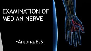
EXAMINATION OF MEDIAN NERVE FOR MBBS UNDERGRADUATES ORTHO
•Download as PPTX, PDF•
3 likes•126 views

EXAMINATION OF MEDIAN NERVE FOR MBBS UNDERGRADUATES- ORTHO

Recommended
More Related Content
What's hot
What's hot (20)
Similar to EXAMINATION OF MEDIAN NERVE FOR MBBS UNDERGRADUATES ORTHO
Similar to EXAMINATION OF MEDIAN NERVE FOR MBBS UNDERGRADUATES ORTHO (20)
More from ANJANA B.S.
More from ANJANA B.S. (9)
Recently uploaded
Recently uploaded (20)
EXAMINATION OF MEDIAN NERVE FOR MBBS UNDERGRADUATES ORTHO
- 2. LEARNING OBJECTIVES HISTORY LOCAL EXAMINATION EXAMINATION OF MUSCLES SUPPLIED BY MEDIAN NERVE CARPAL TUNNEL SYNDROME
- 4. HISTORY History of trauma (incisional wound, penetrating wound, fracture, dislocation) .Supracondylar fracture of humerus- median nerve can be injured. History of malignant growth. History of injecting drugs (direct injury or irritant drugs). History of diabetes/leprosy (can cause peripheral neuropathy) Wound infection may lead to fibrosis and will not allow proper regeneration of the nerve
- 5. LOCAL EXAMINATION INSPECTION Attitude: Complete claw hand seen in combined lesion of median and ulnar nerve(median nerve supplies the first 2 lumbricals) . Also seen in Klumpke’s paralysis. Ape thumb deformity (paralysis of opponens pollicis) and pointing index(paralysis of lateral half of flexor digitorum profundus) Wasting of muscles obvious only in long term paralysis. Skin: Becomes dry, glossy and smooth with loss of cutaneous fold and subcutaneous fat, sometimes trophic ulcers may be seen. Look for scar or wound.
- 6. LOCAL EXAMINATION PALPATION Temperature is cold in paralysis. Muscles wasted and their functions are lost. Injuries in the arm cause paralysis of all muscles innervated by the median nerve, including those arising from the medial epicondyle (flexor carpi radialis, pronator teres, palmaris longus, and part of flexor digitorum superficialis), as well as those innervated by the anterior interosseous nerve (flexor pollicis longus, flexor digitorum profundus to index finger) If sensory supply is lost, skin is anaesthetized. Involvement of the palmar cutaneous nerve is useful in localizing a lesion at the wrist. Anterior interosseous palsy is distinguished from high median injury by the absence of any loss of skin sensation. Hyper aesthesia at the site of nerve regeneration. Palpate the scar for tenderness, indicates of adhesion of nerve to the scar.
- 7. Tinel’s sign In closed injuries, percussion of the skin over a nerve in which axons have been ruptured evokes sensations usually described as a wave or surge of pins and needles into the cutaneous distribution of the nerve. This is Tinel’s sign and it is a most useful aid to diagnosis Importance of Tinel’S SIGN Whether nerve interrupted Whether in process of regeneration Rate of regeneration Success of nerve repair
- 9. EXAMINATION FLEXOR POLLICIS LONGUS The patient is asked to bend the terminal phalanx of the thumb against resistance while the proximal phalax is steadied by the physician.
- 10. Flexor digitorum superficialis and profundus(lateral half): Ochsner’s clasping test- the patient is asked to clasp the hand. The index finger of the affected hand fails to flex and remains as a pointed finger
- 11. Abductor pollicis brevis ask the patient to keep the hand on the table and ask the patient to touch a pen which is kept at a slight higher level than the palm of the hand with the thumb-pen test Opponens pollicis Ask the patient to touch the tips of other fingers with the thumb against resistance
- 12. Flexor Carpi Radialis Normally, palmar flexion at the wrist occurs in the long axis of the forearm. In a patient with paralysed flexor carpi radialis………….?
- 13. Causes of peripheral nerve lesions 1.Traumatic- closed injury; open injury 2.inflammatory – eg.herpes zoster 3.neoplastic – neurofibroma,neurofibrosarcoma 4.miscellaneous – tunnel syndrome , lead poisoning, leprosy 5.metabolic disordes – B complex deficiency
- 14. Carpel tunnel syndrome The median nerve is injured in the carpal tunnel due to its compression and produces a clinical condition called carpal tunnel syndrome. The carpal tunnel is formed by anterior concavity of carpus and flexor retinaculum. The tunnel is tightly packed with nine long flexor tendons of fingers and thumb with their surrounding synovial sheaths and median nerve. The median nerve gets compressed in the tunnel due to its narrowing following a number of pathological conditions such as (a) tenosynovitis of flexor tendons (idiopathic), (b) myxedema (deficiency of thyroxine), (c) retention of fluid in pregnancy, (d) fracture dislocation of lunate bone, and (e) osteoarthritis of the wrist.
- 16. Characteristic clinical features of the carpal tunnel syndrome are as follows: • Feeling of burning pain or ‘pins and needles’ along the sensory distribution of median nerve (i.e., lateral 3½ digits) especially at night. • There is no sensory loss over the thenar eminence because skin over thenar eminence is supplied by the palmar cutaneous branch of the median nerve, which passes superficial to flexor retinaculum. • Weakness of thenar muscles. • ‘Ape-thumb deformity’ may occur, if left untreated, due to paralysis of the thenar muscles. • Positive Tinel’s sign and Phalen’s test • Reduced conduction velocity in the median nerve (<30 m/s) is diagnosis.
- 18. Management Splinting- prevents wrist flexion Corticosteroid/anesthetic injection Surgical decompression- Division of the transverse carpal ligament
- 19. Anterior Interosseous Syndrome Damage to the anterior interosseous nerve Pain in the forearm Weakness of the gripping movement of the thumb and index finger (unable to make ok sign) Causes -Injury to elbow Injury during open / closed reduction Okay or circle sign A Quick way to assess flexor digitorum profundus and flexor policis longus With weakness in these muscles, the distal phalanges cannot flex and instead of fingertips touching, the volar surfaces of each distal phalanx make contact.
- 20. THANK YOU