
Recommended
More Related Content
What's hot
What's hot (20)
Similar to 15-180531111035.pdf
Similar to 15-180531111035.pdf (20)
Recently uploaded
Recently uploaded (20)
15-180531111035.pdf
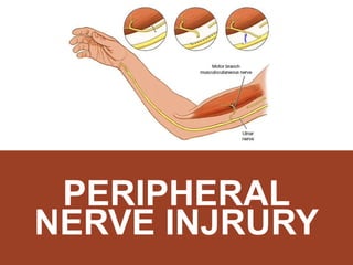
- 3. CLASSIFICATION ULNAR NERVE INJURIES 1. High ulnar nerve lesion 2. Low ulnar nerve lesion 3. Guyon’s tunnel syndrome 4. Cubital tunnel syndrome
- 4. ANATOMY ANATOMY OF ULNAR NERVE Type: Mixed Never (motor & sensory) Root Value: C7, C8 & T1 Origin: Arises in the axilla as the largest branch of median cord of the brachial plexus, at the lower border of pectoralis minor.
- 5. ANATOMY ANATOMY OF ULNAR NERVE Enters the forearm between 2 heads of Flexor Carpi Ulnaris In the upper half, it rest on Flexor Digitorum Profundus & covered by Flexor Carpi Ulnaris In lower half, it runs lateral to Flexor Carpi Ulnaris tendon accompanied by ulnar artery laterally Enters palm on lateral side of pisiform bone above the flexor retinaculum Divides into Superficial & Deep Terminal Branches
- 6. ANATOMY ULNAR NERVE IN THE HAND
- 7. ANATOMY ULNAR NERVE IN THE HAND
- 8. ULNAR NERVE ULNAR INERVATED MUSCLES
- 9. ULNAR NERVE SUMMARY OF BRANCHES OF ULNAR NERVE MOTOR BRANCHES: To 1 muscles only: Flexor carpi ulnaris Medial half of flexor digiti profundus Superficial Terminal Branches supplies one muscle only: palmaris brevis Deep Terminal Branch supplies: 8 interossei + medial 2 lumbricals 3 hypothenar muscle Adductor Pollicis & may be FPB FEMORAL HAND
- 10. ULNAR NERVE SUMMARY OF BRANCHES OF ULNAR NERVE SENSORY BRANCHES: Palmar cutaneous branch: Medial 1/3 of palm Dorsal cutaneous branches: Medial 1/3 of dorsum of hand & dorsum of medial 1 Superficial terminal branches gives 3 palmar digital nerves to the palmar surfaces of the medial 1fingers FEMORAL HAND
- 11. 1. ABOVE ELBOW (HIGH ULNAR LESION) ULNAR NERVE INJURY Injuries lead to complete loss of all the functions of nerve. The injury may be caused by: A. Penetrating wounds B. Gun Shots C. Fracture of medial epicondyle D. Cubitus Valgus (a deformity of elbow joint causing stretch of ulnar nerve)
- 12. 1. ABOVE ELBOW (HIGH ULNAR LESION) ULNAR NERVE INJURY 1. Paralysis of all muscles supplied: 1 muscles in the forearm 15 muscles in the hand 2. Weak flexion of the wrist, with radial deviation of the hand – due to paralysis of FCU 3. Inability to flex the terminal phalanges of the medial 2 fingers – due to paralysis of medial ½ of the FDP 4. Inability to put the hand in the writing position – due to paralysis of interossei & medial 2 lumbricals 5. Loss of adduction of the thumb – paralysis of adductor pollicis MOTOR AFFECTION
- 13. MANIFESTATIONS: ABOVE ELBOW ULNAR NERVE INJURY Partial Claw Hand – characterized by: I. Extension of the metacarpophalangeal & flexion of the interphalangeal joints – due to paralysis of lumbricals & interossei I. Flat hypothenar eminence – due to paralysis of it’s muscles Sensory loss from the skin of palmar & dorsal surfaces of medial 1 of fingers. DEFORMITY SENSORY LOSS
- 14. 2. AT OR ABOVE THE WRIST (LOW ULNAR LESION) ULNAR NERVE INJURY Wrist laceration Fracture of the carpal bones Malunion of Colles fracture Handcuffs Motor: Limited to hand muscles only. The forearm muscles are intact because they receive supply very close to the elbow. CAUSED BY MANIFESTATIONS
- 15. 2. AT OR ABOVE THE WRIST (LOW ULNAR LESION) ULNAR NERVE INJURY Complete Claw Hand (in combined with median nerve injury) More severe than in injury above the wrist, this is called the ulnar paradox. Sensory Loss: Loss of sensation from the palmar surfaces of the medial 1 fingers only – because the palmar & dorsal cutaneous branches are intact. DEFORMITY
- 16. SIGNS OF LOW LESION ULNAR NERVE INJURY Hypothenar muscle wasting Claw hand FROMENT’S SIGN POSITIVE
- 17. WEAKNESS OF FINGERS ABDUCTION AND ADDUCTION ULNAR NERVE INJURY
- 18. SIGNS OF LOW LESION ULNAR NERVE INJURY Loss of sensation over ulnar 1 ½ digits Tinel’s sign
- 19. COMPRESSION AT GUYON’S CANAL ULNAR NERVE INJURY Anatomy of Guyon’s canal : Floor = transverse carpal ligament to pisiform Ulnar wall = pisiform Radial distal wall = hook of hamate Roof = volar carpal ligament Contains only ulnar nerve and artery ULNAR TUNNEL SYNDROME
- 20. COMPRESSION AT GUYON’S CANAL ULNAR NERVE INJURY Repetitive indirect trauma most common Tumours- ganglion, lipoma Pisiform instability Pisotriquetral arthritis Fractured hook of hamate / pisiform Ulnar artery thrombosis CAUSES
- 21. COMPRESSION AT GUYON’S CANAL ULNAR NERVE INJURY Symptoms could be: 1. Pure motor 2. Pure sensory 3. Mixed (depending on exact site of entrapment) CLINICAL FEATURES
- 22. COMPRESSION AT CUBITAL TUNNEL ULNAR NERVE INJURY Compression of ulnar nerve as it passes behind elbow (behind medial epicondyle) Intrinsic muscle wasting Waternberg’s sign CUBITAL TUNNEL SYNDROME
- 23. COMPRESSION AT CUBITAL TUNNEL ULNAR NERVE INJURY Conservative Modification of posture and avoidance of repetitive trauma Splint Surgical Operative decompression if symptoms persist Tendon transfer to recover hand function TREATMENT
- 25. CLASSIFICATION MEDIAN NERVE INJURIES 1. High median nerve lesion 2. Low median nerve lesion 3. Carpal tunnel syndrome
- 26. ANATOMY ANATOMY OF MEDIAN NERVE Type: Mixed nerve (contains motor & sensory fibres) Root Values: C5, 6, 7, 8 & T1 Origin: Arises in the axilla by 2 roots Lateral root – lateral cord of the brachial plexus Medial root – medial cord of the brachial plexus The medial root crosses in front of the 3rd part of axillary artery to join the lateral root.
- 27. ANATOMY ANATOMY OF MEDIAN NERVE
- 28. ANATOMY ANATOMY OF MEDIAN NERVE ARM Descends medial side at upper ½ then lateral side of brachial artery at lower ½ arm Cross bicipital aponeurosis then enters cubital fossa FOREARM Supply PT, FCR, PL, FDS Between two head of PT, give anterior interosseous nerve branches and supply FPL, FDP, PQ HAND Passes to the flexor retinaculum Supply AbdPB, OP, FPB, 2 lumbrical and skin
- 29. ANATOMY BRANCHES OF MEDIAN NERVE
- 30. HIGH MEDIAN NERVE LESION MEDIAN NERVE INJURY AXILLA Crutch compression Anterior shoulder dislocation Stabs wound Fracture of humerus shaft Fracture humerus supracondylar in children Fracture medial epicondylar Elbow dislocation UPPER ARM ELBOW
- 31. EVERYTHING IS AFFECTED!! MEDIAN NERVE INJURY
- 32. SIGNS Wasting of muscles of forearm Wasting of thenar eminence Weakness of thumb abduction and opposition o Loss of abductor pollicis brevis + flexor pollicis brevis The hand is held with ulnar fingers flexed and index finger straight (pointing sign) o Loss of FDP, FDS, FPL Lost sensation at radial three and half digits Weak Ok sign Ape hand deformity
- 33. SIGNS
- 34. LOW MEDIAN NERVE LESION MEDIAN NERVE INJURY INJURY TO DISTAL THIRD OF THE FOREARM I. Cuts in front of the wrist II. Carpal dislocation I. Wasting of thenar muscle II. forearm muscle spared III. Paralyzed muscle of the hand IV. Weakness of thumb abduction and opposition V. Loss of abductor pollicis brevis + flexor pollicis brevis VI. Lost sensation at radial three and half digits SIGNS
- 35. COMPARISON MEDIAN NERVE INJURY Low lesion High lesion Wasting of thenar eminence Weak thumb abduction Weak thumb opposition Loss of sensation over lateral 3 and half digits All the signs of low lesion Wasting of lateral forearm Weakened OK sign (AIN – FPL & FDP) Pointing finger (2nd and 3rd finger remains partially extended in an attempt to make a fist)
- 36. PHYSICAL EXAMINATION MEDIAN NERVE PE a. Thenar wasting b. Atrophy of pulp of index, cracking of nails and other trophic changes c. Cigarette burns or other loss of sensory deprivation d. Pointing finger in high nerve lesion Flexor carpi radialis and palmaris longus. Patients hand is placed on a flat surface, palm upwards. Ask patient to attempt to flex the wrist with examiners hand putting pressure on top. The (pl) and (fcr) tendon will be prominent INSPECTION
- 37. PHYSICAL EXAMINATION MEDIAN NERVE PE Pronator teres. Extend the patients elbow and give resistance as patient attempt to pronate
- 38. PE OF MEDIAN NERVE MUSCLE DISTRIBUTION MEDIAN NERVE PE Flexor pollicis longus Flex the terminal phalanx of the thumb against resistance while the proximal phalanx is kept steady by examiner Flexor carpi radialis The wrist deviates to the ulnar side
- 39. PE OF MEDIAN NERVE MUSCLE DISTRIBUTION MEDIAN NERVE PE Flexor digitorum superficialis Patient asked to clasp his hand, the index finger will remain straight (Pointing index) Muscle of thenar eminence Abductor pollicis brevis (Pen test) Lay the hand flat, a pen is held above the thumb, try to touch the pen with tip of thumb
- 40. PE OF MEDIAN NERVE MUSCLE DISTRIBUTION MEDIAN NERVE PE Opponence pollicis Appose the tip of thumb to other finger
- 41. SCREENING MEDIAN NERVE REMEMBER LOAF! Lumbricals Opposition Abduction Flexion
- 42. CARPAL TUNNEL SYNDROME MEDIAN NERVE INJURY ANATOMY OF CARPAL TUNNEL
- 43. CARPAL TUNNEL SYNDROME MEDIAN NERVE INJURY ROOF Flexor retinaculum Ulnar artery, ulnar nerve, palmaris longus Carpal bone (hamate, capitat, trapezoid, trapezium) Lateral- tendon FCR Medial- tendon FPL, FDS,FDP, median nerve FLOOR CONTENTS
- 44. CARPAL TUNNEL SYNDROME MEDIAN NERVE INJURY Common in middle age group 40-50 years More common in female Causes: i. Inflammatory - RA, wrist OA, ii. Post traumatic – Dislocation of one of the carpal bones inside the carpal tunnel iii. Endocrine – Myxoedema, Cushing, Acromegaly (thickening of the tendons passing) iv. Tumour inside the carpal tunnel pressing on the median nerve v. Idiopathic
- 45. CARPAL TUNNEL SYNDROME MEDIAN NERVE INJURY HISTORY Pain and paresthesia at distribution of median nerve in the hand Wake up at night due to burning pain, tingling and numbness Relieve by shaking the arm Dropping object
- 46. CARPAL TUNNEL SYNDROME MEDIAN NERVE INJURY PHYSICAL EXAMINATION Hands may look normal/ wasting in severe cases TINEL SIGN Percussing over the median nerve causing sensation of current/ hyperesthesia at median nerve distribution PHALEN SIGN Flexed the wrist fully for one or two minutes causing paresthesia
- 47. CARPAL TUNNEL SYNDROME MEDIAN NERVE INJURY
- 48. CARPAL TUNNEL SYNDROME MEDIAN NERVE INJURY INVESTIGATIONS ELECTRODIAGNOSTIC TEST I. Nerve conduction studies Measure Median motor and sensory latencies and conduction velocities across the wrist Sensory latency of greater than 3.5 millisecond or a motor latency of greater than 4.5 millisecond is considered an abnormal finding Distal compound muscle action potential (CMAP) and sensory nerve action potential (SNAP) amplitudes may be decreased
- 49. CARPAL TUNNEL SYNDROME MEDIAN NERVE INJURY NERVE CONDUCTION STUDY
- 50. CARPAL TUNNEL SYNDROME MEDIAN NERVE INJURY INVESTIGATIONS ELECTRODIAGNOSTIC TEST II. Electromyography To determine completeness of a nerve injury Technique: I. Very small needle is inserted into various muscle II. Then, the signal is magnified by high gain amplifier III. Finally, the reading are monitored via oscilloscope and recorded on the magnetic tape or paper recording IV. Should performed 3-7 days after peripheral nerve injury V. It may show low amplitude evoked compound muscle potential (CMAP)
- 51. CARPAL TUNNEL SYNDROME MEDIAN NERVE INJURY TREATMENT Conservatives Painkiller - PCM,NSAIDS Corticosteroid injection - to reduce edema Splint - prevent wrist flexion Physiotherapy Modifying activities - avoid repetitive or strenuous work Surgical Open surgical division of transverse carpal ligament
- 53. ANATOMY ANATOMY OF RADIAL NERVE
- 54. ANATOMY ANATOMY OF RADIAL NERVE
- 55. ANATOMY ANATOMY OF RADIAL NERVE
- 56. ANATOMY ANATOMY OF RADIAL NERVE
- 57. ANATOMY ANATOMY OF RADIAL NERVE
- 58. ANATOMY ANATOMY OF RADIAL NERVE
- 59. ANATOMY ANATOMY OF RADIAL NERVE
- 60. ANATOMY ANATOMY OF RADIAL NERVE
- 61. ANATOMY ANATOMY OF RADIAL NERVE
- 62. ANATOMY ANATOMY OF RADIAL NERVE
- 63. ANATOMY ANATOMY OF RADIAL NERVE
- 64. ANATOMY ANATOMY OF RADIAL NERVE
- 65. RADIAL NERVE LESION 1. Very high lesion (in the axilla) 2. High lesion (humeral shaft level) 3. Low lesion(around the elbow ) 4. Wartenberg’s Syndrome (at the wrist)
- 66. RADIAL NERVE LESIONS VERY HIGH LESION (IN THE AXILLA) 1. Pressure from badly fitting crutch (‘crutch palsy’) 2. Saturday night’s palsy 3. Honeymoon palsy CAUSES
- 67. RADIAL NERVE LESIONS VERY HIGH LESION (IN THE AXILLA) SATURDAY NIGHT’S PALSY
- 68. RADIAL NERVE LESIONS VERY HIGH LESION (IN THE AXILLA) CRUTCH’S PALSY
- 69. RADIAL NERVE LESIONS VERY HIGH LESION (IN THE AXILLA) HONEYMOON PALSY
- 70. RADIAL NERVE LESIONS VERY HIGH LESION (IN THE AXILLA) 1. Weakness of wrist, fingers and thumb extension – wrist, fingers and thumb drop 2. Weakness of elbow extension – due to paralysis of the triceps 3. Absent of triceps reflex 4. Sensory loss in the distribution of the more proximal cutaneous branches CLINICAL FEATURES
- 71. RADIAL NERVE LESIONS VERY HIGH LESION (IN THE AXILLA) CLINICAL FEATURES
- 72. RADIAL NERVE LESIONS HIGH LESION (HUMERAL SHAFT) CAUSES 1. Fracture of shaft of humerus 2. Pressure on the back of the arm on the edge of table in an unconscious patient 3. Prolong application of tourniquet to the arm 4. Drunken man who fall asleep with the arm dangling over the back of a chair ( Saturday night palsy)
- 73. RADIAL NERVE LESIONS HIGH LESION (HUMERAL SHAFT) CLINICAL FEATURES 1. Wrist drop / weakness of wrist extension – due to paralysis of the Extensor Carpi Radialis Longus and Brevis 1. Finger drop / weakness of fingers extension at the MCPJ – due to paralysis of the Extensor Digitorum 1. Thumb drop / weakness of the whole thumb extension – due to paralysis of the Extensor Pollicis longus and brevis)
- 74. RADIAL NERVE LESIONS HIGH LESION (HUMERAL SHAFT) CLINICAL FEATURES 4. Paralysis of the brachioradialis muscle. The patients is asked to hold his forearm in 90 degree flexion and midprone position. Ask him to flex the elbow against resistance applied at the wrist. The brachio radialis does not stand out prominently as it is paralysed. Sensory signs: Sensory loss is minimal and is confined to a small area in the dorsum of the hand over the metacarpal bones of the thumb and index fingers (the distribution of the s/f radial nerve)
- 75. RADIAL NERVE LESIONS HIGH LESION (HUMERAL SHAFT) 1 = ulnar nerve 2 = median nerve 3 = radial nerve
- 76. RADIAL NERVE LESIONS LOW LESION (AROUND THE ELBOW) CAUSES The posterior interosseous branch of the radial nerve is injured in :- o Dislocation of the head of radius o Accidently injured during surgical excision of the head of the radius
- 77. RADIAL NERVE LESIONS LOW LESION (AROUND THE ELBOW) CAUSES The wrist extension is preserved No wrist drop because branch to ECRL arise proximal to the elbow. Weakness of the fingers and thumb extension at the MCPJ No sensory loss
- 78. RADIAL NERVE LESIONS WARTENBERG’S SYNDROME Wartenberg syndrome, described in 1932, is essentially entrapment of the superficial sensory branch of the radial nerve. Many factors may contribute to the development of Wartenberg syndrome. In patients with de Quervain tenosynovitis, secondary irritation of the RSN is frequent. Other common causes include postoperative injury, external compression, and trauma.
- 79. TREATMENT CONSERVSTIVE TREATMENT The wrist and fingers are splinted in a position of extension at the wrist and M.P. Joints by ‘cock up’ splint To prevent overstretching of the paralysed muscles. Disadvantage : prevent activity of unparalysed flexor muscles of the wrist and m.P.Joints.
- 80. TREATMENT CONSERVSTIVE TREATMENT The modern splint : Dynamic or Lively splint Applied on the dorsal aspect Keeps the wrist and fingers extended by elastic bands or springs attached to it Allows active flexion of the fingers and wrist.
- 81. TREATMENT CONSERVSTIVE TREATMENT Passive movements to the wrist and finger joints. Electrical stimulation is given to the paralyzed muscles to prevent wasting of muscles. Progressive active exercises are given to the muscles showing recovery. As most of the lesions are neuoparaxia or axonotmesis, recovery occurs in about 4 to 6 weeks.
- 82. TREATMENT SURGICAL TREATMENT When there is evidence of Neuronotmesis, exploration and repair of the nerve gives good results as it is mostly a motor nerve. When the radial nerve is irreparably damaged, tendon transfer operations are done to restore the extensor functions at the wrist, fingers and thumb.
- 84. ANATOMY ANATOMY OF TIBIAL NERVE
- 85. ANATOMY ANATOMY OF TIBIAL NERVE
- 86. ANATOMY ANATOMY OF TIBIAL NERVE The tibial nerve is derived from the L4 – S3 nerve roots as part of the sciatica nerve. The tibial nerve runs through the popliteal fossa to pass below the arch of soleus. In the popliteal fossa the nerve gives off branches to gastrocnemius, popliteus, soleus and plantaris muscles, an articular branch to the knee joint, and a cutaneous branch that will become the sural nerve. After passing deep to the soleus, it continues in the posterior compartment between the tibialis posterior and the soleus muscles. At the medial ankle, the nerve becomes superficial, before passing into the foot through the tarsal tunnel. Within the tunnel it splits into the medial and lateral plantar nerves. The medial plantar nerve divides into muscular and cutaneous branches. The lateral plantar nerve passes between the quadrates plantae and flexor digitorum brevis.
- 87. 1. TRAUMATIC Can be direct or indirect trauma, (commonly to distal tibia, ankle ) such as open fractures, deep laceration in the popliteal fossa, dislocation of the knee and injuries to the ankle as well MECHANISM OF INJURY Can be caused by any mass, abscess, bleeding into the knee, unreleased compartment syndrome. 2. COMPRESSION Such as rheumatoid arthritis, diabetes or vascular disease. Thus, any trauma or pressure will destroy the myelin sheath that protects and insulates the nerve, or part of the nerve cell (the axon). This damage reduces or prevents the movement of impulses through the nerve. 3. SYSTEMIC DISEASE
- 88. Sensation changes in the bottom of the foot and toes, including burning sensation, numbness, tingling, or other abnormal sensation, or pain. Weakness of foot muscles. Weakness of the toes or ankle. Ankle that rolls inwards. Muscle atrophy SIGNS AND SYMPYTOMS
- 89. 1. Walking and running, ( active in sports ) 2. High impact sports, ( rugby, football ) 3. Hiking 4. Climbing stairs 5. Obesity 6. Diabetes and hypertension RISK FACTORS
- 90. INSPECTION PHYSICAL EXAMINATION Look for signs of injury, wound, scars Look for any ulcers or pressure points ( commonly in toes and medial malleolus ) Clawing of toes ( posterior tibial nerve affected ) Muscle atrophy ( calf and foot ) PALPATION Swelling or dryness Sensory test Motor function test ( plantar flex and toe flex ) INVESTIGATIONS Electromyography Nerve conduction test Nerve biopsy
- 91. MANAGEMENT The treatment of tibial nerve injury is undertaken based on the severity of the condition and assessment of the signs and symptoms. The condition is often treatable without surgery. Applying ice to the sore area. ( Cold therapy ) Taking non-steroidal anti-inflammatory medications. Resting; avoiding running or playing high impact sports until the affected leg heels. If Tibial nerve injury is severe and does not resolve using the above non- surgical treatment methods, then the following invasive surgical procedures may be necessary: a. Lengthening of the calf muscle. b. Removing damaged tendon areas. c. Osteotomy Physiotherapy Electric Stimulation
- 93. ANATOMY The common peroneal nerve, about one-half the size of the tibial nerve, arises from the dorsal branches of L4,L5 and S1,S2 It descends obliquely along the lateral side of the popliteal fossa to the head of the fibula, close to the medial margin of the biceps femoris muscle. Where the common peroneal nerve winds round the head of the fibula, it is palpable. Between the peroneus longus and the bone, it divides beneath the muscle into the superficial peroneal nerve and deep peroneal nerve. A peroneal nerve injury (also called foot drop), is a peripheral nerve injury that affects a patient’s ability to lift the foot at the ankle. While foot drop injury is a neuromuscular disorder, it can also be a symptom of a more serious injury, such as a nerve compression or herniated disc.
- 94. ANATOMY
- 95. 1. TRAUMATIC Caused by any fractures around the knee, specifically to the head of fibula, supracondylar even knee dislocation. MECHANISM OF INJURY Caused by any plaster usage or cast, swelling, mass, or abscess. Certain cases habitual leg crossing as well. 2. COMPRESSION Such as rheumatoid arthritis, diabetes or hypertension 3. SYSTEMIC DISEASE
- 96. Decreased sensation and numbness on the outer half of the leg or dosum of the foot. Weakness of the ankles or feet, Foot drop Toes drag while walking High stepping gait SIGNS AND SYMPYTOMS
- 97. INSPECTION PHYSICAL EXAMINATION 1. High stepping gait and foot drop 2. Look for any injuries or previous scars 3. Muscle atrophy PALPATION 1. Sensory test 2. Motor function test INVESTIGATIONS 1. EMG 2. Nerve conduction test 3. Nerve biopsy
- 98. MANAGEMENT Resting from any activities that cause the symptoms to get worse. Applying ice to the sore area. Use of ankle or foot braces to support the foot Analgesics and NSAIDS If severe, surgery is done to reduce decompression or to fix the underlying cause. ( peroneal nerve repair