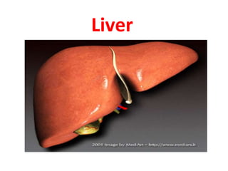
2. Liver
• Largest gland in the body
• Weight: 1.4-1.8 kg in males
• 1.2-1.4 in females
• 150 gm at birth
• Function of the liver
• Secretion of bile & bile salt
• Metabolism of carbohydrate, fat and
protein
• Formation of heparin & anticoagulant
substances
• Detoxication
• Storage of glycogen and vitamins
• Activation of vita .D
4. Surface anatomy of the liver
-The greater part of the
liver is situated under cover
of the right costal margin
- Diaphragm separates it
from the pleura, lungs,
pericardium, and heart.
5.
6. Ant. View of the liver(External features
)
• Right lobe
• Cut edge of the
Falciform ligament
left lobe
• Diverging cut edges
of the superior part
of the coronary
ligament
• Fundus of the gall
bladder
7. Surfaces of the liver, their relations &
impressions
• Diaphragmatic
surface
Superior surface
Anterior surface
Posterior surface
Right Lateral surface
• Postero - inferior
surface= visceral
surface
8. Postero- infero surface= visceral
surface
Relations
• I.V.C
• the esophagus
• the stomach
• the duodenum
• the right colic flexure
• the right kidney
• Rt. Suprarenal gland
• the gallbladder.
• Porta hepatic( bile duct,H.a.H.V)
• Fissure for lig. Venoosum &
lesser omentum
• Tubular omentum
• Lig.teres
9.
10. Lobes of the liver
• Rt. Lobe
• Lt .lobe
• Quadrate lobe
• Caudate lobe
11. Separation of the four lobes of the liver
• Right sagittal
fissure - groove
for IVC and GB
• left sagittal
fissure -
contains the
Lig. Venosum
and Lig. Teres
• Transverse
fissure -porta
hepatis
13. Left Lobe
– Varied in size
– Lies in the epigastric
and left
hypochondriac
regions
14. Lobes of the liver…..cont
Rt. & Lt lobe separated by
• Falciform ligament
• Ligamentum Venoosum
• Ligamentum teres
15. Caudate Lobe
-present in the posterior
surface from the Rt.
Lobe
Relations of caudate
lobe
- Inf. the porta
hepatis
- The right the fossa
for the inferior vena cava
- The left the fossa
for the lig.venosum.
16. Quadrate lobe
Present on the inferior
surface from the Rt. Lobe
Relation
- Sup. porta hepatis
- Rt. fossa for the
gallbladder
- Lt by the fossa for
lig.teres
17. Porta hepatis
-It is the hilum of the liver
-It is found on the
posteroinferior surface
- lies between the caudate
and quadrate lobes
-Lesser omentum attach to
its margin
Contents
Hepatic artery
Portal vein
Rt &ly hepatic duct leaving
the porta hepatis
18. Peritoneum of the liver
• The liver is covered by
peritoneum
(intraperitoneal
organ)except at bare
area(it is origin from
septum transversum)
• Inferior surface covered
with peritoneum of
greater sac except porta
hepatis, G.B & Lig.teres
fissure
• Rt. Lateral surface
covered by peritoneum,
related to diaphragm
which separate it from
Rt. Pleura , lung and the
Rt Ribs (6-11)
19. Postero- infero surface= visceral
surface
Relations
• I.V.C
• the esophagus
• the stomach
• the duodenum
• the right colic flexure
• the right kidney
• Rt. Suprarenal gland
• the gallbladder.
• Porta hepatic( bile duct,H.a.H.V)
• Fissure for lig. Venoosum &
lesser omentum
• Tubular omentum
• Lig.teres
20.
21. Sup. Surface of the liver
• Right & left lobes
• Cut edge of the
Falciform ligament
• The cut edges of
the superior and
inferior parts of
the coronary
ligament
• The left triangular
ligament
• The right
triangular
ligament
22. Relations of Sup . surface of liver
• Diaphragm
• Pleura & lung
• Pericardium &
heart
23. Relations of the liver Anteriorly
• Diaphragm
• Rt & Lt pleura and
lung
• Costal cartilage
• Xiphoid process
• Ant. abdominal wall
24.
25. 1- The Falciform ligament of liver
2- The Ligamentum teres hepatis
3- The coronary ligament
4- The right triangular ligament
5- The left triangular ligament
6- The Hepatogastric ligament
7- The hepatoduonedenal ligament
8- The Ligamentum Venoosum
1. The ligaments of the liver
26. Falciform Ligament
A sickle-shaped fold of
peritoneum connects the AAW
with the liver slightly to the right
of the median plane.
• Ant border: Attached to under
surface of diaphragm & AAW
• Post border: Attached to sup &
ant surfaces of liver
• Free margin connects the
umbilicus to liver it contains the
round ligament of the liver or
Ligamentum teres.
27. • Coronary ligament
the area between upper and
lower layer of the coronary
ligament is the bare area of
liver which contract with the
diaphragm;
• Left and right triangular
ligaments formed by left and
right extremity of coronary
ligament
30. Segmental anatomy of the liver
• Anatomical Lobes
Rt .& Lt. lobes -no
morphological
significance.
Separation by ligaments
(Falciform, lig. Venoosum
& Lig.teres)
• True morphological and
physiological Lobes -line
extends from fossa of GB
to fossa of I.V.C each has
its own arterial blood
supply, venous drainage
and biliary drainage
33. Blood supply of the liver
• Two sources
• Hepatic artery-which deliver oxygenated blood
from general circulation
• Portal vein- delivering deoxygenated blood from
small intestine
34. Vein drainage of the liver
• The portal vein divides
into right and left
terminal branches that
enter the porta hepatis
behind the arteries.
• The hepatic veins (three
or more) emerge from
the posterior surface of
the liver and drain into
the inferior vena cava.
35. Lymphatic drainage of the liver
• Superficial lymphatics-
• from post. Aspect of liver drain in to the post.
Mediastinal lymph node
• From ant. Aspect- drain in to hepatic node
• Deep lymphatics
• Terminate in the nodes around IVC
36. • Nerve supply
• Sympathetic celiac plexuses
• Parasympathetic vagus nerve( anterior part)
• The anterior vagal trunk gives rise to a large hepatic
branch, which passes directly to the liver
37. Liver cirrhosis- hepatocytes sometimes may undergo necrosis
The dead hepatocytes are replaced by fibrous tissue so hepatic fibrosis
clinically termed as LC
38. Fact
• The liver is the only
internal human organ
capable of natural
regeneration of lost
tissue; as little as 25% of a
liver can regenerate into a
whole liver.