
Recommended
More Related Content
Similar to tracheo-oesophagealfistula
Similar to tracheo-oesophagealfistula (20)
Recently uploaded
Recently uploaded (20)
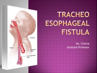
tracheo-oesophagealfistula
- 3. Composed of the nose, the pharynx, and the larynx, the organs of the upper respiratory tract are located outside the chest cavity. Nasal cavity: Inside the nose, the sticky mucous membrane lining the nasal cavity traps dust particles, and tiny hairs called cilia help move them to the nose to be sneezed or blown out. Sinuses: These air-filled spaces along side the nose help make the skull lighter.
- 4. Pharynx: Both food and air pass through the pharynx before reaching their appropriate destinations. The pharynx also plays a role in speech. Larynx: The larynx is essential to human speech.
- 5. Composed of the trachea, the lungs, and all segments of the bronchial tree (including the alveoli), the organs of the lower respiratory tract are located inside the chest cavity. Trachea: Located just below the larynx, the trachea is the main airway to the lungs.
- 6. Lungs: Together the lungs form one of the body’s largest organs. They’re responsible for providing oxygen to capillaries and exhaling carbon dioxide. Bronchi: The bronchi branch from the trachea into each lung and create the network of intricate passages that supply the lungs with air. Diaphragm: The diaphragm is the main respiratory muscle that contracts and relaxes to allow air into the lungs.
- 8. Tracheo-oesophageal fistula and Oesophagal Atresia are the malformation of digestive system, In which oesophagus does not develop properly. The oesophagus is a tube that normally carries food from the mouth to stomach.
- 9. Oesophagal Atresia Oesophagal Atresia is the failure of oesophagus to form a continuous passage from the pharynx to the stomach. Tracheo-oesophageal fistula Tracheo-oesophageal fistula is an abnormal connection between the trachea and the oesophagus.
- 10. Tracheo-oesophageal fistula occurs in 1 in 3500 births, with slight male dominance. Approximately 50% of neonates with oesophagus atresia or Tracheo-oesophageal fistula have other anomalies also. Usually cardiac anomalies are seen in 14.7 – 28 % cases of TEF.
- 11. CONGENITAL TEF The cause of Tracheo-oesophageal fistula and Oesophagal Atresia is still unknown.
- 12. ACQUIRED TEF Iatrogenic injury Blunt chest or neck trauma Prolonged mechanical ventilation via endotracheal or tracheotomy tube
- 13. VATER V- Vertebral defect A- Imperforate Anus T- Tracheoesophageal fistula R- Renal dysplasia
- 14. VACTERAL V- Vertebral A- Anal C- Cardiac T- Tracheal E- Esophageal R- Renal L- Limb
- 15. Type A:Esophageal atresia without fistula In this type, there is Oesophagal Atresia and proximal and distal segments of oesophagus are blind. There is no communication between trachea and oesophagus. This type is present in 3-7 % of cases.
- 16. Types B: Esophageal atresia with fistula In this type, Oesophagal Atresia is present and the blind proximal segment of oesophagus connects with trachea by a fistula. The distal end of oesophagus is blind. This type is present in 0.8 % cases.
- 17. Type C:Distel esophageal fistula with atresia In this type, Oesophagal Atresia is present. The proximal end of oesophagus is a blind pouch and distal segment of oesophagus is connected by fistula to trachea. This is the commonest type, present in about 87 % cases.
- 18. Type D:Proximal and distel esophageal fistula with atresia It is the rarest type that occurs in 0.7 % cases. In this type, both upper and lower segments of oesophagus communicate with trachea.
- 19. Type E: H’ type In this type, oesophagus and trachea are normal and completely formed but are connected by a fistula.
- 20. Three Cs Coughing Choking Cyanosis Apnea Increased respiratory distress during feeding Abdominal distension
- 21. Ultrasound examination:- reveals polyhydramnios, absence of a fluid-filled stomach, a small abdomen, lower-than-expected fetal weight, and a distended esophageal pouch. Fetal MRI:- may be used to confirm the presence of EA/TEF
- 22. X-ray taken with radiopaque catheter placed in esophagus to check for obstruction; standard chest X- ray shows a dilated air-filled upper esophageal pouch and can demonstrate pneumonia. Inability to pass a NG tube into stomach because it meets resistance:; Bronchoscopy visualizes fistula between trachea and esophagus; Abdominal ultrasound and echocardiogram to check for cardiac abnormalities.
- 23. The management of trachea-oesophageal fistula is mainly surgical. Surgical intervention depends on the distance between proximal and distal pouch of oesophagus, type of defect, condition of neonate and his weight.
- 24. If distance between upper and lower oesophageal segments is less than 2.5 cm if the condition of infant is good, primary repair is done by division and ligation of the fistula along with end-to-end anastomosis of proximal and distal segments of oesophagus.
- 28. Pre-Operative Risk for aspiration related to structural abnormality. Risk for deficient fluid volume related to inability to take oral feeds. Impaired breathing related to frequent laryngospasm and excessive secretions in the trachea.
- 29. Post-Operative Ineffective airway clearance related to disease process. Impaired nutrition related to surgery. Altered comfort related to surgical process.
- 30. A nasogastric tube is put in the upper oesophageal segment and is aspirated frequently, to prevent collection and aspiration of secretion into the trachea. Intravenous fluids are administered to maintain the hydration status of infant. Place the infant in semi-upright position and administer oxygen if cyanosis is present.
- 31. A cervical oesophagostomy is made to drain out secretions from blind oesophageal pouch and administer oxygen to infant. Feed the infant orally or by gastrostomy. Position the baby comfortably in semi-fowler’s position and administer analgesics.
- 32. Tracheomalacia (weakness of tracheal wall) Anastomotic leak (tension) Strictures (narrowing, esophageal dilation) Dysphagia (esophageal motility disorder) Respiratory distress Gastro-esophageal reflux.