Female breast anatomy for nursing students. Female breast anatomy for nursing students. Female breast anatomy for nursing students. Female breast anatomy for nursing students Female breast anatomy for nursing students Female breast anatomy for nursing students Female breast anatomy for nursing students Female breast anatomy for nursing students Female breast anatomy for nursing students Female breast anatomy for nursing students Female breast anatomy for nursing students Female breast anatomy for nursing students Female breast anatomy for nursing students Female breast anatomy for nursing students Female breast anatomy for nursing students Female breast anatomy for nursing students Female breast anatomy for nursing students Female breast anatomy for nursing students Female breast anatomy for nursing students Female breast anatomy for nursing students Female breast anatomy for nursing students Female breast anatomy for nursing students Female breast anatomy for nursing students Female breast anatomy for nursing students Female breast anatomy for nursing students Female breast anatomy for nursing students Female breast anatomy for nursing students Female breast anatomy for nursing students Female breast anatomy for nursing students Female breast anatomy for nursing students Female breast anatomy for nursing students Female breast anatomy for nursing students Female breast anatomy for nursing students Female breast anatomy for nursing students Female breast anatomy for nursing students Female breast anatomy for nursing students Female breast anatomy for nursing students Female breast anatomy for nursing students Female breast anatomy for nursing students Female breast anatomy for nursing students Female breast anatomy for nursing students Female breast anatomy for nursing students Female breast anatomy for nursing students Female breast anatomy for nursing students Female breast anatomy for nursing students Female breast anatomy for nursing students Female breast anatomy for nursing students Female breast anatomy for nursing students Female breast anatomy for nursing students Female breast anatomy for nursing students Female breast anatomy for nursing students Female breast anatomy for nursing students Female breast anatomy for nursing students Female breast anatomy for nursing students Female breast anatomy for nursing students Female breast anatomy for nursing students Female breast anatomy for nursing students Female breast anatomy for nursing students Female breast anatomy for nursing students Female breast anatomy for nursing students Female breast anatomy for nursing students Female breast anatomy for nursing students Female breast anatomy for nursing students Female breast anatomy for nursing students Female breast anatomy for nursing students Female breast anatomy for nursing students Female breast anatomy for nursing students Female breast anatomy for nursing students Female breast anatomy for nursing students Female breast anatomy for nurs
6. • Modified sweat gland- modified apocrine gland
• Fat accounts for its smooth contour and most of its bulk.
• The resting (non-lactating) breast, however consists mostly of
fibrous & fatty tissue; variations in size are due to variations in
fat content, not glandular tissue which is very sparse.
• During pregnancy alveoli bud off from the smaller ducts & the
organ usually enlarges significantly, & more so in preparation
for lactation.
• When lactation ceases there is involution of secretory tissue.
• After menopause progressive atrophy of lobes & ducts takes
place.
• MALE BREAST
• Resembles the rudimentary female breast
• has NO lobules or alveoli.
• The small nipple and areola lie over the 4th intercostal space
8. Parts, Shape & position of the Gland
• It is conical in shape.
• It lies in superficial
fascia of the front of
chest.
• It has a base, apex
and tail.
• Its base extends from
2nd to 6th ribs.
• It extends from the
sternum to the
midaxillary line
laterally.
• It has no capsule.
9. SHAPE AND POSITION OF FEMALE BREAST
• 2/3 of its base lies
on the pectoralis
major muscle, while
its inferolateral 1/3
lies on:
• Serratus anterior &
• External oblique
muscles.
• Its superolateral
part sends a process
into the axilla called
the axillary tail or
axillary process
up to the 3ed rib
10. • Nipple:
• It is a conical eminence that
projects forwards from the
anterior surface of the breast.
• The nipple lies opposite 4th
intercostal space.
• It carries 15-20 narrow pores
of the lactiferous ducts.
SHAPE AND POSITION OF FEMALE BREAST
11. SHAPE AND POSITION OF FEMALE BREAST
• Areola :
• It is a dark pink brownish
circular area of skin that
surrounds the nipple.
• Lubricated by the areolar
glands of Montgomery
• large,
• modified sebaceous glands
→may form sebaceous cysts
→ may infected
• The subcutaneous tissues of
nipple & areola are devoid of
fat.
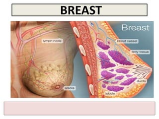
12. STRUCTURE OF MAMMARY GLAND
• It is non capsulated gland.
• It consists of lobes and lobules
which are embedded in the
subcutaneous fatty tissue of
superficial fascia.
• It has fibrous strands
(ligaments of cooper) which
connect the skin with deep
fascia of pectoralis major.
• It is separated from the deep
fascia covering the underlying
muscles by a layer of loose
areolar tissue which forms the
retromammary space. (allows
the breast to move freely).
13. STRUCTURE OF MAMMARY GLAND
• It is formed of 15-20 lobes.
• Each lobe is formed of a
number of lobules.
• The lobes and lobules are
separated by interlobar and
interlobular fibrous & fatty
tissue, called ligaments of
Cooper. These ligaments give the
breasts support by connecting the
skin of the breasts to the pectoralis
muscles below them.
• It has from 15-20 lactiferous
ducts which open by the
same number of openings on
the summit of the nipple.
14. ARTERIAL SUPPLY
• 1. Perforating
branches of internal
thoracic (internal
mammary) artery.
• 2. Mammary
branches of lateral
thoracic artery.
• 3. Mammary
branches of
Intercostal arteries.
15. VENOUS SUPPLY
• Veins are
corresponding to
the arteries.
• Circular venous
plexus are found
at the base of
nipple.
• Finally, veins of
this plexus drain
into axillary &
internal thoracic
veins.
16. AXILLARY LYMPH NODES
• They are arranged into 5 groups which
lie in axillary fat :
• Pectoral (Anterior) group : which lies
on the pectoralis minor along lateral
thoracic vessels.
• Subscapular (Posterior) group : which
lies on posterior wall of axilla on lower
border of subscapularis along
subscapular vessels.
• Brachial (Lateral) group : lies on lateral
wall of axilla along 3rd part of axillary
vessels.
• Central group : lies in axillary fat at the
base of axilla.
• Apical group : lies at apex of axilla.
• Subclavian lymph trunk:
• it is formed by union of efferent lymph
vessels of apical group. It usually
opens in subclavian vein. On the left
side it usually opens into thoracic duct.
17. LYMPHATIC DRAINAGE
• Subareolar lymphatic
plexus :
• Lies beneath the areola
(the plexus of Sappey).
• Deep lymphatic plexus:
• Lies on the deep fascia
covering pectoralis
major.
• Both plexuses radiate in
many directions and
drain into different
lymph nodes.
18. LYMPHATIC DRAINAGE
• Central & lateral parts of the
gland (75%) drain into pectoral
group of axillary lymph nodes.
• Upper part of the gland drains
into apical group of axillary
lymph nodes.
• Medial part drains into internal
thoracic (parasternal) lymph
nodes, forming a chain along the
internal thoracic vessels.
• Some lymphatics from the medial
part of the gland pass across the
front of sternum to anastomose
with that of opposite side.
• Lymphatics from the inferomedial
part anastomose with lymphatics
of rectus sheath & linea alba, and
some vessels pass deeply to
anastomose with the sub
diaphragmatic lymphatics.
19.
20. APPLIED ANATOMY- CANCER BREAST
• It is a common surgical condition.
• 60% of carcinomas of breast occur
in the upper lateral quadrant.
• 75% of lymph from the breast
drains into the axillary lymph
nodes.
• In case of carcinoma of one
breast, the other breast and the
opposite axillary lymph nodes are
affected because of the
anastomosing lymphatics between
both breasts.
• In patients with localized cancer
breast, a simple mastectomy,
followed by radiotherapy to the
axillary lymph nodes is the
treatment of choice.
21. Applied Anatomy
• The lactiferous
ducts are radially
arranged from the
nipple, so incision
of the gland should
be made in a radial
direction to avoid
cutting through the
ducts.
• Infiltration of the
ligaments of
Cooper by breast
cancer leads to its
shortening giving
peau de’orange
appearance of the
breast.
22. Mammary ridge
• Mammary ridge
extends from the axilla
to the inguinal region.
• In human, the ridge
disappears EXCEPT for a
small part in the
pectoral region.
• In animals, several
mammary glands are
formed along this ridge.
23.
24. • Fibrocystic Breast
-Breast tissue responds to fluctuating
levels of hormones,
especially estrogen
• Cause;
-Changes in the breasts may include:
• an overgrowth of cells that line
the milk ducts
• an increase in fibrous tissue
• the formation of cyst
• People who develop fibrocystic
changes may be more sensitive to
hormonal fluctuations during the
menstrual cycle. It is common for
symptoms to become more
bothersome right before or
during a menstrual period.
• Common cause of ‘lumpy breast’