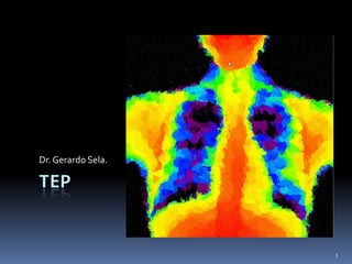
Tromboembolia pulmonar
•
3 likes•1,461 views

Minipresentacion de Tromboembolia pulmonar, algunos casos, y citas.

Recommended
More Related Content
What's hot
What's hot (20)
Viewers also liked
Viewers also liked (13)
Similar to Tromboembolia pulmonar
Similar to Tromboembolia pulmonar (20)
More from gerardo sela
More from gerardo sela (20)
Recently uploaded
Recently uploaded (20)
Tromboembolia pulmonar
- 2. Signos radiologicos Disnea subita Asintomatica 2
- 3. 3
- 4. Rxlucidez unilateral Mastectomia Polland Amastia Enfisema lobar unil. C. extrano 4
- 5. HAP Dilatacion del tronco de la pulmonar Engrosamiento de la Arteria interlobar derecha Disminucion de la vasculirdad periferica Crecimiento de VD 5
- 6. Masc 75 disnea y taquicardia 6
- 7. 7
- 8. 8
- 9. 9
- 10. Chest x-ray: The lungs are clear. No focal infiltrate or pleural effusion. V/Q scan: Ventilation: There is delayed washout of radiotracer in the left lung base. There is normal ventilation and washout in the right lung. Perfusion: There is a wedge shaped defect in the lateral segment of the right middle lobe. 10
- 11. The presenting signs and symptoms of pulmonary embolism are nonspecific and mimic that of other cardiopulmonary disease. CXR has low sensitivity and specificity in diagnosing PE and is used to rule out other processes that mimic PE. Mismatched, wedge shaped, segmental ventilation-perfusion defects raises the suspicion of PE. Probability of PE is based on PIOPED II criteria. COPD, pleural effusion, hilar masses, 1.Mettler, F.A., Guiberteau, M.J. Essentials of Nuclear Medicine Imaging. New York: Saunders, 2006. pulmonary hypoplasia and many other 2.Han, D., et al. Thrombotic and Nonthrombotic Pulmonary Arterial Embolism: Spectrum of Imaging. Findings. RadioGraphics 2003; 23:1521–1539. 3.Sabatine, M., et al. Pocket Medicine. New York: Lippincott, 2004. 11
- 12. M 70 12
- 13. 13
- 14. EVP TEP 14
- 15. Fem sobrepeso, disnea ANGIOTAC PULMONAR 30 DIAS DESPUES 15
- 16. Dual-Energy CT: Clinical Applications in Various Pulmonary Diseases1 RadioGraphics 2010; 30:685–698 16
- 19. Aquilion PRIME, the next generation 160- slice multi detector CT scanner 19