2. 2476
FUJIWARA ET AL.
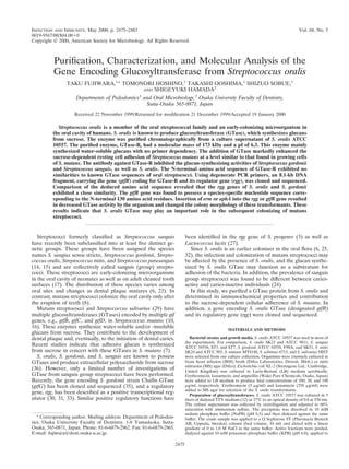
INFECT. IMMUN.
FIG. 2. Physical characteristics of GTase-R. (A) SDS-PAGE of GTase preparations at different stages of purification. Lanes: 1, culture supernatant; 2,
ammonium sulfate precipitate; 3, pooled active fractions from Q-ion-exchange
chromatography; 4, pooled active fraction from CHT-10 hydroxylapatite chromatography; M, molecular mass markers. (B) Isoelectric focusing-PAGE of
GTase-R. Lane: 1, pI markers (3.50 to 8.65); 2, Purified GTase-R visualized by
PAS staining. (C) Effect of pH on GTase activity.
FIG. 1. Chromatographic purification of GTase-R from S. oralis ATCC
10557. (A) Separation of ammonium sulfate-precipitated GTase (60% saturation) by anion-exchange chromatography on a Q Sepharose FF column (bed
volume, 10 ml). GTase was eluted with a linear gradient of 0 to 0.3 M NaCl. (B)
Further purification of GTase-R containing fractions from the elution profile
shown in panel A on a Bio-Scale CHT10-I column (bed volume, 10 ml). Elution
was done with a 10 to 500 mM KPB linear gradient. A280, optical density at 280
nm.
a Bio-Scale CHT10-I column (bed volume, 10 ml; Bio-Rad Laboratories, Hercules, Calif.), and then eluted with a 10 to 500 mM KPB linear gradient.
GTase samples from other streptococci were obtained from the culture supernatants of test strains by 50% saturation ammonium sulfate precipitation.
Cell-associated GTase (CA-GTase) was extracted from centrifuged cells of S.
mutans with 8 M urea followed by ammonium sulfate precipitation (11).
Generation of antiserum. Antisera were prepared by repeated intramuscular
injections of rabbits with the purified GTase from S. oralis ATCC 10557 suspended in Freund’s complete adjuvant (Difco) followed by immunization with
the antigen suspended in Fruend’s incomplete adjuvant (Difco). The antibody to
S. oralis GTase was purified from rabbit antiserum by repeated 33% saturation
with ammonium sulfate.
Glucan synthesis assay. GTase activity was determined using [glucose-14C]
sucrose with or without primer dextran T10, as described previously (11). Briefly,
reaction mixtures composed of GTase, 10 mM [glucose-14C]sucrose (11.47 GBq/
mmol), and 0 or 20 M dextran T10 in 20 l of 50 mM KPB (pH 6.0) were
incubated for 1 h at 37°C, spotted on a filter paper square (1.5 by 1.5 cm), and
dried in air. The filters were washed with methanol or distilled water and then
immersed in scintillation fluid to estimate the amount of total [14C]glucan or
water-insoluble [14C]glucan. Kinetic constants were determined by LineweaverBurk analyses of the glucan synthesis rates.
Determination of pI and optimum pH. The pI was determined by analytical
isoelectric focusing using a PhastSystem (Pharmacia) with a PhastGel IEF3-9
(Pharmacia). After electrophoresis, the gel was incubated for 1 h at 37°C in 10
mM NaPB (pH 6.5) containing 5% sucrose, 2% Triton X-100, and 0.05% NaN3.
The enzyme activity was visualized by periodic acid-Schiff staining. The optimum
pH of GTase was determined by measuring the GTase activity in 50 mM KPB
(pH 5.0 to 7.5).
SDS-PAGE and Western blotting. Sodium dodecyl sulfate-polyacrylamide gel
electrophoresis (SDS-PAGE) and Western blot analyses were carried out as
described previously (9). Briefly, GTase samples and E. coli cells carrying the
recombinant plasmid were suspended in SDS gel-loading buffer (26) and boiled
for 5 min. Proteins separated by SDS-PAGE were transferred onto a polyvinylidene difluoride membrane (Immobilon; Millipore). After being blocked with 5%
bovine serum albumin, the membrane was reacted with the rabbit antibody to S.
oralis GTase at 37°C for 1 h, and the antibody which was bound to the protein
band(s) was detected by a solid-phase immunoassay.
Effects of S. oralis GTase on the sucrose-dependent adhesion of S. mutans
resting cells. S. mutans strain MT8148 cells grown in BHI broth were washed at
0°C with 0.1 M KPB (pH 6.0) containing 0.05% NaN3. The centrifuged cells were
resuspended in the same buffer containing 1% sucrose and then adjusted to an
optical density of 1.0 at 550 nm. Aliquots (3 ml) of the cell suspension were mixed
with various amount of S. oralis GTase and incubated at 37°C for 18 h at a 30°
angle. Next, the culture tubes were vigorously vibrated with a Vortex mixer for
3 s. The degree of cell adhesion was determined by reading the optical density at
550 nm and expressed as the percentage of total cell mass. To assess the adhesion
of S. mutans growing cells, the organism was grown at 37°C for 18 h at a 30° angle
in BHI broth containing 1% sucrose. The percent adhesion was determined as
described above.
Amino acid sequence. S. oralis GTase was subjected to SDS-PAGE and blotted
onto ProBlott membranes (Applied Biosystems, Foster City, Calif.). The GTase
band was excised from several lanes and subjected to sequencing using an ABI
477A/120A protein sequencer (Applied Biosystems).
TABLE 1. Purification of S. oralis GTase
Preparationa step
Total amt of protein
(mg)
Total GTase activity
(U)
GTase sp act
(U/mg)
Recovery
(%)
Purification
(fold)
Culture supernatant
Ammonium sulfate precipitation
Q Sepharose fraction
CHT-I fraction
736
180
5.5
0.3
140
117
4.0
2.4
0.19
0.65
0.72
8.00
100
84.0
2.9
1.7
1
3.4
3.8
42.0
a
S. oralis ATCC 10557 was grown in 5 liters of dialyzed TTY medium to an optical density of 0.8 at 550 nm. The culture supernatant was concentrated by a 60%
saturation of ammonium sulfate. The enzyme fraction was purified on a Q Sepharose FF column followed by a Bio-Scale CHT10-I column.
3. GLUCOSYLTRANSFERASE OF S. ORALIS
VOL. 68, 2000
2477
TABLE 2. Effects of anti-S. oralis ATCC 10557 GTase antibody on
the GTase activities of various oral streptococci
GTase activity (dpm)b
GTase origina
Without antiGTase-R
With antiGTase-R
% Inhibition
S. oralis
ATCC 10557
SK23
ATCC 9811
4,930.3 Ϯ 12.8
4,997.9 Ϯ 49.7
5,022.0 Ϯ 37.6
831.8 Ϯ 14.2
1,065.1 Ϯ 42.2
1,128.8 Ϯ 36.7
83.1
78.7
77.5
S. sanguis
ATCC 10556
ST3
ST7
5,000.0 Ϯ 100.0 1,305.4 Ϯ 59.5
5,027.1 Ϯ 25.8 1,244.4 Ϯ 36.1
5,053.5 Ϯ 21.8 1,243.8 Ϯ 29.6
73.9
75.3
75.4
S. gordonii
ATCC 10558
90A
SK51
4,975.8 Ϯ 37.9
4,950.2 Ϯ 32.9
4,956.5 Ϯ 40.9
949.4 Ϯ 23.3
1,468.6 Ϯ 30.9
1,007.3 Ϯ 58.9
80.9
70.3
79.7
S. mutans
MT8148 cell-associated
MT8148 cell-free
4,996.4 Ϯ 53.3
4,964.7 Ϯ 72.5
4,687.9 Ϯ 63.8
3,431.3 Ϯ 61.6
6.2
30.9
S. sobrinus
6715
5,006.9 Ϯ 138.4 3,732.1 Ϯ 194.9
25.5
S. sobrinus
HHT
4,989.9 Ϯ 66.9
26.6
3,662.2 Ϯ 76.5
a
GTase was from the concentrates of test strain culture supernatants, except
for the cell-associated GTase from S. mutans. The cell-associated GTase was
extracted from the cells of S. mutans with 8 M urea.
b
The GTase fraction (1 mU) was reacted with the antibody to GTase-R (32 g
of protein) at 37°C for 30 min or left unreacted. The reaction mixture was then
incubated with [glucose-14C]sucrose at 37°C for 1 h, and the amount of synthesized [14C]glucan was measured. Data are expressed as means Ϯ standard deviations of triplicate experiments.
DNA manipulations. Restriction enzymes, ligase, and other DNA-modifying
enzymes were purchased from New England Biolabs (Beverly, Mass.) or Takara
(Kyoto, Japan). Manipulations of DNA with these enzymes were performed as
recommended by the manufacturers. All other DNA manipulations were carried
out using standard protocols (26).
Chromosomal DNA isolation and Southern blot analysis. Organisms were
grown in BHI broth for 18 h at 37°C, collected, and then washed by centrifugation. Cells (750 mg [wet weight]) were suspended in 5 ml of 50 mM NaCl–10 mM
Tris-HCl (pH 7.4) and then digested with mutanolysin (0.25 mg/ml; Dainippon
Pharmaceutical Co., Osaka, Japan) for 1 h at 50°C, and N-lauroyl sarcosine (final
concentration, 1.5%) and EDTA (final concentration, 10 mM) were added to
lyse the cells. The lysate was treated with RNase (0.3 mg/ml; Wako) and proteinase K (0.3 mg/ml; Merck, Darmstadt, Germany). The DNA was purified from
the cell lysate by phenol and phenol-chloroform extractions and then collected by
ethanol precipitation.
Southern blot analysis was carried out as a standard procedure. Briefly, chromosomal DNA from the test organisms was digested with EcoRI, separated by
electrophoresis on a 0.8% agarose gel, and transferred onto a nylon membrane
(Hybond-N; Amersham, Little Chalfont, United Kingdom). Next, the DNA was
cross-linked to the membrane by UV radiation. A 397-bp DNA fragment corresponding to positions 54 to 186 in the deduced amino acid of the gtfR gene was
amplified by PCR and used as a probe. The membrane was then hybridized
stringently with the 32P labeled probe.
PCR. PCR was performed in reaction mixtures containing 50 mM KCl, 10 mM
Tris-HCl (pH 8.3), 1.5 mM MgCl2, 200 M deoxyribonucleoside triphosphate,
1.0 M primer, template DNA (Ͻ10 ng/l), and AmpliTaq Gold DNA polymerase (0.025 U/l; Applied Biolystems). Amplification was performed in a
Gene AmpPCR System 2400 apparatus (Perkin-Elmer) as specified by the manufacturer. Degenerate PCR was performed as follows: a preincubation step at
95°C for 9 min followed by 30 cycles of a denaturation step at 94°C for 30 s, a
primer-annealing step at 36°C for 30 s, and an extension step at 60°C for 30 s.
Long PCR was performed using a TaKaPa LA PCR kit Ver 2.1 (Takara), as
recommended by the manufacturer.
Cloning and sequencing of the GTase gene. Two sets of genomic libraries were
constructed by cloning EcoRI- or KpnI-digested S. oralis ATCC 10557 chromosomal DNA into plasmid pMW119 (Nippon Gene) or pUC19 (Takara) and then
transforming them into competent E. coli. In addition, a vector named pGEM-T
Easy (Promega, Madison, Wis.) was used for cloning of the PCR products.
FIG. 3. Effects of GTase-R on the cellular adhesion of S. mutans. S. mutans
cells grown in BHI broth were resuspended at 0°C in 0.1 M KPB (pH 6.0) with
0.05% NaN3 containing 1% sucrose. The cell suspension was mixed with increasing amounts of GTase-R and incubated at 37°C for 18 h at a 30° angle (resting
cells). As a positive control, S. mutans was grown in BHI broth with 1% sucrose
at 37°C for 18 h at a 30° angle (growing cells). Numbers of adhesive cells were
then determined. Data are expressed as means and standard deviations of triplicate experiments. The asterisks indicate statistical significance (P Ͻ 0.01) from
the sucrose value for resting cells incubated in sucrose containing KPB without
GTase-R.
For a DNA-sequencing template, plasmid DNA and PCR products were
prepared using a Wizard Plus Minipreps DNA purification system (Promega)
and a Centricon 100 spin column (Millipore, Bedford, Mass.), respectively. The
dideoxy dye termination reaction was performed with an ABI PRISM cyclesequencing kit (Perkin Elmer) in a GeneAmp 2400 thermal cycler. The products
were then analyzed using an automated DNA sequencer model 373 (Applied
Biosystems). The homology search, multiple-sequence alignment, and phylogenetic tree creation were performed with the BLAST, FASTA, and CLUSTAL W
programs on the DDBJ “supernig” computer system.
Expression of recombinant GTase. E. coli carrying a recombinant plasmid was
grown in LB broth (3 ml) to an optical density of 0.6 at 550 nm. Cells were
collected by centrifugation, suspended in 100 l of 10 mM NaPB (pH 6.0), and
disrupted by sonication. The sonic supernatant was then separated and examined
for glucan synthesis.
Transformation of S. oralis. S. oralis was subjected to transformation as reported previously (9). Briefly, the recipient organisms were cultured in ToddHewitt broth (Difco) supplemented with 10% heat-inactivated horse serum
(Gibco, Grand Island, N.Y.) for 18 h. The culture was diluted 1:40 with the broth
(10 ml) and then incubated for another 1.5 h at 37°C, and the donor DNA was
added to a final concentration of 25 g/ml. The culture was further incubated for
2 h, concentrated approximately 10-fold by centrifugation, and then spread on
MS agar plates containing antibiotics. The plates were incubated in a CO2
incubator for 2 to 3 days at 37°C, and possible transformant colonies were picked
up for further examinations.
Construction of the insertional mutants. Recombinant plasmid pYT303 or
pYT311 carrying the 830- or 1,070-bp fragment of the erythromycin resistance
gene (erm) from pVA838 (20) or the kanamycin resistance gene (aphA) from
transposon Tn1545 (2) was used. A subclone, pTHR8, carrying a 1.5-kb SphI-PstI
insert containing the rgg gene was generated from pTH171. A 2.5-kb DNA
fragment containing the center portion of the gtfR gene was amplified by PCR
and cloned to generate pTH808. pTHR8 or pTH808 was restricted with ApaI or
HindIII to be linear at a unique site. The linear plasmid was then blunted and
ligated with the erm or aphA cassette to yield pTHR805 or pTH818. After being
made linear at the unique PstI site, the plasmid was introduced into S. oralis
ATCC 10557 by transformation to allow an allelic exchange.
Statistical analysis. Differences between S. oralis GTase concentrations and S.
mutans resting-cell adhesion were determined by analysis of variance with subsequent use of the Tukey-Kramer multiple-comparisons test. Significance levels
were taken at P Ͻ 0.01.
4. 2478
FUJIWARA ET AL.
INFECT. IMMUN.
FIG. 4. Genetic map and cloning strategy of the rgg and gtfR genes.
Nucleotide sequence accession numbers. The nucleotide sequences of the rgg
and gtfR gene have been deposited in the DDBJ database under accession no.
AB025228.
RESULTS
Purification of S. oralis GTase. GTase was purified from the
culture supernatant of S. oralis ATCC 10557 by ammonium
sulfate precipitation followed by anion-exchange and hydroxylapatite chromatography (Fig. 1). The recovery of the purified
GTase preparation was 1.7%, and the degree of purification
was 42-fold. The specific activity of the purified enzyme,
FIG. 5. Western blot analysis and glucan synthesis activity of recombinant
GTase-R. (A) The recombinant proteins and native GTase-R were separated by
SDS-PAGE and blotted onto a polyvinylidene difluoride membrane. The blot
was reacted with the antibody to GTase-R. (B) The sonic supernatants of E. coli
cells carrying the recombinant plasmid were used for the glucan synthesis assay.
GTase activity was determined as the amount of [14C]glucan synthesized from
[glucose-14C]sucrose. Data are expressed as means and standard deviations of
triplicate experiments.
GTase-R, was 8.0 mU/g of protein (Table 1). SDS-PAGE of
GTase-R gave a single protein band with a molecular mass of
173 kDa. The optimum pH and pI values were 6.5 and 6.3,
respectively (Fig. 2). The Km value was determined to be 2.49
mM. Glucan synthesized by GTase-R from sucrose was largely
water soluble (89.7%), and its production was not enhanced in
the presence of the primer dextran T10.
Immunological properties of GTase-R. Western blot analyses revealed that the rabbit antibody to GTase-R reacted
strongly with GTase preparations from other sanguis streptococci and cell-free GTase (CF-GTase) but not with CA-GTase
from S. mutans. Further, the enzyme activity of GTase-R was
markedly inhibited by the antibody to GTase-R. The antibody
strongly inhibited S. sanguis and S. gordonii GTase, as well as
S. mutans CF-GTase. S. sobrinus and S. salivarius GTases were
only weakly inhibited, while S. mutans CA-GTase was not
affected by the antibody (Table 2). The inhibition of glucan
synthesis exhibited a similar pattern to the reactivity when
analyzed by Western blotting.
Effects of GTase-R on the adhesion of S. mutans. The effects
of GTase-R on the sucrose-dependent adhesion of S. mutans
resting cells are shown in Fig. 3. Cells incubated without
GTase-R adhered to the glass surface only loosely, and approximately 60% of the cells were easily removed by vibration.
However, the addition of a small amount of GTase-R (1 mU/
ml) resulted in firm adhesion of the S. mutans cells. This
adhesion was as strong as that of the S. mutans cells grown in
sucrose-containing medium.
Amino acid sequencing of GTase-R. The N-terminal amino
acid sequence of GTase-R was determined to be DDVKQVV
VQEPATAQTSGPGQQ. This sequence did not show any
similarity to other reported sequences, including GTases from
S. gordonii and other oral streptococci, by BLAST and FASTA
homology searching.
Cloning of the GTase-R gene (gtfR) by PCR with degenerate
primers. A schematic diagram of the GTase-R gene and cloning strategy is shown in Fig. 4. PCR was done using the degenerated oligonucleotides corresponding to the N-terminal
amino acid sequence VKQVVV (5ЈGTNAARCARGTNGTN
GT3Ј, forward primer) and GPGQQ (5ЈYTGYTGNCCNG
GNCC3Ј, reverse primer). A 60-bp gene fragment was cloned
into a pGEM-T Easy vector, resulting in the recombinant plasmid pTHN01. The sequence of the insert of pTHN01 was
consistent with the N-terminal amino acid sequence. Then a
60-mer oligonucleotide primer (5ЈGTAAAGCAGGTTGT
AGTTCAAGAACCTGCTACAGCTCAGACTAGTGGTC
CCGGTCAGCAA3Ј) was synthesized and used for hybridization. The E. coli transformants carrying the EcoRI-digested
5. VOL. 68, 2000
GLUCOSYLTRANSFERASE OF S. ORALIS
2479
FIG. 6. DNA sequence and deduced amino acid sequence of the rgg and gtfR genes. The putative promoter (Ϫ10 and Ϫ35) and ribosome binding sites (SD) are
shown. The sequence corresponding to the identified N-terminal amino acid sequence is marked. Regions of dyad symmetry are indicated by arrows.
insert were screened by colony hybridization with this primer.
The recombinant plasmids pTH121 and pTH171, with 1.4- and
6.1-kb S. oralis chromosomal inserts, respectively, were isolated.
A sequenced analysis of pTH171 revealed that an open
reading frame (ORF) composed of the 861-bp nucleotide was
present, and an incomplete reading frame without the termination codon was identified 89 bp downstream of the ORF.
This frame encoded the N-terminal amino acid sequence of
GTase-R; however, the molecular mass of the recombinant
protein expressed by pTH171 was only 128 kDa (Fig. 5). To
clone the C-terminal region, the KpnI-digested library was
screened and pTH181, carrying a 1.8-kb insert with a termination codon, was obtained. The complete nucleotide sequence
of the gtfR gene was determined by reconstruction of the DNA
sequences from the inserts of pTH171 and pTH181. To confirm the sequence, primers for amplification of the whole gtfR
gene were synthesized and long PCR amplification was performed. The PCR product was cloned into a pGEM-T Easy
vector to yield pTH275. The recombinant protein expressed by
pTH275 had a molecular mass of 175 kDa and glucan-synthesizing activity (Fig. 5).
Nucleotide sequences. The nucleotide sequence determined
in this study is shown in Fig. 6. The ORF in pTH171 was
composed of 861 bp and encoded a polypeptide of 287 amino
acids. This gene exhibited a high degree of homology (74%) to
the rgg gene of S. gordonii (accession no. M89776), which has
been reported to be a regulatory gene of GTase. We therefore
designated it rgg. A multiple alignment of the deduced amino
acid sequence of rgg revealed that the rgg gene of S. oralis
exhibited a 76% homology to the S. gordonii rgg gene, whereas
the homology of S. oralis rgg to the S. pyogenes and L. lactis rgg
genes was only 21%.
The gtfR gene was composed of 4,728 bp, which encoded a
polypeptide composed of 1,575 amino acids with a predicted
molecular mass of 177 kDa and a pI of 5.58. The alignment of
the deduced amino acid sequence of the gtfR gene and the gtfG
gene encoding S. gordonii GTase is shown in Fig. 7. The gtfR
gene displayed 79.9% homology to the gtfG gene. The Nterminal sequence from amino acids 55 to 186, deduced from
the nucleotide sequence data of gtfR, was completely different
from that of other streptococcal species. These findings were
supported by Southern hybridization analyses, which indicated
that no hybridized bands were detected, except for strains of S.
oralis, when the PCR-amplified DNA fragment corresponding
to the N-terminal 140 amino acid residues of gtfR was used as
a probe (Fig. 8).
Inactivation of the rgg or gtfR gene. Inactivation of the chromosomal rgg or gtfR gene of S. oralis ATCC 10557 was performed by insertion of the erm or aphA gene. The colony
morphology and glucan synthesis activity of representative
transformants are shown in Fig. 9. The rgg mutant had a flat
and dull appearance without the zooglealic zone, while the gtfR
mutant had smaller colonies with a more transparent appear-
6. 2480
FUJIWARA ET AL.
INFECT. IMMUN.
FIG. 7. Alignment of the deduced amino acid sequences of GtfR and GtfG of S. gordonii (accession no. U12643). Identical amino acid sequences are indicated by
shaded boxes.
7. GLUCOSYLTRANSFERASE OF S. ORALIS
VOL. 68, 2000
2481
FIG. 7—Continued.
ance. The GTase activity of both mutants had decreased to
about 10% that of the parent strain.
DISCUSSION
Several methods of comparing the amino acid sequences of
GTases from various oral streptococci have revealed that
GTase possesses two functional domains. One is a catalytic
domain that is composed of approximately 800 amino acid
residues located in the N-terminal region, while the other is a
glucan binding domain that includes a large number of repeated units in the C-terminal region. The former contains
common putative active-site peptides involved in sucrose hydrolysis (22, 28). Molecular modeling analyses have suggested
that the core region contains a cyclically permuted form of the
(␣/)8-barrel structure (4, 19). Recently, circular dichroism
analysis has verified the presence of that structure (21). Direct
repeats of the glucan binding domain are also found in the
C-terminal region of the ligand binding proteins in some grampositive organisms, including S. mutans glucan binding protein,
Clostridium difficile ToxA and ToxB, and lysins from S. pneumoniae and its bacteriophage (37). Structure-function relationship studies have revealed that GTases synthesizing waterinsoluble and/or water-soluble glucans exhibit an almost
identical amino acid sequence at the putative active sites. The
8. 2482
FUJIWARA ET AL.
FIG. 8. Southern blot analysis of chromosomal DNA from various oral streptococci digested with EcoRI. As a probe, the PCR-amplified DNA fragment
corresponding to the N-terminal sequence of gtfR was used. Lanes: 1, S. mutans
MT8148; 2, S. sobrinus 6715; 3, S. salivarius HHT; 4, S. sanguis ATCC 10556; 5,
S. sanguis ST3; 6, S. sanguis ST7; 7, S. oralis ATCC 10557; 8, S. oralis SK23; 9, S.
oralis ATCC 9811; 10, S. gordonii ATCC 10558; 11, S. gordonii F90A; 12, S.
gordonii SK51; 13, S. mitis SK24; and 14, S. mitis ATCC 903.
putative active sites of gtfR are the same as those of gene
encoding GTases that synthesize water-soluble glucan (28)
(Fig. 7). Deletion of glucan binding domain direct repeats
affects the activity and localization of GTase (13), as well as its
glucan production (1). Our finding that the recombinant protein of pTH171 lacking the C-terminal region of gtfR did not
exhibit GTase activity (Fig. 5) accords with the finding reported by Vickerman et al. (34) using S. gordonii GTase.
It seems quite clear from the GTase antibody inhibition data
that there are three groups of organisms with similar levels of
inhibition (Table 2). The enzymes from S. oralis, S. sanguis, and
S. gordonii strains are all inhibited by 75 to 80%; those from S.
sobrinus, S. salivarius, and the cell-free enzyme from S. mutans
are all inhibited by 25 to 30%; and the cell-associated enzyme
from S. mutans is inhibited by 6%. These differences in inhibition should reflect differences in the nucleotide sequence of
GTase gene.
Southern blot analysis indicated that the N-terminal 130
amino acid residues were conserved exclusively in S. oralis (Fig.
8). Thus, this region is thought to be a species-specific sequence for S. oralis. Classification of sanguis streptococci has
been difficult. In fact, DNA-DNA hybridization studies have
revealed that many strains which had previously been identified phenotypically as S. mitis, S. oralis, or S. sanguis were not
correctly classified (5). Moreover, 16S rRNA sequencing anal-
INFECT. IMMUN.
ysis has indicated that S. mitis, S. pneumoniae, and S. oralis
exhibited Ͼ99% homology in nucleotide sequencing (14).
Thus, the 5Ј region of gtfR can be used as a useful probe in
PCR amplification for the rapid and exact classification of S.
oralis.
S. oralis also possessed the rgg gene immediately upstream of
gtfR. The presence of an rgg-like gene in strains of S. oralis and
S. sanguis was previously reported (33). Moreover, the deduced amino acid sequences of the rgg gene from S. oralis and
S. gordonii were very similar. An S. oralis mutant strain in
which the rgg gene was inactivated displayed a soft-colony
phenotype and markedly reduced GTase activity (Fig. 9).
These results indicate that the rgg gene of S. oralis is a positive
transcriptional regulator of gtfR. Similar findings have been
reported for S. gordonii (31). However, the putative RNA
secondary structures at the junction of the rgg and gtf genes in
S. oralis were different from those in S. gordonii. These results
clearly indicate that the regulatory mechanism of the rgg gene
is different for S. gordonii and S. oralis.
It is interesting that the colony morphologies of our rgg and
gtfR mutants were different, even though both mutants exhibited minimal GTase activity (Fig. 9). Recently, rgg-like genes
have been identified in some bacterial species; the rgg gene in
S. pyogenes positively regulates the expression of cysteine proteinase (3, 18), while that of L. lactis has been claimed to be
glutamate-␥-aminobutyrate antiporter and glutamate decarboxylase (27). Those two test strains have no GTase, and it is
of interest to know if the rgg-like genes regulated the expression of proteins other than GTase. These findings, coupled
with the results of the present study, may suggest that the rgg
gene of S. oralis can regulate a gene(s) other than gtfR.
Sucrose-dependent adhesion is an important pathogenic
trait of mutans streptococci. S. mutans produces three GTases:
GTase-I, GTase-SI, and GTase-S, which are encoded by the
gtfB, gtfC, and gtfD genes, respectively. GTase-I is present in
association with the cell surface, whereas GTase-S is released
extracellularly. GTase-SI can be coextracted from cells by 8 M
urea treatment and is more likely to play an important role in
cellular adhesion (9). S. mutans adheres firmly to solid surfaces
by the cooperative action of these GTases in the presence of
sucrose in vivo. However, firm cellular adhesion was obtained
only when S. mutans was grown in a sucrose-containing broth
medium. In this study, we revealed that S. mutans resting cells
firmly adhered to a glass surface in the presence of S. oralis
GTase and sucrose (Fig. 3). The maximum adhesion was almost equivalent to that of growing S. mutans cells in terms of
adhesion strength and macroscopic features. However, the
presence of excess amounts of GTase-R resulted in a surfeit of
soluble-glucan synthesis, which in turn may interfere with the
cell adhesion of S. mutans and cariogenic dental-plaque formation. These results indicate that S. oralis GTase may play a
significant role in the formation of dental plaque in vivo.
Further, the evidence reported here suggests that S. oralis
GTase strongly contributes to the establishment of oral bacterial biofilms, and therefore a more precise description of its
mechanism should be sought.
ACKNOWLEDGMENTS
FIG. 9. Colonial morphology and GTase activity of S. oralis ATCC 10557
(A), and its erythromycin-resistant rgg-deficient (B) and kanamycin-resistant
gtfR-deficient (C) mutants. GTase activity was determined by the synthesis of
[14C]glucan from [glucose-14C]sucrose. Data are expressed as means Ϯ standard
deviations of triplicate experiments.
We thank Toshiyuki Miyata (National Cardiovascular Center Research Institute, Suita-Osaka, Japan) for technical assistance with the
amino acid sequence.
This work was supported in part by a grant-in-aid from the Ministry
of Education, Science and Sports of Japan (11470451).
REFERENCES
1. Abo, H., T. Matsumura, T. Kodama, H. Ohta, K. Fukui, K. Kato, and H.
Kagawa. 1991. Peptide sequences for sucrose splitting and glucan binding
9. GLUCOSYLTRANSFERASE OF S. ORALIS
VOL. 68, 2000
2.
3.
4.
5.
6.
7.
8.
9.
10.
11.
12.
13.
14.
15.
16.
17.
18.
19.
within Streptococcus sobrinus glucosyltransferase (water-insoluble glucan
synthetase). J. Bacteriol. 173:989–996.
Caillaud, F., C. Carlier, and P. Courvalin. 1987. Physical analysis of the
conjugative shuttle transposon Tn1545. Plasmid 17:58–60.
Chaussee, M. S., D. Ajdic, and J. J. Ferretti. 1999. The rgg gene of Streptococcus pyogenes NZ131 positively influences extracellular SPE B production.
Infect. Immun. 67:1715–1722.
Devulapalle, K. S., S. D. Goodman, Q. Gao, A. Hemsley, and G. Mooser.
1997. Knowledge-based model of a glucosyltransferase from the oral bacterial group of mutans streptococci. Protein Sci. 6:2489–2493.
Ezaki, T., Y. Hashimoto, N. Takeuchi, H. Yamamoto, S. L. Liu, H. Miura, K.
Matsui, and E. Yabuuchi. 1988. Simple genetic method to identify viridans
group streptococci by colorimetric dot hybridization and fluorometric hybridization in microdilution wells. J. Clin. Microbiol. 26:1708–1713.
Frandsen, E. V., V. Pedrazzoli, and M. Kilian. 1991. Ecology of viridans
streptococci in the oral cavity and pharynx. Oral Microbiol. Immunol. 6:129–
133.
Fujiwara, T., S. Kawabata, and S. Hamada. 1992. Molecular characterization and expression of the cell-associated glucosyltransferase gene from
Streptococcus mutans. Biochem. Biophys. Res. Commun. 187:1432–1438.
Fujiwara, T., E. Sasada, N. Mima, and T. Ooshima. 1991. Caries prevalence
and salivary mutans streptococci in 0-2-year-old children of Japan. Community Dent. Oral Epidemiol. 19:151–154.
Fujiwara, T., M. Tamesada, Z. Bian, S. Kawabata, S. Kimura, and S.
Hamada. 1996. Deletion and reintroduction of glucosyltransferase genes of
Streptococcus mutans and role of their gene products in sucrose dependent
cellular adherence. Microb. Pathog. 20:225–233.
Fujiwara, T., Y. Terao, T. Hoshino, S. Kawabata, T. Ooshima, S. Sobue, S.
Kimura, and S. Hamada. 1998. Molecular analyses of glucosyltransferase
genes among strains of Streptococcus mutans. FEMS Microbiol. Lett. 161:
331–336.
Hamada, S., T. Horikoshi, T. Minami, N. Okahashi, and T. Koga. 1989.
Purification and characterization of cell-associated glucosyltransferase synthesizing water-insoluble glucan from serotype c Streptococcus mutans.
J. Gen. Microbiol. 135:335–344.
Hamada, S., and M. Torii. 1978. Effect of sucrose in culture media on the
location of glucosyltransferase of Streptococcus mutans and cell adherence to
glass surfaces. Infect. Immun. 20:592–599.
Kato, C., and H. K. Kuramitsu. 1991. Molecular basis for the association of
glucosyltransferases with the cell surface of oral streptococci. FEMS Microbiol. Lett. 63:153–157.
Kawamura, Y., X. G. Hou, F. Sultana, H. Miura, and T. Ezaki. 1995. Determination of 16S rRNA sequences of Streptococcus mitis and Streptococcus
gordonii and phylogenetic relationships among members of the genus Streptococcus. Int. J. Syst. Bacteriol. 45:406–408.
Kilian, M., L. Mikkelsen, and J. Henrichsen. 1989. Taxonomic study of
viridans streptococci: description of Streptococcus gordonii sp. nov. and
emended descriptions of Streptococcus sanguis (White and Niven 1946),
Streptococcus oralis (Bridge and Sneath 1982), and Streptococcus mitis (Andrewes and Horder 1906). Int. J. Syst. Bacteriol. 39:471–484.
Kuramitsu, H. K. 1993. Virulence factors of mutans streptococci: role of
molecular genetics. Crit. Rev. Oral Biol. Med. 4:159–176.
Long, S. S., and R. M. Swenson. 1976. Determinants of the developing oral
flora in normal newborns. Appl. Environ. Microbiol. 32:494–497.
Lyon, W. R., C. M. Gibson, and M. G. Caparon. 1998. A role for trigger
factor and an rgg-like regulator in the transcription, secretion and processing
of the cysteine proteinase of Streptococcus pyogenes. EMBO J. 17:6263–6275.
MacGregor, E. A., H. M. Jespersen, and B. Svensson. 1996. A circularly
Editor: E. I. Tuomanen
20.
21.
22.
23.
24.
25.
26.
27.
28.
29.
30.
31.
32.
33.
34.
35.
36.
37.
2483
permuted ␣-amylase-type ␣/-barrel structure in glucan-synthesizing glucosyltransferases. FEBS Lett. 378:263–266.
Macrina, F. L., R. P. Evans, J. A. Tobian, D. L. Hartley, D. B. Clewell, and
K. R. Jones. 1983. Novel shuttle plasmid vehicles for Escherichia-Streptococcus transgeneric cloning. Gene 25:145–150.
Monchois, V., J. H. Lakey, and R. R. B. Russel. 1999. Secondary structure of
Streptococcus downei GTF-I glucansucrase. FEMS Microbiol. Lett. 177:273–
248.
Mooser, G., S. A. Hefta, R. J. Paxton, J. E. Shively, and T. D. Lee. 1991.
Isolation and sequence of an active-site peptide containing a catalytic aspartic acid from two Streptococcus sobrinus alpha-glucosyltransferases. J. Biol.
Chem. 266:8916–8922.
Nyvad, B., and M. Kilian. 1987. Microbiology of the early colonization of
human enamel and root surfaces in vivo. Scand. J. Dent. Res. 95:369–380.
Nyvad, B., and M. Kilian. 1990. Microflora associated with experimental root
surface caries in humans. Infect. Immun. 58:1628–1633.
Pearce, C., G. H. Bowden, M. Evans, S. P. Fitzsimmons, J. Johnson, M. J.
Sheridan, R. Wientzen, and M. F. Cole. 1995. Identification of pioneer
viridans streptococci in the oral cavity of human neonates. J. Med. Microbiol.
42:67–72.
Sambrook, J., E. F. Fritsch, and T. Maniatis. 1989. Molecular cloning: a
laboratory manual, 2nd ed. Cold Spring Harbor Laboratory Press, Cold
Spring Harbor, N.Y.
Sanders, J. W., K. Leenhouts, J. Burghoorn, J. R. Brands, G. Venema, and
J. Kok. 1998. A chloride-inducible acid resistance mechanism in Lactococcus
lactis and its regulation. Mol. Microbiol. 27:299–310.
Shimamura, A., Y. J. Nakano, H. Mukasa, and H. K. Kuramitsu. 1994.
Identification of amino acid residues in Streptococcus mutans glucosyltransferases influencing the structure of the glucan product. J. Bacteriol. 176:
4845–4850.
Simpson, C. L., N. W. Cheetham, P. M. Giffard, and N. A. Jacques. 1995.
Four glucosyltransferases, GtfJ, GtfK, GtfJ and GtfM, from Streptococcus
salivarius ATCC 25975. Microbiology 141:1451–1460.
Sulavik, M. C., and D. B. Clewell. 1996. Rgg is a positive transcriptional
regulator of the Streptococcus gordonii gtfG gene. J. Bacteriol. 178:5826–
5830.
Sulavik, M. C., G. Tardif, and D. B. Clewell. 1992. Identification of a gene,
rgg, which regulates expression of glucosyltransferase and influences the Spp
phenotype of Streptococcus gordonii Challis. J. Bacteriol. 174:3577–3586.
Tappuni, A. R., and S. J. Challacombe. 1993. Distribution and isolation
frequency of eight streptococcal species in saliva from predentate and dentate children and adults. J. Dent. Res. 72:31–36.
Vickerman, M. M., M. C. Sulavik, and D. B. Clewell. 1995. Oral streptococci
with genetic determinants similar to the glucosyltransferase regulatory gene,
rgg. Infect. Immun. 63:4524–4527.
Vickerman, M. M., M. C. Sulavik, P. E. Minick, and D. B. Clewell. 1996.
Changes in the carboxyl-terminal repeat region affect extracellular activity
and glucan products of Streptococcus gordonii glucosyltransferase. Infect.
Immun. 64:5117–5128.
Vickerman, M. M., M. C. Sulavik, J. D. Nowak, N. M. Gardner, G. W. Jones,
and D. B. Clewell. 1997. Nucleotide sequence analysis of the Streptococcus
gordonii glucosyltransferase gene, gtfG. DNA Seq. 7:83–95.
Willcox, M. D., M. Patrikakis, and K. W. Knox. 1995. Degradative enzymes
of oral streptococci. Aust. Dent. J. 40:121–128.
Wren, B. W. 1991. A family of clostridial and streptococcal ligand-binding
proteins with conserved C-terminal repeat sequences. Mol. Microbiol.
5:797–803.

![2476
FUJIWARA ET AL.
INFECT. IMMUN.
FIG. 2. Physical characteristics of GTase-R. (A) SDS-PAGE of GTase preparations at different stages of purification. Lanes: 1, culture supernatant; 2,
ammonium sulfate precipitate; 3, pooled active fractions from Q-ion-exchange
chromatography; 4, pooled active fraction from CHT-10 hydroxylapatite chromatography; M, molecular mass markers. (B) Isoelectric focusing-PAGE of
GTase-R. Lane: 1, pI markers (3.50 to 8.65); 2, Purified GTase-R visualized by
PAS staining. (C) Effect of pH on GTase activity.
FIG. 1. Chromatographic purification of GTase-R from S. oralis ATCC
10557. (A) Separation of ammonium sulfate-precipitated GTase (60% saturation) by anion-exchange chromatography on a Q Sepharose FF column (bed
volume, 10 ml). GTase was eluted with a linear gradient of 0 to 0.3 M NaCl. (B)
Further purification of GTase-R containing fractions from the elution profile
shown in panel A on a Bio-Scale CHT10-I column (bed volume, 10 ml). Elution
was done with a 10 to 500 mM KPB linear gradient. A280, optical density at 280
nm.
a Bio-Scale CHT10-I column (bed volume, 10 ml; Bio-Rad Laboratories, Hercules, Calif.), and then eluted with a 10 to 500 mM KPB linear gradient.
GTase samples from other streptococci were obtained from the culture supernatants of test strains by 50% saturation ammonium sulfate precipitation.
Cell-associated GTase (CA-GTase) was extracted from centrifuged cells of S.
mutans with 8 M urea followed by ammonium sulfate precipitation (11).
Generation of antiserum. Antisera were prepared by repeated intramuscular
injections of rabbits with the purified GTase from S. oralis ATCC 10557 suspended in Freund’s complete adjuvant (Difco) followed by immunization with
the antigen suspended in Fruend’s incomplete adjuvant (Difco). The antibody to
S. oralis GTase was purified from rabbit antiserum by repeated 33% saturation
with ammonium sulfate.
Glucan synthesis assay. GTase activity was determined using [glucose-14C]
sucrose with or without primer dextran T10, as described previously (11). Briefly,
reaction mixtures composed of GTase, 10 mM [glucose-14C]sucrose (11.47 GBq/
mmol), and 0 or 20 M dextran T10 in 20 l of 50 mM KPB (pH 6.0) were
incubated for 1 h at 37°C, spotted on a filter paper square (1.5 by 1.5 cm), and
dried in air. The filters were washed with methanol or distilled water and then
immersed in scintillation fluid to estimate the amount of total [14C]glucan or
water-insoluble [14C]glucan. Kinetic constants were determined by LineweaverBurk analyses of the glucan synthesis rates.
Determination of pI and optimum pH. The pI was determined by analytical
isoelectric focusing using a PhastSystem (Pharmacia) with a PhastGel IEF3-9
(Pharmacia). After electrophoresis, the gel was incubated for 1 h at 37°C in 10
mM NaPB (pH 6.5) containing 5% sucrose, 2% Triton X-100, and 0.05% NaN3.
The enzyme activity was visualized by periodic acid-Schiff staining. The optimum
pH of GTase was determined by measuring the GTase activity in 50 mM KPB
(pH 5.0 to 7.5).
SDS-PAGE and Western blotting. Sodium dodecyl sulfate-polyacrylamide gel
electrophoresis (SDS-PAGE) and Western blot analyses were carried out as
described previously (9). Briefly, GTase samples and E. coli cells carrying the
recombinant plasmid were suspended in SDS gel-loading buffer (26) and boiled
for 5 min. Proteins separated by SDS-PAGE were transferred onto a polyvinylidene difluoride membrane (Immobilon; Millipore). After being blocked with 5%
bovine serum albumin, the membrane was reacted with the rabbit antibody to S.
oralis GTase at 37°C for 1 h, and the antibody which was bound to the protein
band(s) was detected by a solid-phase immunoassay.
Effects of S. oralis GTase on the sucrose-dependent adhesion of S. mutans
resting cells. S. mutans strain MT8148 cells grown in BHI broth were washed at
0°C with 0.1 M KPB (pH 6.0) containing 0.05% NaN3. The centrifuged cells were
resuspended in the same buffer containing 1% sucrose and then adjusted to an
optical density of 1.0 at 550 nm. Aliquots (3 ml) of the cell suspension were mixed
with various amount of S. oralis GTase and incubated at 37°C for 18 h at a 30°
angle. Next, the culture tubes were vigorously vibrated with a Vortex mixer for
3 s. The degree of cell adhesion was determined by reading the optical density at
550 nm and expressed as the percentage of total cell mass. To assess the adhesion
of S. mutans growing cells, the organism was grown at 37°C for 18 h at a 30° angle
in BHI broth containing 1% sucrose. The percent adhesion was determined as
described above.
Amino acid sequence. S. oralis GTase was subjected to SDS-PAGE and blotted
onto ProBlott membranes (Applied Biosystems, Foster City, Calif.). The GTase
band was excised from several lanes and subjected to sequencing using an ABI
477A/120A protein sequencer (Applied Biosystems).
TABLE 1. Purification of S. oralis GTase
Preparationa step
Total amt of protein
(mg)
Total GTase activity
(U)
GTase sp act
(U/mg)
Recovery
(%)
Purification
(fold)
Culture supernatant
Ammonium sulfate precipitation
Q Sepharose fraction
CHT-I fraction
736
180
5.5
0.3
140
117
4.0
2.4
0.19
0.65
0.72
8.00
100
84.0
2.9
1.7
1
3.4
3.8
42.0
a
S. oralis ATCC 10557 was grown in 5 liters of dialyzed TTY medium to an optical density of 0.8 at 550 nm. The culture supernatant was concentrated by a 60%
saturation of ammonium sulfate. The enzyme fraction was purified on a Q Sepharose FF column followed by a Bio-Scale CHT10-I column.](data:image/gif;base64,R0lGODlhAQABAIAAAAAAAP///yH5BAEAAAAALAAAAAABAAEAAAIBRAA7)