SlideShare a Scribd company logo
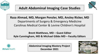
Adult Abdominal Imaging Case Studies
Raza Ahmad, MD, Morgan Penzler, MD, Ansley Ricker, MD
Departments of Surgery & Emergency Medicine
Carolinas Medical Center & Levine Children’s Hospital
Brent Matthews, MD – Guest Editor
Kyle Cunningham, MD & Michael Gibbs MD – Faculty Editors
Abdominal Imaging Mastery Project
December 2021
Disclosures
▪ This ongoing abdominal imaging interpretation series is proudly co-
sponsored by the Emergency Medicine & Surgery Residency Programs at
Carolinas Medical Center.
▪ The goal is to promote widespread interpretation mastery.
▪ There is no personal health information [PHI] within, and ages have been
changed to protect patient confidentiality.
It’s All About The Anatomy!
Systematic Approach to Abdominal CT Interpretation
● Aorta Down - follow the flow of blood!
○ Thoracic Aorta → Abdominal Aorta → Bifurcation → Iliac a.
● Veins Up - again, follow the flow!
○ Femoral v. → IVC → Right Atrium
● Solid Organs Down
○ Heart → Spleen → Pancreas → Liver → Gallbladder → Adrenal →
Kidney/Ureters → Bladder
● Rectum Up
○ Rectum → Sigmoid → Transverse → Cecum → Appendix
● Esophagus Down
○ Esophagus → Stomach → Small bowel
CASE #1:
A 64-year-old male
presenting after being
found down in a
parking lot. He is a
diabetic with a 40 pack-
year smoking history.
The exam reveals
tachycardia and
peritonitis on exam. CT
imaging was obtained.
Diagnosis?
CASE #1:
The patient was
suffering from a gastric
ulcer perforation. On
CT imaging there was
significant
pneumoperitoneum, a
large perforation along
the lesser curvature of
the stomach, and
extravasation of gastric
contents into the
peritoneal cavity.
Pneumoperitoneum
Lesser Curvature Perforation With
Extravasation Of Gastric Content
Gastric Ulcer Perforation
• Pathophysiology: there is a full-thickness injury to the stomach wall. Partial-
thickness injuries from trauma or electrocautery can progress to a full-
thickness injury over time causing perforation.
• History: peptic ulcer disease1, a previous diagnosis of H. pylori, NSAID
or aspirin use, recent instrumentation or surgery, and ingested foreign
body.
• Incidence: 3 to 6.5 per 100,000 individuals
• Presentation: the sudden onset severe abdominal pain, tachycardia,
and abdominal rigidity represent the “classic triad” of peptic ulcer
perforation.
1Peptic ulcer disease is the most common cause of stomach and duodenal perforation.
Back To Our Case!
• Our patient adamantly denied NSAID usage, so peptic ulcer as the cause
of gastric ulcer perforation was assumed.
• The patient was taken emergently to the operating room by General
Surgery for an exploratory laparotomy with subtotal gastrectomy left in
discontinuity, abdominal washout, and ABThera™ placement.
• He was taken back to surgery the following days for completion
antrectomy, vagotomy, Billroth 2 gastrojejunostomy, gastrojejunostomy
feeding tube placement, further washout, and abdominal closure.
Pneumoperitoneum – Imaging Findings
There Are Several
Radiographic Findings And
“Signs” Associated With
Pneumoperitoneum.
4-Year-Old With Abdominal Pain
Bowel Perforation Due To Enteritis
 Subphrenic air (solid white arrow)
 Falciform ligament sign (dashed white
arrows)
 Rigler sign (dashed black arrows)
 Ligamentum teres sign (solid black arrows)
87-Year-Old With Peptic Ulcer Disease
Presents With Three Days Of Abdominal Pain
Duodenal Perforation
A. Falciform ligament sign on plain radiograph
B. Seen as a vertical band on abdominal CT
56-Year-Old With Three Days Of Diarrhea And
Epigastric Abdominal Pain
Perforated Gastric Ulcer
• Small triangular pocket of air outlined by
three adjacent bowel loops
• The telltale triangle sign
69-Year-Old On Dexamethasone For Cerebral
Edema Presents With Abdominal Pain
Bedside Venting Procedure Performed And Then
The Patient Was Made Comfort Care
• Massive pneumoperitoneum
• Centralization of abdominal organs
• Air outlining the liver and gallbladder
(arrow)
65-Year-Old Becomes Unstable During An
Elective Colonoscope
Perforation Of The Ascending Colon. The Patient
Recovered After Decompressive Laparotomy
• Massive pneumoperitoneum
• Centralization of abdominal contents,
consistent with tension pneumoperitoneum
Two-Year-Old Receives CPR Briefly Following A
Witnessed Seizure
Posterior Gastric Perforation. The Child
Recovered After Surgical Repair
• Massive pneumoperitoneum
• Centralization of abdominal contents
• Football sign representing air outlining the
entire abdominal wall
IMAGES IN CLINICAL MEDICINE
Pneumoperitoneum from aGastricPerforation
Alexandra Masson, M.D., and Gerard Cheron, M.D., Ph.D.
JANUARY 2, 2022
PHYSICIAN J
OBS
J
uly 4, 2019
N Engl JMed 2019; 381:75
DOI: 10.1056/NEJ
Micm1814352
Metrics
Cambridge, Massachusetts
EmergencyMedicine
post-marketing period, non-serious and serious cases
of DILI were reported. Cases of severe liver injury with
fatal outcome have been reported in the post-
marketing period. The majority of hepatic events occur
within the first three months of treatment. OFEV was
associated with elevations of liver enzymes (ALT, AST,
ALKP, and GGT) and bilirubin. Liver enzyme and
bilirubin increases were reversible with dose
modification or interruption in the majority of cases.
DOWNLOAD THE FULL PRESCRIBING INFORMATION.
ADVERTISEMENT PHYSICIAN J
OBS
A two-and-a-half-year-old boywasbrought to theemergencydepartment a! er hehad aseizureduring a
N Engl JMed 2019;
DOI: 10.1056/NEJ
M
Metrics
EmergencyMedicin
Emergency Physici
EmergencyMedicin
Board Certi ed/Bo
Physician
EmergencyMedicin
Emergency Medici
EmergencyMedicin
Emergency Medici
Earning | Nebraska
EmergencyMedicin
Duke Emergency M
EmergencyMedicin
Pediatric Emergen
post-mar
of DILI w
fatal outc
marketing
within the
associate
ALKP, an
bilirubin i
modificat
DOWNLOA
Subdiaphragmatic Free Air
Free Air On Lateral Decubitus Film
Subdiaphragmatic Free Air, Lucent Liver Sign (LLS), And Cupola Sign (➤)
LLS
➤
➤
Subdiaphragmatic Free Air
Subdiaphragmatic Free Air And Rigler Sign (➤)
Free Air On Lateral Decubitus Film
Rigler Sign, Also Known As The Double Wall Sign
Subdiaphragmatic Free Air And Cupula Sign (➤)
Free Air On Lateral Decubitus Film
Falciform Ligament Sign
Reappraisal of radiographic signs of pneumoperitoneum
at emergency department
Yu-Hui C
hiu MDa,b
, Jen-Dar C
hen MDb,c,
, C
hui-Mei Tiu MDb,c
, Yi-Hong C
hou MDb,c
,
David Hung-Tsang Yen MD, PhDa,b
, C
hun-I Huang MDa,b
, C
heng-Yen C
hang MDb,c
a
Department of Emergency Medicine, Taipei Veterans General Hospital, Taiwan, ROC
b
National Yang-Ming University School of Medicine, Taiwan
c
Department of Radiology, Taipei Veterans General Hospital, Taipei, Taiwan, ROC
Received 4 January 2008; revised 14 February 2008; accepted 1 March 2008
Abstract
Purpose: Thisstudy aimed to evaluate the sensitivities of thereported free air signs on supine chest and
abdominal radiographs of hollow organ perforation. We also verified the value of supine radiographic
images as compared with erect chest and decubitus abdominal radiographs in detection of
pneumoperitoneum.
Methods: Two hundred fifty cases with surgically proven hollow organ perforation were included. Five
hundred twenty-seven radiographs were retrospectively reviewed on the picture archiving and
communication system. Medical charts were reviewed for operative findings of upper gastrointestinal
tract, small bowel, or colon perforations. The variable free air signs on both supine abdominal
radiographs (KUB) and supine chest radiographs (CXR) were evaluated and determined by consensus
without knowledge of initial radiographic reports or final diagnosis. Erect CXR and left decubitus
abdominal radiographs were evaluated for subphrenic free air or air over nondependent part of the right
abdomen.
Result: Upper gastrointestinal tract perforation was proven in 91.2%; small bowel perforation, in 6.8%;
and colon perforation, in 2.0%. Thepositiverateof freeair was80.4% on supineKUB, 78.7% on supine
CXR, 85.1% on erect CXR, and 98.0% on left decubitus abdominal radiograph. Anterior superior oval
sign was the most common radiographic sign on supine KUB (44.0%) and supine CXR (34.0%). Other
free air signs ranged from 0% to 30.4%.
C
onclusions: Most free air signs on supine radiographs are located over the right upper abdomen.
Familiarity with free air signs on supine radiographs is very important to emergency physicians and
American Journal of Emergency Medicine (2009) 27, 320–327
a
Department of Emergency Medicine, Taipei Veterans General Hospital, Taiwan, ROC
b
National Yang-Ming University School of Medicine, Taiwan
c
Department of Radiology, Taipei Veterans General Hospital, Taipei, Taiwan, ROC
Received 4 January 2008; revised 14 February 2008; accepted 1 March 2008
Abstract
Purpose: This study aimed to evaluate the sensitivities of the reported free air signs on supine chest and
abdominal radiographs of hollow organ perforation. We also verified the value of supine radiographic
images as compared with erect chest and decubitus abdominal radiographs in detection of
pneumoperitoneum.
Methods: Two hundred fifty cases with surgically proven hollow organ perforation were included. Five
hundred twenty-seven radiographs were retrospectively reviewed on the picture archiving and
communication system. Medical charts were reviewed for operative findings of upper gastrointestinal
tract, small bowel, or colon perforations. The variable free air signs on both supine abdominal
radiographs (KUB) and supine chest radiographs (CXR) were evaluated and determined by consensus
without knowledge of initial radiographic reports or final diagnosis. Erect CXR and left decubitus
abdominal radiographs were evaluated for subphrenic free air or air over nondependent part of the right
abdomen.
Result: Upper gastrointestinal tract perforation was proven in 91.2%; small bowel perforation, in 6.8%;
and colon perforation, in 2.0%. Thepositiverateof freeair was80.4% on supineKUB, 78.7% on supine
CXR, 85.1% on erect CXR, and 98.0% on left decubitus abdominal radiograph. Anterior superior oval
at emergency department
Yu-Hui C
hiu MDa,b
, Jen-Dar C
hen MDb,c,
, C
hui-Mei Tiu MDb,c
, Yi-Hong C
hou M
David Hung-Tsang Yen MD, PhDa,b
, C
hun-I Huang MDa,b
, C
heng-Yen C
hang MD
a
Department of Emergency Medicine, Taipei Veterans General Hospital, Taiwan, ROC
b
National Y
ang-Ming University School of Medicine, Taiwan
c
Department of Radiology, Taipei Veterans General Hospital, Taipei, Taiwan, ROC
Received 4 January 2008; revised 14 February 2008; accepted 1 March 2008
Abstract
Purpose: Thisstudy aimed to evaluatethesensitivitiesof thereported freeair signson supinechest and
abdominal radiographs of hollow organ perforation. We also verified the value of supine radiographic
images as compared with erect chest and decubitus abdominal radiographs in detection of
pneumoperitoneum.
Methods: Two hundred fifty caseswith surgically proven hollow organ perforation wereincluded. Five
hundred twenty-seven radiographs were retrospectively reviewed on the picture archiving and
communication system. Medical charts were reviewed for operative findings of upper gastrointestinal
tract, small bowel, or colon perforations. The variable free air signs on both supine abdominal
radiographs (KUB) and supine chest radiographs (CXR) were evaluated and determined by consensus
without knowledge of initial radiographic reports or final diagnosis. Erect CXR and left decubitus
abdominal radiographs wereevaluated for subphrenic freeair or air over nondependent part of theright
abdomen.
KUB Findings
CXR Findings
hundred twenty-seven radiographs were retrospectively reviewed on the picture archiving and
communication system. Medical charts were reviewed for operative findings of upper gastrointestinal
tract, small bowel, or colon perforations. The variable free air signs on both supine abdominal
radiographs (KUB) and supine chest radiographs (CXR) were evaluated and determined by consensus
without knowledge of initial radiographic reports or final diagnosis. Erect CXR and left decubitus
abdominal radiographs were evaluated for subphrenic free air or air over nondependent part of the right
abdomen.
Result: Upper gastrointestinal tract perforation was proven in 91.2%; small bowel perforation, in 6.8%;
and colon perforation, in 2.0%. Thepositiverateof freeair was80.4% on supineKUB, 78.7% on supine
CXR, 85.1% on erect CXR, and 98.0% on left decubitus abdominal radiograph. Anterior superior oval
sign was the most common radiographic sign on supine KUB (44.0%) and supine CXR (34.0%). Other
free air signs ranged from 0% to 30.4%.
C
onclusions: Most free air signs on supine radiographs are located over the right upper abdomen.
Familiarity with free air signs on supine radiographs is very important to emergency physicians and
radiologists for detection of hollow organ perforation.
© 2009 Elsevier Inc. All rights reserved.
1. Introduction
American Journal of Emergency Medicine (2009) 27, 320–327
CASE #2:
A 79-year-old female
with a history of
diverticulosis and atrial
fibrillation (on
apixaban) presents to
the ED with maroon-
colored stools. The
patient’s vital signs and
hemoglobin are
normal.
Diagnosis?
CASE #2:
CT angiography of the
abdomen and pelvis
revealed an acute
diverticular bleed in the
mid descending colon.
Active Diverticular
Bleeding With A
Contrast Blush
Lower GI Bleed
• Occurs in 20-35 per 100,000 adults per year, mortality rate 2-5%
• More common in patients who are female and elderly
• Caused by diverticular disease, colitis, polyps, AVM, angiodysplasia,
malignancy, brisk upper gastrointestinal bleeding, hemorrhoids
• Diverticulosis represents 30% of lower GI bleeding:
• Usually painless, typically results from penetrating artery erosion
• Some require embolization by interventional radiology for control
• Clinician can consider using the Oakland Score to identify patients
who may be able to be discharged home safely
Back To Our Case!
• The patient was transferred to interventional radiology for
embolization.
• She had intermittent bleeding of the distal left colic branch, which
was embolized.
• Hemoglobin on admission was 11.2, it trended down to 9.3 where it
remained stable. She did not require transfusion.
• She was instructed to restart her apixaban (Eliquis®) once her stools
had returned to normal.
CASE #3:
45-year-old male with
1 day of no ostomy
output, nausea,
abdominal pain, and
feculent vomiting. The
patient is tachycardic
and hypotensive. He
has abdominal
tenderness and a
parastomal hernia.
Diagnosis?
Small Bowel Obstruction Transition Point -
Proximally Dilated & Distally Decompressed
Questionable Transition Point, But Notice
That The Bowel Is Not Dilated
Parastomal Hernia
Parastomal Hernia
• Parastomal hernias occur up to 50% of patients after construction of
a colostomy or ileostomy.
• A parastomal hernia is a type of incisional hernia that allows
protrusion of abdominal contents through the abdominal wall defect
created during ostomy formation.
• Surgical repair is generally avoided due to the propensity for
parastomal hernia to recur.
• Patients with incarcerated or strangulated bowel within the hernia
sac can have symptoms of bowel obstruction, and these patients
require an operation.
Back To Our Case!
• The patient was taken to the operating room after failing
conservative management with nasogastric decompression and a
Gastrograffin challenge.
• A lysis of adhesions was performed followed by revision of
parastomal hernia. The adhesive band was found deep in the pelvis,
and not in the parastomal hernia sac.
• Postoperatively the patient recovered bowel function, however
developed an entero-cutaneous (EC) fistula.
• He was discharged to long-term-care with TPN to optimize nutrition
and aid with wound healing and EC fistula closure.
See You Next Month!

More Related Content

What's hot

Drs. Lena, Avery, and Davis’s CMC Abdominal Imaging Mastery Project: May Cases
Drs. Lena, Avery, and Davis’s CMC Abdominal Imaging Mastery Project: May CasesDrs. Lena, Avery, and Davis’s CMC Abdominal Imaging Mastery Project: May Cases
Drs. Lena, Avery, and Davis’s CMC Abdominal Imaging Mastery Project: May Cases
Sean M. Fox
 
Drs. Rossi and Shreve’s CMC Abdominal Imaging Mastery Project: June Cases
Drs. Rossi and Shreve’s CMC Abdominal Imaging Mastery Project: June CasesDrs. Rossi and Shreve’s CMC Abdominal Imaging Mastery Project: June Cases
Drs. Rossi and Shreve’s CMC Abdominal Imaging Mastery Project: June Cases
Sean M. Fox
 
Drs. Penzler, Ricker, and Ahmad’s CMC Abdominal Imaging Mastery Project: Nove...
Drs. Penzler, Ricker, and Ahmad’s CMC Abdominal Imaging Mastery Project: Nove...Drs. Penzler, Ricker, and Ahmad’s CMC Abdominal Imaging Mastery Project: Nove...
Drs. Penzler, Ricker, and Ahmad’s CMC Abdominal Imaging Mastery Project: Nove...
Sean M. Fox
 
Drs. Rossi and Shreve’s CMC Abdominal Imaging Mastery Project: May Cases
Drs. Rossi and Shreve’s CMC Abdominal Imaging Mastery Project: May CasesDrs. Rossi and Shreve’s CMC Abdominal Imaging Mastery Project: May Cases
Drs. Rossi and Shreve’s CMC Abdominal Imaging Mastery Project: May Cases
Sean M. Fox
 
Drs. Rossi and Shreve’s CMC Abdominal Imaging Mastery Project: December Cases
Drs. Rossi and Shreve’s CMC Abdominal Imaging Mastery Project: December CasesDrs. Rossi and Shreve’s CMC Abdominal Imaging Mastery Project: December Cases
Drs. Rossi and Shreve’s CMC Abdominal Imaging Mastery Project: December Cases
Sean M. Fox
 
CMC Pediatric X-Ray Mastery: February Case
CMC Pediatric X-Ray Mastery: February CaseCMC Pediatric X-Ray Mastery: February Case
CMC Pediatric X-Ray Mastery: February Case
Sean M. Fox
 
Dr. Kelsey Lena’s CMC Pediatric Orthopedic X-Ray Mastery Project: October Cases
Dr. Kelsey Lena’s CMC Pediatric Orthopedic X-Ray Mastery Project: October CasesDr. Kelsey Lena’s CMC Pediatric Orthopedic X-Ray Mastery Project: October Cases
Dr. Kelsey Lena’s CMC Pediatric Orthopedic X-Ray Mastery Project: October Cases
Sean M. Fox
 
Drs. Rossi and Shreve’s CMC Abdominal Imaging Mastery Project: January Cases
Drs. Rossi and Shreve’s CMC Abdominal Imaging Mastery Project: January CasesDrs. Rossi and Shreve’s CMC Abdominal Imaging Mastery Project: January Cases
Drs. Rossi and Shreve’s CMC Abdominal Imaging Mastery Project: January Cases
Sean M. Fox
 
Drs. Lena, Avery, and Davis’s CMC Abdominal Imaging Mastery Project: December...
Drs. Lena, Avery, and Davis’s CMC Abdominal Imaging Mastery Project: December...Drs. Lena, Avery, and Davis’s CMC Abdominal Imaging Mastery Project: December...
Drs. Lena, Avery, and Davis’s CMC Abdominal Imaging Mastery Project: December...
Sean M. Fox
 
Drs. Rossi and Shreve’s CMC Abdominal Imaging Mastery Project: October Cases
Drs. Rossi and Shreve’s CMC Abdominal Imaging Mastery Project: October CasesDrs. Rossi and Shreve’s CMC Abdominal Imaging Mastery Project: October Cases
Drs. Rossi and Shreve’s CMC Abdominal Imaging Mastery Project: October Cases
Sean M. Fox
 
CMC Pediatric X-Ray Mastery: October Cases
CMC Pediatric X-Ray Mastery: October CasesCMC Pediatric X-Ray Mastery: October Cases
CMC Pediatric X-Ray Mastery: October Cases
Sean M. Fox
 
Drs. Rossi and Shreve’s CMC Abdominal Imaging Mastery Project: November Cases
Drs. Rossi and Shreve’s CMC Abdominal Imaging Mastery Project: November CasesDrs. Rossi and Shreve’s CMC Abdominal Imaging Mastery Project: November Cases
Drs. Rossi and Shreve’s CMC Abdominal Imaging Mastery Project: November Cases
Sean M. Fox
 
Drs. Lena, Avery, and Davis’s CMC Abdominal Imaging Mastery Project: August C...
Drs. Lena, Avery, and Davis’s CMC Abdominal Imaging Mastery Project: August C...Drs. Lena, Avery, and Davis’s CMC Abdominal Imaging Mastery Project: August C...
Drs. Lena, Avery, and Davis’s CMC Abdominal Imaging Mastery Project: August C...
Sean M. Fox
 
EMGuideWire's Radiology Reading Room: Diaphragm Injury Cases
EMGuideWire's Radiology Reading Room: Diaphragm Injury CasesEMGuideWire's Radiology Reading Room: Diaphragm Injury Cases
EMGuideWire's Radiology Reading Room: Diaphragm Injury Cases
Sean M. Fox
 
Drs. Lena, Avery, and Davis’s CMC Abdominal Imaging Mastery Project: April Cases
Drs. Lena, Avery, and Davis’s CMC Abdominal Imaging Mastery Project: April CasesDrs. Lena, Avery, and Davis’s CMC Abdominal Imaging Mastery Project: April Cases
Drs. Lena, Avery, and Davis’s CMC Abdominal Imaging Mastery Project: April Cases
Sean M. Fox
 
Dr. Kelsey Lena’s CMC Pediatric Orthopedic X-Ray Mastery Project: January Cases
Dr. Kelsey Lena’s CMC Pediatric Orthopedic X-Ray Mastery Project: January CasesDr. Kelsey Lena’s CMC Pediatric Orthopedic X-Ray Mastery Project: January Cases
Dr. Kelsey Lena’s CMC Pediatric Orthopedic X-Ray Mastery Project: January Cases
Sean M. Fox
 
Drs. Olson’s and Jackson’s CMC Pediatric X-Ray Mastery: September Cases
Drs. Olson’s and Jackson’s CMC Pediatric X-Ray Mastery: September CasesDrs. Olson’s and Jackson’s CMC Pediatric X-Ray Mastery: September Cases
Drs. Olson’s and Jackson’s CMC Pediatric X-Ray Mastery: September Cases
Sean M. Fox
 
Drs. Lena, Avery, and Davis’s CMC Abdominal Imaging Mastery Project: March Cases
Drs. Lena, Avery, and Davis’s CMC Abdominal Imaging Mastery Project: March CasesDrs. Lena, Avery, and Davis’s CMC Abdominal Imaging Mastery Project: March Cases
Drs. Lena, Avery, and Davis’s CMC Abdominal Imaging Mastery Project: March Cases
Sean M. Fox
 
Dr. Michael Gibbs's CMC X-Ray Mastery Project - Week #4 Cases
Dr. Michael Gibbs's CMC X-Ray Mastery Project - Week #4 CasesDr. Michael Gibbs's CMC X-Ray Mastery Project - Week #4 Cases
Dr. Michael Gibbs's CMC X-Ray Mastery Project - Week #4 Cases
Sean M. Fox
 
Drs. Milam and Thomas's CMC X-Ray Mastery Project: February Cases
Drs. Milam and Thomas's CMC X-Ray Mastery Project: February CasesDrs. Milam and Thomas's CMC X-Ray Mastery Project: February Cases
Drs. Milam and Thomas's CMC X-Ray Mastery Project: February Cases
Sean M. Fox
 

What's hot (20)

Drs. Lena, Avery, and Davis’s CMC Abdominal Imaging Mastery Project: May Cases
Drs. Lena, Avery, and Davis’s CMC Abdominal Imaging Mastery Project: May CasesDrs. Lena, Avery, and Davis’s CMC Abdominal Imaging Mastery Project: May Cases
Drs. Lena, Avery, and Davis’s CMC Abdominal Imaging Mastery Project: May Cases
 
Drs. Rossi and Shreve’s CMC Abdominal Imaging Mastery Project: June Cases
Drs. Rossi and Shreve’s CMC Abdominal Imaging Mastery Project: June CasesDrs. Rossi and Shreve’s CMC Abdominal Imaging Mastery Project: June Cases
Drs. Rossi and Shreve’s CMC Abdominal Imaging Mastery Project: June Cases
 
Drs. Penzler, Ricker, and Ahmad’s CMC Abdominal Imaging Mastery Project: Nove...
Drs. Penzler, Ricker, and Ahmad’s CMC Abdominal Imaging Mastery Project: Nove...Drs. Penzler, Ricker, and Ahmad’s CMC Abdominal Imaging Mastery Project: Nove...
Drs. Penzler, Ricker, and Ahmad’s CMC Abdominal Imaging Mastery Project: Nove...
 
Drs. Rossi and Shreve’s CMC Abdominal Imaging Mastery Project: May Cases
Drs. Rossi and Shreve’s CMC Abdominal Imaging Mastery Project: May CasesDrs. Rossi and Shreve’s CMC Abdominal Imaging Mastery Project: May Cases
Drs. Rossi and Shreve’s CMC Abdominal Imaging Mastery Project: May Cases
 
Drs. Rossi and Shreve’s CMC Abdominal Imaging Mastery Project: December Cases
Drs. Rossi and Shreve’s CMC Abdominal Imaging Mastery Project: December CasesDrs. Rossi and Shreve’s CMC Abdominal Imaging Mastery Project: December Cases
Drs. Rossi and Shreve’s CMC Abdominal Imaging Mastery Project: December Cases
 
CMC Pediatric X-Ray Mastery: February Case
CMC Pediatric X-Ray Mastery: February CaseCMC Pediatric X-Ray Mastery: February Case
CMC Pediatric X-Ray Mastery: February Case
 
Dr. Kelsey Lena’s CMC Pediatric Orthopedic X-Ray Mastery Project: October Cases
Dr. Kelsey Lena’s CMC Pediatric Orthopedic X-Ray Mastery Project: October CasesDr. Kelsey Lena’s CMC Pediatric Orthopedic X-Ray Mastery Project: October Cases
Dr. Kelsey Lena’s CMC Pediatric Orthopedic X-Ray Mastery Project: October Cases
 
Drs. Rossi and Shreve’s CMC Abdominal Imaging Mastery Project: January Cases
Drs. Rossi and Shreve’s CMC Abdominal Imaging Mastery Project: January CasesDrs. Rossi and Shreve’s CMC Abdominal Imaging Mastery Project: January Cases
Drs. Rossi and Shreve’s CMC Abdominal Imaging Mastery Project: January Cases
 
Drs. Lena, Avery, and Davis’s CMC Abdominal Imaging Mastery Project: December...
Drs. Lena, Avery, and Davis’s CMC Abdominal Imaging Mastery Project: December...Drs. Lena, Avery, and Davis’s CMC Abdominal Imaging Mastery Project: December...
Drs. Lena, Avery, and Davis’s CMC Abdominal Imaging Mastery Project: December...
 
Drs. Rossi and Shreve’s CMC Abdominal Imaging Mastery Project: October Cases
Drs. Rossi and Shreve’s CMC Abdominal Imaging Mastery Project: October CasesDrs. Rossi and Shreve’s CMC Abdominal Imaging Mastery Project: October Cases
Drs. Rossi and Shreve’s CMC Abdominal Imaging Mastery Project: October Cases
 
CMC Pediatric X-Ray Mastery: October Cases
CMC Pediatric X-Ray Mastery: October CasesCMC Pediatric X-Ray Mastery: October Cases
CMC Pediatric X-Ray Mastery: October Cases
 
Drs. Rossi and Shreve’s CMC Abdominal Imaging Mastery Project: November Cases
Drs. Rossi and Shreve’s CMC Abdominal Imaging Mastery Project: November CasesDrs. Rossi and Shreve’s CMC Abdominal Imaging Mastery Project: November Cases
Drs. Rossi and Shreve’s CMC Abdominal Imaging Mastery Project: November Cases
 
Drs. Lena, Avery, and Davis’s CMC Abdominal Imaging Mastery Project: August C...
Drs. Lena, Avery, and Davis’s CMC Abdominal Imaging Mastery Project: August C...Drs. Lena, Avery, and Davis’s CMC Abdominal Imaging Mastery Project: August C...
Drs. Lena, Avery, and Davis’s CMC Abdominal Imaging Mastery Project: August C...
 
EMGuideWire's Radiology Reading Room: Diaphragm Injury Cases
EMGuideWire's Radiology Reading Room: Diaphragm Injury CasesEMGuideWire's Radiology Reading Room: Diaphragm Injury Cases
EMGuideWire's Radiology Reading Room: Diaphragm Injury Cases
 
Drs. Lena, Avery, and Davis’s CMC Abdominal Imaging Mastery Project: April Cases
Drs. Lena, Avery, and Davis’s CMC Abdominal Imaging Mastery Project: April CasesDrs. Lena, Avery, and Davis’s CMC Abdominal Imaging Mastery Project: April Cases
Drs. Lena, Avery, and Davis’s CMC Abdominal Imaging Mastery Project: April Cases
 
Dr. Kelsey Lena’s CMC Pediatric Orthopedic X-Ray Mastery Project: January Cases
Dr. Kelsey Lena’s CMC Pediatric Orthopedic X-Ray Mastery Project: January CasesDr. Kelsey Lena’s CMC Pediatric Orthopedic X-Ray Mastery Project: January Cases
Dr. Kelsey Lena’s CMC Pediatric Orthopedic X-Ray Mastery Project: January Cases
 
Drs. Olson’s and Jackson’s CMC Pediatric X-Ray Mastery: September Cases
Drs. Olson’s and Jackson’s CMC Pediatric X-Ray Mastery: September CasesDrs. Olson’s and Jackson’s CMC Pediatric X-Ray Mastery: September Cases
Drs. Olson’s and Jackson’s CMC Pediatric X-Ray Mastery: September Cases
 
Drs. Lena, Avery, and Davis’s CMC Abdominal Imaging Mastery Project: March Cases
Drs. Lena, Avery, and Davis’s CMC Abdominal Imaging Mastery Project: March CasesDrs. Lena, Avery, and Davis’s CMC Abdominal Imaging Mastery Project: March Cases
Drs. Lena, Avery, and Davis’s CMC Abdominal Imaging Mastery Project: March Cases
 
Dr. Michael Gibbs's CMC X-Ray Mastery Project - Week #4 Cases
Dr. Michael Gibbs's CMC X-Ray Mastery Project - Week #4 CasesDr. Michael Gibbs's CMC X-Ray Mastery Project - Week #4 Cases
Dr. Michael Gibbs's CMC X-Ray Mastery Project - Week #4 Cases
 
Drs. Milam and Thomas's CMC X-Ray Mastery Project: February Cases
Drs. Milam and Thomas's CMC X-Ray Mastery Project: February CasesDrs. Milam and Thomas's CMC X-Ray Mastery Project: February Cases
Drs. Milam and Thomas's CMC X-Ray Mastery Project: February Cases
 

Similar to Drs. Penzler, Ricker, and Ahmad’s CMC Abdominal Imaging Mastery Project: December Cases

Abdominal Imaging Case Studies #27.pptx
Abdominal Imaging Case Studies #27.pptxAbdominal Imaging Case Studies #27.pptx
Abdominal Imaging Case Studies #27.pptx
Sean M. Fox
 
Annular Pancreas: An Unusual Presentation
Annular Pancreas: An Unusual PresentationAnnular Pancreas: An Unusual Presentation
Annular Pancreas: An Unusual Presentation
Apollo Hospitals
 
Minimally Invasive Esophagectomy
Minimally Invasive EsophagectomyMinimally Invasive Esophagectomy
Minimally Invasive Esophagectomyguest87d35b
 
International Journal of Reproductive Medicine & Gynecology
International Journal of Reproductive Medicine & GynecologyInternational Journal of Reproductive Medicine & Gynecology
International Journal of Reproductive Medicine & Gynecology
SciRes Literature LLC. | Open Access Journals
 
Case History of Dedifferentiated Liposarcoma
Case History of Dedifferentiated LiposarcomaCase History of Dedifferentiated Liposarcoma
Case History of Dedifferentiated Liposarcoma
Victor Effiom
 
Drs. Brooks, Hambright, Holland, and Lorenz’s CMC Abdominal Imaging Mastery P...
Drs. Brooks, Hambright, Holland, and Lorenz’s CMC Abdominal Imaging Mastery P...Drs. Brooks, Hambright, Holland, and Lorenz’s CMC Abdominal Imaging Mastery P...
Drs. Brooks, Hambright, Holland, and Lorenz’s CMC Abdominal Imaging Mastery P...
Sean M. Fox
 
A Rare Case of Choledochal Cyst Connecting Intra- And ExtraHepatic Duct
A Rare Case of Choledochal Cyst Connecting Intra- And ExtraHepatic DuctA Rare Case of Choledochal Cyst Connecting Intra- And ExtraHepatic Duct
A Rare Case of Choledochal Cyst Connecting Intra- And ExtraHepatic Duct
semualkaira
 
Drs. Lorenzen and Barlock’s CMC X-Ray Mastery Project: May Cases
Drs. Lorenzen and Barlock’s CMC X-Ray Mastery Project: May CasesDrs. Lorenzen and Barlock’s CMC X-Ray Mastery Project: May Cases
Drs. Lorenzen and Barlock’s CMC X-Ray Mastery Project: May Cases
Sean M. Fox
 
Adult Intussusception A Retrospective Review
Adult Intussusception  A Retrospective ReviewAdult Intussusception  A Retrospective Review
Adult Intussusception A Retrospective Review
Deja Lewis
 
Laparoscopic Sleeve Gastrectomy in Situs Inversus Totalis
Laparoscopic Sleeve Gastrectomy in Situs Inversus TotalisLaparoscopic Sleeve Gastrectomy in Situs Inversus Totalis
Laparoscopic Sleeve Gastrectomy in Situs Inversus Totalis
submissionclinmedima
 
Colopericardial fistula following colonic interposition can primary repair be...
Colopericardial fistula following colonic interposition can primary repair be...Colopericardial fistula following colonic interposition can primary repair be...
Colopericardial fistula following colonic interposition can primary repair be...
Georges Khalifeh
 
Gastrointestinal Cancer: Research & Therapy
Gastrointestinal Cancer: Research & TherapyGastrointestinal Cancer: Research & Therapy
Gastrointestinal Cancer: Research & Therapy
Austin Publishing Group
 
Gastrointestinal Cancer: Research & Therapy
Gastrointestinal Cancer: Research & TherapyGastrointestinal Cancer: Research & Therapy
Gastrointestinal Cancer: Research & Therapy
Austin Publishing Group
 
Successful Repeated CT-Guided Drainage Of Rectal Mucocele After L
Successful Repeated CT-Guided Drainage Of Rectal Mucocele After LSuccessful Repeated CT-Guided Drainage Of Rectal Mucocele After L
Successful Repeated CT-Guided Drainage Of Rectal Mucocele After LAleksandr Reznichenko
 
COMPARATIVE STUDY BETWEEN LAPAROSCOPIC AND OPEN CHOLECYSTECTOMY (STUDY OF 50 ...
COMPARATIVE STUDY BETWEEN LAPAROSCOPIC AND OPEN CHOLECYSTECTOMY (STUDY OF 50 ...COMPARATIVE STUDY BETWEEN LAPAROSCOPIC AND OPEN CHOLECYSTECTOMY (STUDY OF 50 ...
COMPARATIVE STUDY BETWEEN LAPAROSCOPIC AND OPEN CHOLECYSTECTOMY (STUDY OF 50 ...
KETAN VAGHOLKAR
 
Enterocutaneous Umbilical Fistula: An Uncommon Manifestation of Abdominal Tub...
Enterocutaneous Umbilical Fistula: An Uncommon Manifestation of Abdominal Tub...Enterocutaneous Umbilical Fistula: An Uncommon Manifestation of Abdominal Tub...
Enterocutaneous Umbilical Fistula: An Uncommon Manifestation of Abdominal Tub...
KETAN VAGHOLKAR
 

Similar to Drs. Penzler, Ricker, and Ahmad’s CMC Abdominal Imaging Mastery Project: December Cases (16)

Abdominal Imaging Case Studies #27.pptx
Abdominal Imaging Case Studies #27.pptxAbdominal Imaging Case Studies #27.pptx
Abdominal Imaging Case Studies #27.pptx
 
Annular Pancreas: An Unusual Presentation
Annular Pancreas: An Unusual PresentationAnnular Pancreas: An Unusual Presentation
Annular Pancreas: An Unusual Presentation
 
Minimally Invasive Esophagectomy
Minimally Invasive EsophagectomyMinimally Invasive Esophagectomy
Minimally Invasive Esophagectomy
 
International Journal of Reproductive Medicine & Gynecology
International Journal of Reproductive Medicine & GynecologyInternational Journal of Reproductive Medicine & Gynecology
International Journal of Reproductive Medicine & Gynecology
 
Case History of Dedifferentiated Liposarcoma
Case History of Dedifferentiated LiposarcomaCase History of Dedifferentiated Liposarcoma
Case History of Dedifferentiated Liposarcoma
 
Drs. Brooks, Hambright, Holland, and Lorenz’s CMC Abdominal Imaging Mastery P...
Drs. Brooks, Hambright, Holland, and Lorenz’s CMC Abdominal Imaging Mastery P...Drs. Brooks, Hambright, Holland, and Lorenz’s CMC Abdominal Imaging Mastery P...
Drs. Brooks, Hambright, Holland, and Lorenz’s CMC Abdominal Imaging Mastery P...
 
A Rare Case of Choledochal Cyst Connecting Intra- And ExtraHepatic Duct
A Rare Case of Choledochal Cyst Connecting Intra- And ExtraHepatic DuctA Rare Case of Choledochal Cyst Connecting Intra- And ExtraHepatic Duct
A Rare Case of Choledochal Cyst Connecting Intra- And ExtraHepatic Duct
 
Drs. Lorenzen and Barlock’s CMC X-Ray Mastery Project: May Cases
Drs. Lorenzen and Barlock’s CMC X-Ray Mastery Project: May CasesDrs. Lorenzen and Barlock’s CMC X-Ray Mastery Project: May Cases
Drs. Lorenzen and Barlock’s CMC X-Ray Mastery Project: May Cases
 
Adult Intussusception A Retrospective Review
Adult Intussusception  A Retrospective ReviewAdult Intussusception  A Retrospective Review
Adult Intussusception A Retrospective Review
 
Laparoscopic Sleeve Gastrectomy in Situs Inversus Totalis
Laparoscopic Sleeve Gastrectomy in Situs Inversus TotalisLaparoscopic Sleeve Gastrectomy in Situs Inversus Totalis
Laparoscopic Sleeve Gastrectomy in Situs Inversus Totalis
 
Colopericardial fistula following colonic interposition can primary repair be...
Colopericardial fistula following colonic interposition can primary repair be...Colopericardial fistula following colonic interposition can primary repair be...
Colopericardial fistula following colonic interposition can primary repair be...
 
Gastrointestinal Cancer: Research & Therapy
Gastrointestinal Cancer: Research & TherapyGastrointestinal Cancer: Research & Therapy
Gastrointestinal Cancer: Research & Therapy
 
Gastrointestinal Cancer: Research & Therapy
Gastrointestinal Cancer: Research & TherapyGastrointestinal Cancer: Research & Therapy
Gastrointestinal Cancer: Research & Therapy
 
Successful Repeated CT-Guided Drainage Of Rectal Mucocele After L
Successful Repeated CT-Guided Drainage Of Rectal Mucocele After LSuccessful Repeated CT-Guided Drainage Of Rectal Mucocele After L
Successful Repeated CT-Guided Drainage Of Rectal Mucocele After L
 
COMPARATIVE STUDY BETWEEN LAPAROSCOPIC AND OPEN CHOLECYSTECTOMY (STUDY OF 50 ...
COMPARATIVE STUDY BETWEEN LAPAROSCOPIC AND OPEN CHOLECYSTECTOMY (STUDY OF 50 ...COMPARATIVE STUDY BETWEEN LAPAROSCOPIC AND OPEN CHOLECYSTECTOMY (STUDY OF 50 ...
COMPARATIVE STUDY BETWEEN LAPAROSCOPIC AND OPEN CHOLECYSTECTOMY (STUDY OF 50 ...
 
Enterocutaneous Umbilical Fistula: An Uncommon Manifestation of Abdominal Tub...
Enterocutaneous Umbilical Fistula: An Uncommon Manifestation of Abdominal Tub...Enterocutaneous Umbilical Fistula: An Uncommon Manifestation of Abdominal Tub...
Enterocutaneous Umbilical Fistula: An Uncommon Manifestation of Abdominal Tub...
 

More from Sean M. Fox

Implanted Devices - VP Shunts: EMGuidewire's Radiology Reading Room
Implanted Devices - VP Shunts: EMGuidewire's Radiology Reading RoomImplanted Devices - VP Shunts: EMGuidewire's Radiology Reading Room
Implanted Devices - VP Shunts: EMGuidewire's Radiology Reading Room
Sean M. Fox
 
Sternal Fractures & Dislocations - EMGuidewire Radiology Reading Room
Sternal Fractures & Dislocations - EMGuidewire Radiology Reading RoomSternal Fractures & Dislocations - EMGuidewire Radiology Reading Room
Sternal Fractures & Dislocations - EMGuidewire Radiology Reading Room
Sean M. Fox
 
Diaphragmatic Injuries - Radiology Reading Room
Diaphragmatic Injuries - Radiology Reading RoomDiaphragmatic Injuries - Radiology Reading Room
Diaphragmatic Injuries - Radiology Reading Room
Sean M. Fox
 
Acute Chest Syndrome - EMGuidewire's Radiology Reading Room
Acute Chest Syndrome - EMGuidewire's Radiology Reading RoomAcute Chest Syndrome - EMGuidewire's Radiology Reading Room
Acute Chest Syndrome - EMGuidewire's Radiology Reading Room
Sean M. Fox
 
Adult Orthopedic Imaging Series: Presentation #2 Native Hip Dislocations
Adult Orthopedic Imaging Series: Presentation #2 Native Hip DislocationsAdult Orthopedic Imaging Series: Presentation #2 Native Hip Dislocations
Adult Orthopedic Imaging Series: Presentation #2 Native Hip Dislocations
Sean M. Fox
 
Neuroimaging Mastery Project: Presentation #5 Subdural Hematomas
Neuroimaging Mastery Project: Presentation #5 Subdural HematomasNeuroimaging Mastery Project: Presentation #5 Subdural Hematomas
Neuroimaging Mastery Project: Presentation #5 Subdural Hematomas
Sean M. Fox
 
Neuroimaging Mastery Project Presentation #4: Acute Epidural Hematomas
Neuroimaging Mastery Project Presentation #4: Acute Epidural HematomasNeuroimaging Mastery Project Presentation #4: Acute Epidural Hematomas
Neuroimaging Mastery Project Presentation #4: Acute Epidural Hematomas
Sean M. Fox
 
Pediatric Orthopedic Imaging Case Studies #7 Pediatric Elbow Fractures
Pediatric Orthopedic Imaging Case Studies #7 Pediatric Elbow FracturesPediatric Orthopedic Imaging Case Studies #7 Pediatric Elbow Fractures
Pediatric Orthopedic Imaging Case Studies #7 Pediatric Elbow Fractures
Sean M. Fox
 
Adult Orthopedic Imaging Mastery Project - Pelvic Ring Fractures
Adult Orthopedic Imaging Mastery Project - Pelvic Ring FracturesAdult Orthopedic Imaging Mastery Project - Pelvic Ring Fractures
Adult Orthopedic Imaging Mastery Project - Pelvic Ring Fractures
Sean M. Fox
 
Neurosurgical Intracranial Infections - FINAL 10-17-23.pptx
Neurosurgical Intracranial Infections - FINAL 10-17-23.pptxNeurosurgical Intracranial Infections - FINAL 10-17-23.pptx
Neurosurgical Intracranial Infections - FINAL 10-17-23.pptx
Sean M. Fox
 
CMC Neuroimaging Case Studies - Cerebral Venous Sinus Thrombosis
CMC Neuroimaging Case Studies - Cerebral Venous Sinus ThrombosisCMC Neuroimaging Case Studies - Cerebral Venous Sinus Thrombosis
CMC Neuroimaging Case Studies - Cerebral Venous Sinus Thrombosis
Sean M. Fox
 
Blood Can Be Very Very Bad - CMC Neuroimaging Case Studies
Blood Can Be Very Very Bad - CMC Neuroimaging Case StudiesBlood Can Be Very Very Bad - CMC Neuroimaging Case Studies
Blood Can Be Very Very Bad - CMC Neuroimaging Case Studies
Sean M. Fox
 
Adult Chest X-Rays Of The Month - #46
Adult Chest X-Rays Of The Month - #46Adult Chest X-Rays Of The Month - #46
Adult Chest X-Rays Of The Month - #46
Sean M. Fox
 
Medical Device Imaging Mastery Project #4: Extracorporeal Membrane Oxygenation
Medical Device Imaging Mastery Project #4: Extracorporeal Membrane OxygenationMedical Device Imaging Mastery Project #4: Extracorporeal Membrane Oxygenation
Medical Device Imaging Mastery Project #4: Extracorporeal Membrane Oxygenation
Sean M. Fox
 
Drs. Pikus, Blackwell, Baumgarten, and Malloy-Posts’s CMC X-Ray Mastery Proje...
Drs. Pikus, Blackwell, Baumgarten, and Malloy-Posts’s CMC X-Ray Mastery Proje...Drs. Pikus, Blackwell, Baumgarten, and Malloy-Posts’s CMC X-Ray Mastery Proje...
Drs. Pikus, Blackwell, Baumgarten, and Malloy-Posts’s CMC X-Ray Mastery Proje...
Sean M. Fox
 
CMC Pediatric X-Ray Mastery: 27th Case Series
CMC Pediatric X-Ray Mastery: 27th Case SeriesCMC Pediatric X-Ray Mastery: 27th Case Series
CMC Pediatric X-Ray Mastery: 27th Case Series
Sean M. Fox
 
Dr. Haley Dusek’s CMC Pediatric Orthopedic X-Ray Mastery Project: #6 Presenta...
Dr. Haley Dusek’s CMC Pediatric Orthopedic X-Ray Mastery Project: #6 Presenta...Dr. Haley Dusek’s CMC Pediatric Orthopedic X-Ray Mastery Project: #6 Presenta...
Dr. Haley Dusek’s CMC Pediatric Orthopedic X-Ray Mastery Project: #6 Presenta...
Sean M. Fox
 
Drs. Pikus, Blackwell, Baumgarten, and Malloy-Posts’s CMC X-Ray Mastery Proje...
Drs. Pikus, Blackwell, Baumgarten, and Malloy-Posts’s CMC X-Ray Mastery Proje...Drs. Pikus, Blackwell, Baumgarten, and Malloy-Posts’s CMC X-Ray Mastery Proje...
Drs. Pikus, Blackwell, Baumgarten, and Malloy-Posts’s CMC X-Ray Mastery Proje...
Sean M. Fox
 
Drs. Escobar, Pikus, and Blackwell’s CMC X-Ray Mastery Project: 43rd Case Series
Drs. Escobar, Pikus, and Blackwell’s CMC X-Ray Mastery Project: 43rd Case SeriesDrs. Escobar, Pikus, and Blackwell’s CMC X-Ray Mastery Project: 43rd Case Series
Drs. Escobar, Pikus, and Blackwell’s CMC X-Ray Mastery Project: 43rd Case Series
Sean M. Fox
 
CMC Pediatric X-Ray Mastery: 26th Case Series
CMC Pediatric X-Ray Mastery: 26th Case SeriesCMC Pediatric X-Ray Mastery: 26th Case Series
CMC Pediatric X-Ray Mastery: 26th Case Series
Sean M. Fox
 

More from Sean M. Fox (20)

Implanted Devices - VP Shunts: EMGuidewire's Radiology Reading Room
Implanted Devices - VP Shunts: EMGuidewire's Radiology Reading RoomImplanted Devices - VP Shunts: EMGuidewire's Radiology Reading Room
Implanted Devices - VP Shunts: EMGuidewire's Radiology Reading Room
 
Sternal Fractures & Dislocations - EMGuidewire Radiology Reading Room
Sternal Fractures & Dislocations - EMGuidewire Radiology Reading RoomSternal Fractures & Dislocations - EMGuidewire Radiology Reading Room
Sternal Fractures & Dislocations - EMGuidewire Radiology Reading Room
 
Diaphragmatic Injuries - Radiology Reading Room
Diaphragmatic Injuries - Radiology Reading RoomDiaphragmatic Injuries - Radiology Reading Room
Diaphragmatic Injuries - Radiology Reading Room
 
Acute Chest Syndrome - EMGuidewire's Radiology Reading Room
Acute Chest Syndrome - EMGuidewire's Radiology Reading RoomAcute Chest Syndrome - EMGuidewire's Radiology Reading Room
Acute Chest Syndrome - EMGuidewire's Radiology Reading Room
 
Adult Orthopedic Imaging Series: Presentation #2 Native Hip Dislocations
Adult Orthopedic Imaging Series: Presentation #2 Native Hip DislocationsAdult Orthopedic Imaging Series: Presentation #2 Native Hip Dislocations
Adult Orthopedic Imaging Series: Presentation #2 Native Hip Dislocations
 
Neuroimaging Mastery Project: Presentation #5 Subdural Hematomas
Neuroimaging Mastery Project: Presentation #5 Subdural HematomasNeuroimaging Mastery Project: Presentation #5 Subdural Hematomas
Neuroimaging Mastery Project: Presentation #5 Subdural Hematomas
 
Neuroimaging Mastery Project Presentation #4: Acute Epidural Hematomas
Neuroimaging Mastery Project Presentation #4: Acute Epidural HematomasNeuroimaging Mastery Project Presentation #4: Acute Epidural Hematomas
Neuroimaging Mastery Project Presentation #4: Acute Epidural Hematomas
 
Pediatric Orthopedic Imaging Case Studies #7 Pediatric Elbow Fractures
Pediatric Orthopedic Imaging Case Studies #7 Pediatric Elbow FracturesPediatric Orthopedic Imaging Case Studies #7 Pediatric Elbow Fractures
Pediatric Orthopedic Imaging Case Studies #7 Pediatric Elbow Fractures
 
Adult Orthopedic Imaging Mastery Project - Pelvic Ring Fractures
Adult Orthopedic Imaging Mastery Project - Pelvic Ring FracturesAdult Orthopedic Imaging Mastery Project - Pelvic Ring Fractures
Adult Orthopedic Imaging Mastery Project - Pelvic Ring Fractures
 
Neurosurgical Intracranial Infections - FINAL 10-17-23.pptx
Neurosurgical Intracranial Infections - FINAL 10-17-23.pptxNeurosurgical Intracranial Infections - FINAL 10-17-23.pptx
Neurosurgical Intracranial Infections - FINAL 10-17-23.pptx
 
CMC Neuroimaging Case Studies - Cerebral Venous Sinus Thrombosis
CMC Neuroimaging Case Studies - Cerebral Venous Sinus ThrombosisCMC Neuroimaging Case Studies - Cerebral Venous Sinus Thrombosis
CMC Neuroimaging Case Studies - Cerebral Venous Sinus Thrombosis
 
Blood Can Be Very Very Bad - CMC Neuroimaging Case Studies
Blood Can Be Very Very Bad - CMC Neuroimaging Case StudiesBlood Can Be Very Very Bad - CMC Neuroimaging Case Studies
Blood Can Be Very Very Bad - CMC Neuroimaging Case Studies
 
Adult Chest X-Rays Of The Month - #46
Adult Chest X-Rays Of The Month - #46Adult Chest X-Rays Of The Month - #46
Adult Chest X-Rays Of The Month - #46
 
Medical Device Imaging Mastery Project #4: Extracorporeal Membrane Oxygenation
Medical Device Imaging Mastery Project #4: Extracorporeal Membrane OxygenationMedical Device Imaging Mastery Project #4: Extracorporeal Membrane Oxygenation
Medical Device Imaging Mastery Project #4: Extracorporeal Membrane Oxygenation
 
Drs. Pikus, Blackwell, Baumgarten, and Malloy-Posts’s CMC X-Ray Mastery Proje...
Drs. Pikus, Blackwell, Baumgarten, and Malloy-Posts’s CMC X-Ray Mastery Proje...Drs. Pikus, Blackwell, Baumgarten, and Malloy-Posts’s CMC X-Ray Mastery Proje...
Drs. Pikus, Blackwell, Baumgarten, and Malloy-Posts’s CMC X-Ray Mastery Proje...
 
CMC Pediatric X-Ray Mastery: 27th Case Series
CMC Pediatric X-Ray Mastery: 27th Case SeriesCMC Pediatric X-Ray Mastery: 27th Case Series
CMC Pediatric X-Ray Mastery: 27th Case Series
 
Dr. Haley Dusek’s CMC Pediatric Orthopedic X-Ray Mastery Project: #6 Presenta...
Dr. Haley Dusek’s CMC Pediatric Orthopedic X-Ray Mastery Project: #6 Presenta...Dr. Haley Dusek’s CMC Pediatric Orthopedic X-Ray Mastery Project: #6 Presenta...
Dr. Haley Dusek’s CMC Pediatric Orthopedic X-Ray Mastery Project: #6 Presenta...
 
Drs. Pikus, Blackwell, Baumgarten, and Malloy-Posts’s CMC X-Ray Mastery Proje...
Drs. Pikus, Blackwell, Baumgarten, and Malloy-Posts’s CMC X-Ray Mastery Proje...Drs. Pikus, Blackwell, Baumgarten, and Malloy-Posts’s CMC X-Ray Mastery Proje...
Drs. Pikus, Blackwell, Baumgarten, and Malloy-Posts’s CMC X-Ray Mastery Proje...
 
Drs. Escobar, Pikus, and Blackwell’s CMC X-Ray Mastery Project: 43rd Case Series
Drs. Escobar, Pikus, and Blackwell’s CMC X-Ray Mastery Project: 43rd Case SeriesDrs. Escobar, Pikus, and Blackwell’s CMC X-Ray Mastery Project: 43rd Case Series
Drs. Escobar, Pikus, and Blackwell’s CMC X-Ray Mastery Project: 43rd Case Series
 
CMC Pediatric X-Ray Mastery: 26th Case Series
CMC Pediatric X-Ray Mastery: 26th Case SeriesCMC Pediatric X-Ray Mastery: 26th Case Series
CMC Pediatric X-Ray Mastery: 26th Case Series
 

Recently uploaded

Additional Benefits for Employee Website.pdf
Additional Benefits for Employee Website.pdfAdditional Benefits for Employee Website.pdf
Additional Benefits for Employee Website.pdf
joachimlavalley1
 
Embracing GenAI - A Strategic Imperative
Embracing GenAI - A Strategic ImperativeEmbracing GenAI - A Strategic Imperative
Embracing GenAI - A Strategic Imperative
Peter Windle
 
Cambridge International AS A Level Biology Coursebook - EBook (MaryFosbery J...
Cambridge International AS  A Level Biology Coursebook - EBook (MaryFosbery J...Cambridge International AS  A Level Biology Coursebook - EBook (MaryFosbery J...
Cambridge International AS A Level Biology Coursebook - EBook (MaryFosbery J...
AzmatAli747758
 
The Art Pastor's Guide to Sabbath | Steve Thomason
The Art Pastor's Guide to Sabbath | Steve ThomasonThe Art Pastor's Guide to Sabbath | Steve Thomason
The Art Pastor's Guide to Sabbath | Steve Thomason
Steve Thomason
 
The Challenger.pdf DNHS Official Publication
The Challenger.pdf DNHS Official PublicationThe Challenger.pdf DNHS Official Publication
The Challenger.pdf DNHS Official Publication
Delapenabediema
 
TESDA TM1 REVIEWER FOR NATIONAL ASSESSMENT WRITTEN AND ORAL QUESTIONS WITH A...
TESDA TM1 REVIEWER  FOR NATIONAL ASSESSMENT WRITTEN AND ORAL QUESTIONS WITH A...TESDA TM1 REVIEWER  FOR NATIONAL ASSESSMENT WRITTEN AND ORAL QUESTIONS WITH A...
TESDA TM1 REVIEWER FOR NATIONAL ASSESSMENT WRITTEN AND ORAL QUESTIONS WITH A...
EugeneSaldivar
 
1.4 modern child centered education - mahatma gandhi-2.pptx
1.4 modern child centered education - mahatma gandhi-2.pptx1.4 modern child centered education - mahatma gandhi-2.pptx
1.4 modern child centered education - mahatma gandhi-2.pptx
JosvitaDsouza2
 
Students, digital devices and success - Andreas Schleicher - 27 May 2024..pptx
Students, digital devices and success - Andreas Schleicher - 27 May 2024..pptxStudents, digital devices and success - Andreas Schleicher - 27 May 2024..pptx
Students, digital devices and success - Andreas Schleicher - 27 May 2024..pptx
EduSkills OECD
 
How to Split Bills in the Odoo 17 POS Module
How to Split Bills in the Odoo 17 POS ModuleHow to Split Bills in the Odoo 17 POS Module
How to Split Bills in the Odoo 17 POS Module
Celine George
 
NCERT Solutions Power Sharing Class 10 Notes pdf
NCERT Solutions Power Sharing Class 10 Notes pdfNCERT Solutions Power Sharing Class 10 Notes pdf
NCERT Solutions Power Sharing Class 10 Notes pdf
Vivekanand Anglo Vedic Academy
 
Synthetic Fiber Construction in lab .pptx
Synthetic Fiber Construction in lab .pptxSynthetic Fiber Construction in lab .pptx
Synthetic Fiber Construction in lab .pptx
Pavel ( NSTU)
 
Unit 2- Research Aptitude (UGC NET Paper I).pdf
Unit 2- Research Aptitude (UGC NET Paper I).pdfUnit 2- Research Aptitude (UGC NET Paper I).pdf
Unit 2- Research Aptitude (UGC NET Paper I).pdf
Thiyagu K
 
Home assignment II on Spectroscopy 2024 Answers.pdf
Home assignment II on Spectroscopy 2024 Answers.pdfHome assignment II on Spectroscopy 2024 Answers.pdf
Home assignment II on Spectroscopy 2024 Answers.pdf
Tamralipta Mahavidyalaya
 
50 ĐỀ LUYỆN THI IOE LỚP 9 - NĂM HỌC 2022-2023 (CÓ LINK HÌNH, FILE AUDIO VÀ ĐÁ...
50 ĐỀ LUYỆN THI IOE LỚP 9 - NĂM HỌC 2022-2023 (CÓ LINK HÌNH, FILE AUDIO VÀ ĐÁ...50 ĐỀ LUYỆN THI IOE LỚP 9 - NĂM HỌC 2022-2023 (CÓ LINK HÌNH, FILE AUDIO VÀ ĐÁ...
50 ĐỀ LUYỆN THI IOE LỚP 9 - NĂM HỌC 2022-2023 (CÓ LINK HÌNH, FILE AUDIO VÀ ĐÁ...
Nguyen Thanh Tu Collection
 
Fish and Chips - have they had their chips
Fish and Chips - have they had their chipsFish and Chips - have they had their chips
Fish and Chips - have they had their chips
GeoBlogs
 
Chapter 3 - Islamic Banking Products and Services.pptx
Chapter 3 - Islamic Banking Products and Services.pptxChapter 3 - Islamic Banking Products and Services.pptx
Chapter 3 - Islamic Banking Products and Services.pptx
Mohd Adib Abd Muin, Senior Lecturer at Universiti Utara Malaysia
 
Instructions for Submissions thorugh G- Classroom.pptx
Instructions for Submissions thorugh G- Classroom.pptxInstructions for Submissions thorugh G- Classroom.pptx
Instructions for Submissions thorugh G- Classroom.pptx
Jheel Barad
 
special B.ed 2nd year old paper_20240531.pdf
special B.ed 2nd year old paper_20240531.pdfspecial B.ed 2nd year old paper_20240531.pdf
special B.ed 2nd year old paper_20240531.pdf
Special education needs
 
The Roman Empire A Historical Colossus.pdf
The Roman Empire A Historical Colossus.pdfThe Roman Empire A Historical Colossus.pdf
The Roman Empire A Historical Colossus.pdf
kaushalkr1407
 
UNIT – IV_PCI Complaints: Complaints and evaluation of complaints, Handling o...
UNIT – IV_PCI Complaints: Complaints and evaluation of complaints, Handling o...UNIT – IV_PCI Complaints: Complaints and evaluation of complaints, Handling o...
UNIT – IV_PCI Complaints: Complaints and evaluation of complaints, Handling o...
Sayali Powar
 

Recently uploaded (20)

Additional Benefits for Employee Website.pdf
Additional Benefits for Employee Website.pdfAdditional Benefits for Employee Website.pdf
Additional Benefits for Employee Website.pdf
 
Embracing GenAI - A Strategic Imperative
Embracing GenAI - A Strategic ImperativeEmbracing GenAI - A Strategic Imperative
Embracing GenAI - A Strategic Imperative
 
Cambridge International AS A Level Biology Coursebook - EBook (MaryFosbery J...
Cambridge International AS  A Level Biology Coursebook - EBook (MaryFosbery J...Cambridge International AS  A Level Biology Coursebook - EBook (MaryFosbery J...
Cambridge International AS A Level Biology Coursebook - EBook (MaryFosbery J...
 
The Art Pastor's Guide to Sabbath | Steve Thomason
The Art Pastor's Guide to Sabbath | Steve ThomasonThe Art Pastor's Guide to Sabbath | Steve Thomason
The Art Pastor's Guide to Sabbath | Steve Thomason
 
The Challenger.pdf DNHS Official Publication
The Challenger.pdf DNHS Official PublicationThe Challenger.pdf DNHS Official Publication
The Challenger.pdf DNHS Official Publication
 
TESDA TM1 REVIEWER FOR NATIONAL ASSESSMENT WRITTEN AND ORAL QUESTIONS WITH A...
TESDA TM1 REVIEWER  FOR NATIONAL ASSESSMENT WRITTEN AND ORAL QUESTIONS WITH A...TESDA TM1 REVIEWER  FOR NATIONAL ASSESSMENT WRITTEN AND ORAL QUESTIONS WITH A...
TESDA TM1 REVIEWER FOR NATIONAL ASSESSMENT WRITTEN AND ORAL QUESTIONS WITH A...
 
1.4 modern child centered education - mahatma gandhi-2.pptx
1.4 modern child centered education - mahatma gandhi-2.pptx1.4 modern child centered education - mahatma gandhi-2.pptx
1.4 modern child centered education - mahatma gandhi-2.pptx
 
Students, digital devices and success - Andreas Schleicher - 27 May 2024..pptx
Students, digital devices and success - Andreas Schleicher - 27 May 2024..pptxStudents, digital devices and success - Andreas Schleicher - 27 May 2024..pptx
Students, digital devices and success - Andreas Schleicher - 27 May 2024..pptx
 
How to Split Bills in the Odoo 17 POS Module
How to Split Bills in the Odoo 17 POS ModuleHow to Split Bills in the Odoo 17 POS Module
How to Split Bills in the Odoo 17 POS Module
 
NCERT Solutions Power Sharing Class 10 Notes pdf
NCERT Solutions Power Sharing Class 10 Notes pdfNCERT Solutions Power Sharing Class 10 Notes pdf
NCERT Solutions Power Sharing Class 10 Notes pdf
 
Synthetic Fiber Construction in lab .pptx
Synthetic Fiber Construction in lab .pptxSynthetic Fiber Construction in lab .pptx
Synthetic Fiber Construction in lab .pptx
 
Unit 2- Research Aptitude (UGC NET Paper I).pdf
Unit 2- Research Aptitude (UGC NET Paper I).pdfUnit 2- Research Aptitude (UGC NET Paper I).pdf
Unit 2- Research Aptitude (UGC NET Paper I).pdf
 
Home assignment II on Spectroscopy 2024 Answers.pdf
Home assignment II on Spectroscopy 2024 Answers.pdfHome assignment II on Spectroscopy 2024 Answers.pdf
Home assignment II on Spectroscopy 2024 Answers.pdf
 
50 ĐỀ LUYỆN THI IOE LỚP 9 - NĂM HỌC 2022-2023 (CÓ LINK HÌNH, FILE AUDIO VÀ ĐÁ...
50 ĐỀ LUYỆN THI IOE LỚP 9 - NĂM HỌC 2022-2023 (CÓ LINK HÌNH, FILE AUDIO VÀ ĐÁ...50 ĐỀ LUYỆN THI IOE LỚP 9 - NĂM HỌC 2022-2023 (CÓ LINK HÌNH, FILE AUDIO VÀ ĐÁ...
50 ĐỀ LUYỆN THI IOE LỚP 9 - NĂM HỌC 2022-2023 (CÓ LINK HÌNH, FILE AUDIO VÀ ĐÁ...
 
Fish and Chips - have they had their chips
Fish and Chips - have they had their chipsFish and Chips - have they had their chips
Fish and Chips - have they had their chips
 
Chapter 3 - Islamic Banking Products and Services.pptx
Chapter 3 - Islamic Banking Products and Services.pptxChapter 3 - Islamic Banking Products and Services.pptx
Chapter 3 - Islamic Banking Products and Services.pptx
 
Instructions for Submissions thorugh G- Classroom.pptx
Instructions for Submissions thorugh G- Classroom.pptxInstructions for Submissions thorugh G- Classroom.pptx
Instructions for Submissions thorugh G- Classroom.pptx
 
special B.ed 2nd year old paper_20240531.pdf
special B.ed 2nd year old paper_20240531.pdfspecial B.ed 2nd year old paper_20240531.pdf
special B.ed 2nd year old paper_20240531.pdf
 
The Roman Empire A Historical Colossus.pdf
The Roman Empire A Historical Colossus.pdfThe Roman Empire A Historical Colossus.pdf
The Roman Empire A Historical Colossus.pdf
 
UNIT – IV_PCI Complaints: Complaints and evaluation of complaints, Handling o...
UNIT – IV_PCI Complaints: Complaints and evaluation of complaints, Handling o...UNIT – IV_PCI Complaints: Complaints and evaluation of complaints, Handling o...
UNIT – IV_PCI Complaints: Complaints and evaluation of complaints, Handling o...
 

Drs. Penzler, Ricker, and Ahmad’s CMC Abdominal Imaging Mastery Project: December Cases

  • 1. Adult Abdominal Imaging Case Studies Raza Ahmad, MD, Morgan Penzler, MD, Ansley Ricker, MD Departments of Surgery & Emergency Medicine Carolinas Medical Center & Levine Children’s Hospital Brent Matthews, MD – Guest Editor Kyle Cunningham, MD & Michael Gibbs MD – Faculty Editors Abdominal Imaging Mastery Project December 2021
  • 2. Disclosures ▪ This ongoing abdominal imaging interpretation series is proudly co- sponsored by the Emergency Medicine & Surgery Residency Programs at Carolinas Medical Center. ▪ The goal is to promote widespread interpretation mastery. ▪ There is no personal health information [PHI] within, and ages have been changed to protect patient confidentiality.
  • 3. It’s All About The Anatomy!
  • 4. Systematic Approach to Abdominal CT Interpretation ● Aorta Down - follow the flow of blood! ○ Thoracic Aorta → Abdominal Aorta → Bifurcation → Iliac a. ● Veins Up - again, follow the flow! ○ Femoral v. → IVC → Right Atrium ● Solid Organs Down ○ Heart → Spleen → Pancreas → Liver → Gallbladder → Adrenal → Kidney/Ureters → Bladder ● Rectum Up ○ Rectum → Sigmoid → Transverse → Cecum → Appendix ● Esophagus Down ○ Esophagus → Stomach → Small bowel
  • 5. CASE #1: A 64-year-old male presenting after being found down in a parking lot. He is a diabetic with a 40 pack- year smoking history. The exam reveals tachycardia and peritonitis on exam. CT imaging was obtained. Diagnosis?
  • 6. CASE #1: The patient was suffering from a gastric ulcer perforation. On CT imaging there was significant pneumoperitoneum, a large perforation along the lesser curvature of the stomach, and extravasation of gastric contents into the peritoneal cavity. Pneumoperitoneum Lesser Curvature Perforation With Extravasation Of Gastric Content
  • 7. Gastric Ulcer Perforation • Pathophysiology: there is a full-thickness injury to the stomach wall. Partial- thickness injuries from trauma or electrocautery can progress to a full- thickness injury over time causing perforation. • History: peptic ulcer disease1, a previous diagnosis of H. pylori, NSAID or aspirin use, recent instrumentation or surgery, and ingested foreign body. • Incidence: 3 to 6.5 per 100,000 individuals • Presentation: the sudden onset severe abdominal pain, tachycardia, and abdominal rigidity represent the “classic triad” of peptic ulcer perforation. 1Peptic ulcer disease is the most common cause of stomach and duodenal perforation.
  • 8. Back To Our Case! • Our patient adamantly denied NSAID usage, so peptic ulcer as the cause of gastric ulcer perforation was assumed. • The patient was taken emergently to the operating room by General Surgery for an exploratory laparotomy with subtotal gastrectomy left in discontinuity, abdominal washout, and ABThera™ placement. • He was taken back to surgery the following days for completion antrectomy, vagotomy, Billroth 2 gastrojejunostomy, gastrojejunostomy feeding tube placement, further washout, and abdominal closure.
  • 10. There Are Several Radiographic Findings And “Signs” Associated With Pneumoperitoneum.
  • 11. 4-Year-Old With Abdominal Pain Bowel Perforation Due To Enteritis  Subphrenic air (solid white arrow)  Falciform ligament sign (dashed white arrows)  Rigler sign (dashed black arrows)  Ligamentum teres sign (solid black arrows)
  • 12. 87-Year-Old With Peptic Ulcer Disease Presents With Three Days Of Abdominal Pain Duodenal Perforation A. Falciform ligament sign on plain radiograph B. Seen as a vertical band on abdominal CT
  • 13. 56-Year-Old With Three Days Of Diarrhea And Epigastric Abdominal Pain Perforated Gastric Ulcer • Small triangular pocket of air outlined by three adjacent bowel loops • The telltale triangle sign
  • 14. 69-Year-Old On Dexamethasone For Cerebral Edema Presents With Abdominal Pain Bedside Venting Procedure Performed And Then The Patient Was Made Comfort Care • Massive pneumoperitoneum • Centralization of abdominal organs • Air outlining the liver and gallbladder (arrow)
  • 15. 65-Year-Old Becomes Unstable During An Elective Colonoscope Perforation Of The Ascending Colon. The Patient Recovered After Decompressive Laparotomy • Massive pneumoperitoneum • Centralization of abdominal contents, consistent with tension pneumoperitoneum
  • 16. Two-Year-Old Receives CPR Briefly Following A Witnessed Seizure Posterior Gastric Perforation. The Child Recovered After Surgical Repair • Massive pneumoperitoneum • Centralization of abdominal contents • Football sign representing air outlining the entire abdominal wall IMAGES IN CLINICAL MEDICINE Pneumoperitoneum from aGastricPerforation Alexandra Masson, M.D., and Gerard Cheron, M.D., Ph.D. JANUARY 2, 2022 PHYSICIAN J OBS J uly 4, 2019 N Engl JMed 2019; 381:75 DOI: 10.1056/NEJ Micm1814352 Metrics Cambridge, Massachusetts EmergencyMedicine post-marketing period, non-serious and serious cases of DILI were reported. Cases of severe liver injury with fatal outcome have been reported in the post- marketing period. The majority of hepatic events occur within the first three months of treatment. OFEV was associated with elevations of liver enzymes (ALT, AST, ALKP, and GGT) and bilirubin. Liver enzyme and bilirubin increases were reversible with dose modification or interruption in the majority of cases. DOWNLOAD THE FULL PRESCRIBING INFORMATION. ADVERTISEMENT PHYSICIAN J OBS A two-and-a-half-year-old boywasbrought to theemergencydepartment a! er hehad aseizureduring a N Engl JMed 2019; DOI: 10.1056/NEJ M Metrics EmergencyMedicin Emergency Physici EmergencyMedicin Board Certi ed/Bo Physician EmergencyMedicin Emergency Medici EmergencyMedicin Emergency Medici Earning | Nebraska EmergencyMedicin Duke Emergency M EmergencyMedicin Pediatric Emergen post-mar of DILI w fatal outc marketing within the associate ALKP, an bilirubin i modificat DOWNLOA
  • 18. Free Air On Lateral Decubitus Film
  • 19. Subdiaphragmatic Free Air, Lucent Liver Sign (LLS), And Cupola Sign (➤) LLS ➤ ➤
  • 21. Subdiaphragmatic Free Air And Rigler Sign (➤)
  • 22. Free Air On Lateral Decubitus Film
  • 23. Rigler Sign, Also Known As The Double Wall Sign
  • 24. Subdiaphragmatic Free Air And Cupula Sign (➤)
  • 25. Free Air On Lateral Decubitus Film
  • 27. Reappraisal of radiographic signs of pneumoperitoneum at emergency department Yu-Hui C hiu MDa,b , Jen-Dar C hen MDb,c, , C hui-Mei Tiu MDb,c , Yi-Hong C hou MDb,c , David Hung-Tsang Yen MD, PhDa,b , C hun-I Huang MDa,b , C heng-Yen C hang MDb,c a Department of Emergency Medicine, Taipei Veterans General Hospital, Taiwan, ROC b National Yang-Ming University School of Medicine, Taiwan c Department of Radiology, Taipei Veterans General Hospital, Taipei, Taiwan, ROC Received 4 January 2008; revised 14 February 2008; accepted 1 March 2008 Abstract Purpose: Thisstudy aimed to evaluate the sensitivities of thereported free air signs on supine chest and abdominal radiographs of hollow organ perforation. We also verified the value of supine radiographic images as compared with erect chest and decubitus abdominal radiographs in detection of pneumoperitoneum. Methods: Two hundred fifty cases with surgically proven hollow organ perforation were included. Five hundred twenty-seven radiographs were retrospectively reviewed on the picture archiving and communication system. Medical charts were reviewed for operative findings of upper gastrointestinal tract, small bowel, or colon perforations. The variable free air signs on both supine abdominal radiographs (KUB) and supine chest radiographs (CXR) were evaluated and determined by consensus without knowledge of initial radiographic reports or final diagnosis. Erect CXR and left decubitus abdominal radiographs were evaluated for subphrenic free air or air over nondependent part of the right abdomen. Result: Upper gastrointestinal tract perforation was proven in 91.2%; small bowel perforation, in 6.8%; and colon perforation, in 2.0%. Thepositiverateof freeair was80.4% on supineKUB, 78.7% on supine CXR, 85.1% on erect CXR, and 98.0% on left decubitus abdominal radiograph. Anterior superior oval sign was the most common radiographic sign on supine KUB (44.0%) and supine CXR (34.0%). Other free air signs ranged from 0% to 30.4%. C onclusions: Most free air signs on supine radiographs are located over the right upper abdomen. Familiarity with free air signs on supine radiographs is very important to emergency physicians and American Journal of Emergency Medicine (2009) 27, 320–327 a Department of Emergency Medicine, Taipei Veterans General Hospital, Taiwan, ROC b National Yang-Ming University School of Medicine, Taiwan c Department of Radiology, Taipei Veterans General Hospital, Taipei, Taiwan, ROC Received 4 January 2008; revised 14 February 2008; accepted 1 March 2008 Abstract Purpose: This study aimed to evaluate the sensitivities of the reported free air signs on supine chest and abdominal radiographs of hollow organ perforation. We also verified the value of supine radiographic images as compared with erect chest and decubitus abdominal radiographs in detection of pneumoperitoneum. Methods: Two hundred fifty cases with surgically proven hollow organ perforation were included. Five hundred twenty-seven radiographs were retrospectively reviewed on the picture archiving and communication system. Medical charts were reviewed for operative findings of upper gastrointestinal tract, small bowel, or colon perforations. The variable free air signs on both supine abdominal radiographs (KUB) and supine chest radiographs (CXR) were evaluated and determined by consensus without knowledge of initial radiographic reports or final diagnosis. Erect CXR and left decubitus abdominal radiographs were evaluated for subphrenic free air or air over nondependent part of the right abdomen. Result: Upper gastrointestinal tract perforation was proven in 91.2%; small bowel perforation, in 6.8%; and colon perforation, in 2.0%. Thepositiverateof freeair was80.4% on supineKUB, 78.7% on supine CXR, 85.1% on erect CXR, and 98.0% on left decubitus abdominal radiograph. Anterior superior oval at emergency department Yu-Hui C hiu MDa,b , Jen-Dar C hen MDb,c, , C hui-Mei Tiu MDb,c , Yi-Hong C hou M David Hung-Tsang Yen MD, PhDa,b , C hun-I Huang MDa,b , C heng-Yen C hang MD a Department of Emergency Medicine, Taipei Veterans General Hospital, Taiwan, ROC b National Y ang-Ming University School of Medicine, Taiwan c Department of Radiology, Taipei Veterans General Hospital, Taipei, Taiwan, ROC Received 4 January 2008; revised 14 February 2008; accepted 1 March 2008 Abstract Purpose: Thisstudy aimed to evaluatethesensitivitiesof thereported freeair signson supinechest and abdominal radiographs of hollow organ perforation. We also verified the value of supine radiographic images as compared with erect chest and decubitus abdominal radiographs in detection of pneumoperitoneum. Methods: Two hundred fifty caseswith surgically proven hollow organ perforation wereincluded. Five hundred twenty-seven radiographs were retrospectively reviewed on the picture archiving and communication system. Medical charts were reviewed for operative findings of upper gastrointestinal tract, small bowel, or colon perforations. The variable free air signs on both supine abdominal radiographs (KUB) and supine chest radiographs (CXR) were evaluated and determined by consensus without knowledge of initial radiographic reports or final diagnosis. Erect CXR and left decubitus abdominal radiographs wereevaluated for subphrenic freeair or air over nondependent part of theright abdomen.
  • 30. hundred twenty-seven radiographs were retrospectively reviewed on the picture archiving and communication system. Medical charts were reviewed for operative findings of upper gastrointestinal tract, small bowel, or colon perforations. The variable free air signs on both supine abdominal radiographs (KUB) and supine chest radiographs (CXR) were evaluated and determined by consensus without knowledge of initial radiographic reports or final diagnosis. Erect CXR and left decubitus abdominal radiographs were evaluated for subphrenic free air or air over nondependent part of the right abdomen. Result: Upper gastrointestinal tract perforation was proven in 91.2%; small bowel perforation, in 6.8%; and colon perforation, in 2.0%. Thepositiverateof freeair was80.4% on supineKUB, 78.7% on supine CXR, 85.1% on erect CXR, and 98.0% on left decubitus abdominal radiograph. Anterior superior oval sign was the most common radiographic sign on supine KUB (44.0%) and supine CXR (34.0%). Other free air signs ranged from 0% to 30.4%. C onclusions: Most free air signs on supine radiographs are located over the right upper abdomen. Familiarity with free air signs on supine radiographs is very important to emergency physicians and radiologists for detection of hollow organ perforation. © 2009 Elsevier Inc. All rights reserved. 1. Introduction American Journal of Emergency Medicine (2009) 27, 320–327
  • 31. CASE #2: A 79-year-old female with a history of diverticulosis and atrial fibrillation (on apixaban) presents to the ED with maroon- colored stools. The patient’s vital signs and hemoglobin are normal. Diagnosis?
  • 32. CASE #2: CT angiography of the abdomen and pelvis revealed an acute diverticular bleed in the mid descending colon. Active Diverticular Bleeding With A Contrast Blush
  • 33. Lower GI Bleed • Occurs in 20-35 per 100,000 adults per year, mortality rate 2-5% • More common in patients who are female and elderly • Caused by diverticular disease, colitis, polyps, AVM, angiodysplasia, malignancy, brisk upper gastrointestinal bleeding, hemorrhoids • Diverticulosis represents 30% of lower GI bleeding: • Usually painless, typically results from penetrating artery erosion • Some require embolization by interventional radiology for control • Clinician can consider using the Oakland Score to identify patients who may be able to be discharged home safely
  • 34. Back To Our Case! • The patient was transferred to interventional radiology for embolization. • She had intermittent bleeding of the distal left colic branch, which was embolized. • Hemoglobin on admission was 11.2, it trended down to 9.3 where it remained stable. She did not require transfusion. • She was instructed to restart her apixaban (Eliquis®) once her stools had returned to normal.
  • 35.
  • 36.
  • 37.
  • 38.
  • 39.
  • 40. CASE #3: 45-year-old male with 1 day of no ostomy output, nausea, abdominal pain, and feculent vomiting. The patient is tachycardic and hypotensive. He has abdominal tenderness and a parastomal hernia. Diagnosis?
  • 41. Small Bowel Obstruction Transition Point - Proximally Dilated & Distally Decompressed Questionable Transition Point, But Notice That The Bowel Is Not Dilated Parastomal Hernia
  • 42. Parastomal Hernia • Parastomal hernias occur up to 50% of patients after construction of a colostomy or ileostomy. • A parastomal hernia is a type of incisional hernia that allows protrusion of abdominal contents through the abdominal wall defect created during ostomy formation. • Surgical repair is generally avoided due to the propensity for parastomal hernia to recur. • Patients with incarcerated or strangulated bowel within the hernia sac can have symptoms of bowel obstruction, and these patients require an operation.
  • 43. Back To Our Case! • The patient was taken to the operating room after failing conservative management with nasogastric decompression and a Gastrograffin challenge. • A lysis of adhesions was performed followed by revision of parastomal hernia. The adhesive band was found deep in the pelvis, and not in the parastomal hernia sac. • Postoperatively the patient recovered bowel function, however developed an entero-cutaneous (EC) fistula. • He was discharged to long-term-care with TPN to optimize nutrition and aid with wound healing and EC fistula closure.
  • 44.
  • 45.
  • 46.
  • 47.
  • 48.
  • 49.
  • 50.
  • 51.
  • 52.
  • 53.
  • 54. See You Next Month!