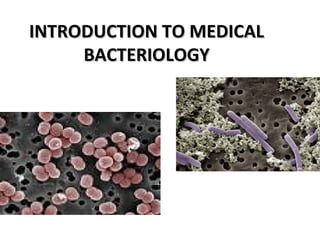
2. DEFINITION of terms:
• Microbiology-Microbiology- is the study of microscopic
organism.
• Medical Microbiology /Clinical Microbiology-Medical Microbiology /Clinical Microbiology- deals with
the diagnosis, treatment, and prevention of
infectious disease caused by all microorganism.
3. What are bacteria?
• Single celled organisms
• Very small
• Need a microscope to see
• Can be found on most
materials and surfaces
– Billions on and in your
body right now
E. Coli O157:H7
can make you
very sick.
Streptococcus
can cause strep
throat.
This E. coli helps
you digest food.
4. What do they look like?
• Three basic shapes
– Rod shaped called bacilli
– Round shaped called
– Spiral shaped
• Some exist as single
cells, others
cluster together
Bacilli
Spiral
Cocci
Cluster of cocci
5. How do bacteria Reproduce?How do bacteria Reproduce?
• Grow in number not in size
– Humans grow in size from child to adult
• Make copies of themselves by dividing in half
– Human parents create a child
6. What is a pathogen?
• Microorganism that can cause disease
– Why do they make you sick?
• To get food they need to survive and reproduce
– How do they make you sick?
• They produce poisons (toxins) that result in fever,
headache, vomiting, and diarrhea and destroy body
tissue
7. Where do you get a
pathogen?
• Contact with people who are sick
– Direct or indirect
• Food, Water, or other Surfaces that are
contaminated
Indirect contact
Direct contact
Foods that
could be
contaminated
8. Are all bacteria Pathogens?Are all bacteria Pathogens?
• No, most are harmless
• Some are even helpful
– Examples of helpful bacteria:
• Lactobacillus: makes cheese, yogurt, &
buttermilk and produces vitamins in your
intestine
• Leuconostoc: makes pickles
• Pediococcus: makes pepperoni, salami, &
summer sausage
10. Structure of BacteriaStructure of Bacteria
• All cells have 3 main components:
– DNA (‘nucleoid”)DNA (‘nucleoid”)
• genetic instructions
– Surrounding membrane (“cytoplasmic membrane”)
• limits access to the cell’s interior
– Cytoplasm, between the DNA and the membrane
• where all metabolic reactions occur
• especially protein synthesis, which occurs on the
ribosomes
• Bacteria also often have these features:Bacteria also often have these features:
– Cell wallCell wall
• resists osmotic pressure
– FlagellaFlagella
• movement
– PiliPili
• attachment
– CapsuleCapsule
• protection and biofilms
11. Cell MembraneCell Membrane
• The cell membrane (often called the plasma
membrane) is composed of 2 layers of
phospholipids.
• Phospholipids have polar heads and non-polar
tails.
– “Polar” implies that the heads are hydrophilic:
they like to stay in an aqueous environment:
facing the outside world and the inside of the
cell.
– “non-polar” means that the tails are
hydrophobic: they want to be away from
water, in an oily environment. The tails are in
the center of the membrane
• A pure phospholipid membrane only allows
water, gases, and a few small molecules to
move freely through it.
12. Membrane ProteinsMembrane Proteins
• Proteins float in the membrane like ships
on the surface of the sea: the fluid-
mosaic model.
• Peripheral membrane proteinsPeripheral membrane proteins are
bound to one surface of the membrane.
– Some attached to the cell membrane by a
fatty acid covalently attached to one of
the protein’s amino acids
– Others are attached by stretches of
hydrophobic amino acids of the protein’s
surface
• Integral membrane proteinsIntegral membrane proteins are
embedded in the membrane by one or
more stretches of hydrophobic amino
acids. Many of these proteins transport
molecules in and out of the cell. The
transport proteins are very selective:
each type of molecule needs its own
transporter.
13. Cell EnvelopeCell Envelope
• The cell envelope is the outer
layer from the cell membrane
it includes the:
- cell wall
- periplasmic space
-outer membrane
-capsule.
• It gives shape to the bacteria
and protects the internal
structure.
14. Cell WallCell Wall
• Osmotic pressureOsmotic pressure is the force generated by water attempting to
move into the cell.
• Bacteria, along with plants and fungi, resist osmotic pressure by
surrounding the cell in a rigid box, the cell wall.
– Composed of peptidoglycan (also called proteoglycan or murein)
– Long chains of polysaccharide cross-linked by short peptides
(amino acid chains).
• The peptides contain the unusual mirror-image amino
acids D-alanine and D-glutamate
• polysaccharide is composed of alternating “amino sugars”:
N-acetylglucosamine and N-acetylmuramic acid
15. Cell Wall
• Gram-positive vs Gram-negativeGram-positive vs Gram-negative are defined
by the structure of the cell wall
– the Gram stain binds to peptidoglycan
• Gram-positive:Gram-positive: many layers of peptidoglycan,
which is anchored to the cell membrane by
teichoic acid.
• Gram-negative:Gram-negative: 1-2 layers of peptidoglycan =
thin
– The periplasmic space is between the cell
membrane and the cell wall. It contains
enzymes and other proteins, such as
chemoreceptors for sensing the
environment.
– Outside the peptidglycan layer is the
“outer membrane”. It is pierced by
porins: protein channels, and its out
surface is covered with lipopolysaccharides
(sugars linked to membrane lipids), which
are often antigenic and or toxic.
16. Capsule
-Outer covering present in
some bacteria.
-Some bacteria (often
pathogens) are surrounded
by a thick polysaccharide
capsule. This is a loose
jelly-like or mucus-like layer.
- It helps prevent immune
system cells from reaching
the bacteria, and it forms
part of biofilms.
17. Membrane Structures
• PiliPili (singular = pilus)(singular = pilus) are hairs projecting from the
surface. They are composed of pilin protein.
There are several types:
– Sex piliSex pili ––involved in sexual conjugation
– Common pili /FimbriaeCommon pili /Fimbriae (singular = fimbria(singular = fimbria) are
pili used to attach the bacteria to target cells
( in infection) or to surfaces, where they form a
biofilm.
• FlagellaFlagella are long hairs used to propel the cells.
They are composed of flagellin protein.
Atrichous-No flagella
Monotrichous- One flagella
Amphtrichous- flagella at 2 poles
Lophotrichous-Tuft of flagella at one/2poles
Peritrichous-Flagella surrounding the bacteria
18. Spores
• Some bacteria can form very tough spores, which are
metabolically inactive and can survive a long time
under very harsh conditions.
• Spores can also survive very high or low temperatures
and high UV radiation for extended periods. This
makes them difficult to kill during sterilization.
– Anthrax
• Spores are produced only by a few genera in the
Firmicutes:
– Bacillus species including anthracis (anthrax) and
cereus (endotoxin causes ~5% of food poisoning)
– Clostridium species including tetani (tetanus),
perfringens (gangrene), and botulinum (botulism:
food poisoning from improperly canned food)
21. Binary fission
1. Prokaryote cells grow
by increasing in cell
number (as opposed to
increasing in size).
2. Replication is by binary
fission, the splitting of
one cell into two
3. Therefore, bacterial
populations increase by
a factor of two (double)
every generation time.
22. Generation time
• The time required for a population of microorganism to
double (doubling time) in number.
• Ex. Escherichia coli (E. coli) double every 20 minutes
• Ex. Mycobacterium tuberculosis double every 12 to 24 hours
4-22
23. 1. Lag phase occurs when bacteria are adjusting to the medium. For
example, with a nutritionally poor medium, several anabolic pathways
need to be turned on, resulting in a lag before active growth begins.
2. In Log or exponential phase, the cells are growing as fast as they can,
limited only by growth conditions and genetic potential. During this
phase, almost all cells are alive, they are most nearly identical, and they
are most affected by outside influences like disinfectants.
3. Due to nutrient depletion and/or accumulation of toxic end products,
replication stops and cells enter a stationary phase where there is no net
change in cell number.
4. Death phase occurs when cells can no longer maintain viability and
numbers decrease as a proportion.
PHASES OF BACTERIAL GROWTH
26. Disinfection Disinfection is the elimination of
pathogens, except spores, from inanimate
objects
Disinfectants are chemical solutions
used to clean inanimate objects (physical
processes, e.g., UV radiation, may also be
employed to effect disinfection)
Germicides are chemicals that can be
applied to both animate (living) and
inanimate objects for the purpose of
eliminating pathogens
27. Sterilization Sterilization is the total elimination of all
microorganisms including spores
Typically the last things to die are the
highly heat- and chemical-resistant
bacterial endospores
Instruments used for invasive procedures
must be sterilized prior to use
Physical Methods(Moist Heat, Dry
Heat), Filtration, Ionizing Radiation, Non
IonizingRadiation.
Chemical Methods
28. OtherTerms
Sanitization: Lowering of microbial counts to prevent
transmission in public setting (e.g., restaurants &
public rest rooms)
Degerming: Mechanical removal of microbes, e.g.,
from hands with washing
Sepsis: Bacterial contamination of blood
Antisepsis: Reduction of or Inhibition of microbes
found on living tissue.
Bacteriocidal -can cause destruction of the cell.
Bacteriolysis Dissolution or lysis of thebacterial cell.
Bacteriostasis -Inhibtion of the growth of bacteria
without destruction.
29. Different Kinds of Bacteria “Death”
1. Bacteriostatic
2. Bacteriocidal
3. Bacteriolytic
LogCell#
Time
Total cell count
Viable cell count
30. Chemical
Antimicrobials
Agent Mechanisms of Action Comments
SurfactantsSurfactants Membrane Disruption;
increased penetration
Soaps; detergents
Quats (cationicQuats (cationic
detergent)detergent)
Denature proteins;
Disrupts lipids
Antiseptic - benzalconium
chloride, Cepacol; Disinfectant
Organic acidsOrganic acids
and basesand bases
High/low pH Mold and Fungi inhibitors; e.g.,
benzoate of soda
Heavy MetalsHeavy Metals Denature protein Antiseptic & Disinfectant; Silver
Nitrate
HalogensHalogens Oxidizing agent
Disrupts cell membrane
Antiseptic - Iodine (Betadine)
Disinfectant - Chlorine (Chlorox)
AlcoholAlcoholss Denatures proteins;
Disrupts lipids
Antiseptic & Disinfectant
Ethanol and isopropyl
PhenolicsPhenolics Disrupts cell membrane Disinfectant
Irritating odor
AldehydesAldehydes Denature proteins Gluteraldehyde - disinfectant
(Cidex); Formaldehyde -
disinfectant
Ethylene OxideEthylene Oxide Denaturing proteins Used in a closed chamber to
sterilize
Oxidizing agentsOxidizing agents Denature proteins Hydrogen peroxide – antiseptic;
Hydrogen peroxide – disinfectan;
Benzoyl peroxide – antiseptic
31. PhysicalAntimicrobials
Agent Mechanisms of Action Comments
Moist Heat,Moist Heat,
boilingboiling
Denatures proteins Kills vegetative bacterial cells and
viruses Endospores survive
Moist Heat,Moist Heat,
AutoclavingAutoclaving
Denatures proteins 121°C at 15 p.s.i. for 30 min kills
everything
Moist Heat,Moist Heat,
PasteurizationPasteurization
Denatures proteins Kills pathogens in food products
Dry Heat, FlamingDry Heat, Flaming Incineration of
contaminants
Used for inoculating loop
Dry Heat, Hot airDry Heat, Hot air
ovenoven
Oxidation & Denatures
proteins
170°C for 2 hours; Used for
glassware & instrument sterilization
FiltrationFiltration Separation of bacteria from
liquid (HEPA: from air)
Used for heat sensitive liquids
Cold,Cold,
LyophilizationLyophilization
(also desiccation)(also desiccation)
Desiccation and low
temperature
Used for food & drug preservation;
Does not necessarily kill so used for
Long-term storage of bacterial cultures
Cold,Cold,
RefrigerationRefrigeration
Decreased chemical
reaction rate
Bacteriostatic
OsmoticOsmotic
Pressure,Pressure,
Addition of salt orAddition of salt or
sugarsugar
Plasmolysis of contaminants Used in food preservation (less
effective against fungi)
Radiation, UVRadiation, UV DNA damage (thymine
dimers)
Limited penetration
Radiation, X-raysRadiation, X-rays DNA damage Used for sterilizing medical supplies