1. Background
Temporal arteritis (TA), also known as giant cell arteritis (GCA), is a common form of systemic
vasculopathy affecting patients older than 50 years. Although typically affecting the superficial
temporal arteries, this inflammatory process has been shown to involve medium- and large-sized
vessels, including the aorta, carotid, subclavian, vertebral, and iliac arteries. Therefore, "giant cell
arteritis" may be more appropriate than "temporal arteritis" to identify this type of vasculitis,
though both terms are used interchangeably. See the images below.
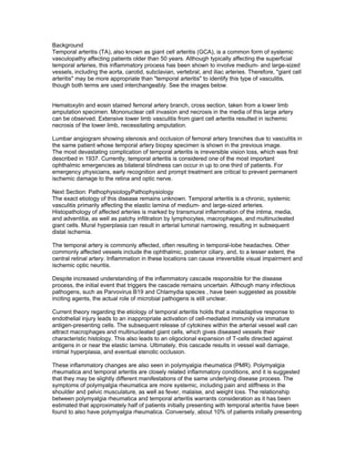
Hematoxylin and eosin stained femoral artery branch, cross section, taken from a lower limb
amputation specimen. Mononuclear cell invasion and necrosis in the media of this large artery
can be observed. Extensive lower limb vasculitis from giant cell arteritis resulted in ischemic
necrosis of the lower limb, necessitating amputation.
Lumbar angiogram showing stenosis and occlusion of femoral artery branches due to vasculitis in
the same patient whose temporal artery biopsy specimen is shown in the previous image.
The most devastating complication of temporal arteritis is irreversible vision loss, which was first
described in 1937. Currently, temporal arteritis is considered one of the most important
ophthalmic emergencies as bilateral blindness can occur in up to one third of patients. For
emergency physicians, early recognition and prompt treatment are critical to prevent permanent
ischemic damage to the retina and optic nerve.
Next Section: PathophysiologyPathophysiology
The exact etiology of this disease remains unknown. Temporal arteritis is a chronic, systemic
vasculitis primarily affecting the elastic lamina of medium- and large-sized arteries.
Histopathology of affected arteries is marked by transmural inflammation of the intima, media,
and adventitia, as well as patchy infiltration by lymphocytes, macrophages, and multinucleated
giant cells. Mural hyperplasia can result in arterial luminal narrowing, resulting in subsequent
distal ischemia.
The temporal artery is commonly affected, often resulting in temporal-lobe headaches. Other
commonly affected vessels include the ophthalmic, posterior ciliary, and, to a lesser extent, the
central retinal artery. Inflammation in these locations can cause irreversible visual impairment and
ischemic optic neuritis.
Despite increased understanding of the inflammatory cascade responsible for the disease
process, the initial event that triggers the cascade remains uncertain. Although many infectious
pathogens, such as Parvovirus B19 and Chlamydia species , have been suggested as possible
inciting agents, the actual role of microbial pathogens is still unclear.
Current theory regarding the etiology of temporal arteritis holds that a maladaptive response to
endothelial injury leads to an inappropriate activation of cell-mediated immunity via immature
antigen-presenting cells. The subsequent release of cytokines within the arterial vessel wall can
attract macrophages and multinucleated giant cells, which gives diseased vessels their
characteristic histology. This also leads to an oligoclonal expansion of T-cells directed against
antigens in or near the elastic lamina. Ultimately, this cascade results in vessel wall damage,
intimal hyperplasia, and eventual stenotic occlusion.
These inflammatory changes are also seen in polymyalgia rheumatica (PMR). Polymyalgia
rheumatica and temporal arteritis are closely related inflammatory conditions, and it is suggested
that they may be slightly different manifestations of the same underlying disease process. The
symptoms of polymyalgia rheumatica are more systemic, including pain and stiffness in the
shoulder and pelvic musculature, as well as fever, malaise, and weight loss. The relationship
between polymyalgia rheumatica and temporal arteritis warrants consideration as it has been
estimated that approximately half of patients initially presenting with temporal arteritis have been
found to also have polymyalgia rheumatica. Conversely, about 10% of patients initially presenting
2. with polymyalgia rheumatica were found to have temporal arteritis upon further investigation.
The etiology of temporal arteritis is multifactorial, as both genetic and environmental associations
have been identified. Some major histocompatibility complex molecules, particularly human
leukocyte antigen HLA-DR4 and HLA-DRB104 alleles, may have a role in a patient’s
susceptibility to temporal arteritis. Additionally, there is a statistically significant increase in the
incidence of temporal arteritis in northern latitudes.
Previous Next Section: EpidemiologyPathophysiology
The exact etiology of this disease remains unknown. Temporal arteritis is a chronic, systemic
vasculitis primarily affecting the elastic lamina of medium- and large-sized arteries.
Histopathology of affected arteries is marked by transmural inflammation of the intima, media,
and adventitia, as well as patchy infiltration by lymphocytes, macrophages, and multinucleated
giant cells. Mural hyperplasia can result in arterial luminal narrowing, resulting in subsequent
distal ischemia.
The temporal artery is commonly affected, often resulting in temporal-lobe headaches. Other
commonly affected vessels include the ophthalmic, posterior ciliary, and, to a lesser extent, the
central retinal artery. Inflammation in these locations can cause irreversible visual impairment and
ischemic optic neuritis.
Despite increased understanding of the inflammatory cascade responsible for the disease
process, the initial event that triggers the cascade remains uncertain. Although many infectious
pathogens, such as Parvovirus B19 and Chlamydia species , have been suggested as possible
inciting agents, the actual role of microbial pathogens is still unclear.
Current theory regarding the etiology of temporal arteritis holds that a maladaptive response to
endothelial injury leads to an inappropriate activation of cell-mediated immunity via immature
antigen-presenting cells. The subsequent release of cytokines within the arterial vessel wall can
attract macrophages and multinucleated giant cells, which gives diseased vessels their
characteristic histology. This also leads to an oligoclonal expansion of T-cells directed against
antigens in or near the elastic lamina. Ultimately, this cascade results in vessel wall damage,
intimal hyperplasia, and eventual stenotic occlusion.
These inflammatory changes are also seen in polymyalgia rheumatica (PMR). Polymyalgia
rheumatica and temporal arteritis are closely related inflammatory conditions, and it is suggested
that they may be slightly different manifestations of the same underlying disease process. The
symptoms of polymyalgia rheumatica are more systemic, including pain and stiffness in the
shoulder and pelvic musculature, as well as fever, malaise, and weight loss. The relationship
between polymyalgia rheumatica and temporal arteritis warrants consideration as it has been
estimated that approximately half of patients initially presenting with temporal arteritis have been
found to also have polymyalgia rheumatica. Conversely, about 10% of patients initially presenting
with polymyalgia rheumatica were found to have temporal arteritis upon further investigation.
The etiology of temporal arteritis is multifactorial, as both genetic and environmental associations
have been identified. Some major histocompatibility complex molecules, particularly human
leukocyte antigen HLA-DR4 and HLA-DRB104 alleles, may have a role in a patient’s
susceptibility to temporal arteritis. Additionally, there is a statistically significant increase in the
incidence of temporal arteritis in northern latitudes.
Epidemiology
Frequency
United States
Temporal arteritis is typically seen in female patients older than 50 years. Incidence increases
with age and can range from 1 in 10,000 to 5 in 10,000.
International
3. Rates are significantly higher in northern latitudes.
Mortality/Morbidity
Temporal arteritis does not appear to affect long-term survival.
Permanent visual loss is the most devastating consequence of temporal arteritis. The incidence
of ocular involvement varies greatly in the literature, ranging from 8-50%. Bilateral visual loss can
occur in up to 33% of patients.
Rarely, patients can experience neurologic manifestations such as transient ischemic attacks
(TIAs) or cerebral vascular accidents (CVAs)[1] . However, the exact relationship between
temporal arteritis and TIA/CVA is uncertain.
Race
Temporal arteritis occurs more frequently in white patients, especially those of northern European
descent.
Sex
Women develop temporal arteritis 2-3 times more frequently than men.
Age
Temporal arteritis rarely occurs in patients younger than 50 years. The mean age of onset is 72
years. Incidence of the disease increases significantly with increasing age.
PreviousHistory
Headache is the most common chief complaint and presents in over two thirds of patients with
temporal arteritis. The headache tends to be new or different in character than previous
headaches and is typically sudden in onset, localizing to the temporal region. However, pain with
temporal arteritis can occur diffusely through the occipital, frontal, or parietal regions as well.
Therefore, any new headache in patients older than 50 years warrants a consideration of
temporal arteritis.
Because temporal arteritis tends to affect the branches of the carotid artery, clinical
manifestations vary depending on the distribution of the ischemic vessel. For example, superficial
temporal artery involvement can lead to severe scalp tenderness during such simple acts as
resting the head on a pillow, combing hair, or wearing hats and eyeglasses. Patients may also
present with visible areas of scalp necrosis. Similarly, jaw claudication while speaking or chewing
is observed in patients with involvement of the maxillary artery, which can occur in half of patients
with temporal arteritis.
Visual loss may also be a presenting symptom and can be sudden and painless. Initial visual
symptoms are usually transient and intermittent, typically manifesting as unilateral visual loss or
occasionally diplopia. However, if left untreated, permanent blindness frequently results.
Constitutional symptoms due to systemic inflammation are common. These nonspecific
symptoms include fever, malaise, memory impairment, anorexia, weight loss, fatigue, and
depression. Additionally, polymyalgia rheumatica symptoms are present in about half of all cases
of temporal arteritis and may be the initial complaint in many patients.
Based on the 1990 American College of Rheumatology criteria for classification of temporal
arteritis, at least 3 of the following 5 items must be present (sensitivity 93.5%, specificity 91.2%)
[2] :
Age of onset older than 50 years
New-onset headache or localized head pain
Temporal artery tenderness to palpation or reduced pulsation
4. Erythrocyte sedimentation rate (ESR) greater than 50 mm/hPhysical
A thorough physical and neurological examination should be performed to exclude other possible
causes of headache and visual disturbances.
The head and face should be examined for inflamed and thickened arteries, tenderness to
palpation, tender scalp nodules, or necrotic areas of the scalp. Inflamed vessels may be tender
and warm. They may also appear thickened and dilated, such that the examiner may be able to
roll the artery between the fingers and the skull. Cranial nerve palsies, particularly of the sixth
nerve, should also be noted.
A complete eye examination should be performed, including visual acuity, visual field check, and
funduscopic as well as a slit lamp examinations. Special attention should be paid to the retinal
vessels, as other causes of loss of vision such as central retinal artery or vein occlusion can
cause a markedly abnormal funduscopic examination result. In temporal arteritis, the funduscopic
examination result may be normal or can show dilated retinal veins.
Previous
Proceed to Differential Diagnoses
Abnormal arterial biopsy (necrotizing vasculitis with granulomatous proliferation and infiltration)
Next Section: PhysicalPhysical
A thorough physical and neurological examination should be performed to exclude other possible
causes of headache and visual disturbances.
The head and face should be examined for inflamed and thickened arteries, tenderness to
palpation, tender scalp nodules, or necrotic areas of the scalp. Inflamed vessels may be tender
and warm. They may also appear thickened and dilated, such that the examiner may be able to
roll the artery between the fingers and the skull. Cranial nerve palsies, particularly of the sixth
nerve, should also be noted.
A complete eye examination should be performed, including visual acuity, visual field check, and
funduscopic as well as a slit lamp examinations. Special attention should be paid to the retinal
vessels, as other causes of loss of vision such as central retinal artery or vein occlusion can
cause a markedly abnormal funduscopic examination result. In temporal arteritis, the funduscopic
examination result may be normal or can show dilated retinal veins.
Previous
Proceed to Differential Diagnoses
Previous Next Section: EpidemiologyDifferentials
Glaucoma, Acute Angle-Closure
Headache, Migraine
Iritis and Uveitis
Orbital Infections
Polymyalgia Rheumatica
Retinal Artery Occlusion
Retinal Vein Occlusion
Stroke, Ischemic
Temporal Arteritis
Transient Ischemic AttackLaboratory Studies
Erythrocyte sedimentation rate (ESR) is a nonspecific marker of inflammation. It is the most
commonly used laboratory test in diagnosing temporal arteritis. Most patients with temporal
arteritis have an ESR greater than 80 mm/h. However, up to 20% of patients with temporal
arteritis may have a normal or low ESR, and thus a normal ESR level can not exclude a diagnosis
of temporal arteritis.
5. Normal ESR levels vary according to a patient’s age and sex. A general guide for estimating
normal ESR values uses the following formulas:
Males: (0.5 X age)
Females: (0.5 X age) + 5
C-reactive protein (CRP) is an acute-phase protein released by hepatocytes in inflammatory
states. 8CRP has been found to be elevated (>2.45 mg/dL) in patients with temporal arteritis,
even in patients with a normal ESR. An advantage to CRP is that the normal value range does
not vary with age or sex. A normal CRP is less than 0.5 mg/dL. An elevated CRP may help to
make the diagnosis when taken under consideration with a normal ESR.
Complete blood cell count (CBC) may reveal leukocytosis, anemia, or thrombocytosis. Several
studies have documented an association between an elevated platelet count >400 X 103/L and
temporal arteritis. However, this test is not sufficiently sensitive or specific to be useful in the
diagnosis of temporal arteritis.
Elevated liver function test (LFT) results, particularly alkaline phosphatase, are obtained in about
one half of patients with temporal arteritis
Next Section: Imaging Studies
Ultraviolet KeratitisColor duplex sonography of temporal arteries revealing a sonographic "halo
sign" around the temporal artery is specific although not very sensitive for temporal arteritis.[3]
Authors of a meta-analysis have concluded that with appropriate expertise, ultrasonography
should become a first-line investigation with biopsy only in patients with negative ultrasonography
findings.[4] Such a strategy remains investigational.
Previous Next Section: Procedures
Proceed to WorkupDefinitive diagnosis relies on temporal artery biopsy. Biopsy should be
performed as an outpatient procedure within 1 week after the initiation of corticosteroid therapy in
the emergency department. Although prompt follow-up is optimal, biopsy results have been useful
even 3-4 weeks after the initiation of steroid therapy. Since affected areas of vessels can be
patchy or segmental, multiple biopsy sites may be required. If clinical suspicion remains high after
an initial negative biopsy result, bilateral biopsies may be required. See the image below.
Hematoxylin and eosin stained superficial temporal artery biopsy specimen, cross section. The
hallmark histologic features of giant cell arteritis shown here include intimal thickening with
luminal stenosis, mononuclear inflammatory cell infiltrate with media invasion and necrosis, and
giant cell formation in the media.
PreviousPrehospital Care
Patients generally do not present via emergency medical services, and no particular prehospital
interventions are warranted.
Next Section: Emergency Department CareEmergency Department Care
Optimal care of patients with temporal arteritis in the emergency department involves maintaining
a high index of suspicion and a low threshold to treat.
Treatment consists of corticosteroids. Although corticosteroids are the only proven treatment of
temporal arteritis, few studies exist regarding dosing protocols. It is generally agreed that patients
with suspected temporal arteritis should be started on oral prednisone 60 mg/day in the
emergency department, with a temporal artery biopsy performed as an outpatient procedure
scheduled within 1 week.
Improvement of systemic symptoms typically occurs within 72 hours of initiation of therapy.
Patients should be counseled that corticosteroid therapy may be lengthy (1-2 y) and can lead to
the typical complications associated with long-term steroid use. Recent data suggest that initial
high-dose intravenous corticosteroid administration is beneficial in reducing temporal arteritis
remission rates.[5] However, further study is warranted before this is routinely practiced.
6. Previous Next Section: ConsultationsConsultations
An ophthalmologist should be consulted for a complete, dilated ocular examination to rule out
other causes of vision loss, particularly when the diagnosis is uncertain.
A rheumatologist or internist should direct follow-up care for these patients, monitor remissions
and recurrence, and manage complications associated with long-term corticosteroid therapy.
Medication Summary
Systemic steroids should be started. Oral steroids are effective. Intravenous steroids may be
administered if visual deficit is established or if the patient requires admission for other
reasons.Glucocorticoids
Class Summary
These agents have anti-inflammatory properties and cause profound and varied metabolic
effects.
View full drug information
Prednisone (Deltasone, Sterapred, Orasone)
DOC in treatment of temporal arteritis. Useful in treatment of inflammatory and allergic reactions
by reversing increased capillary permeability and suppressing PMN activity.
In prolonged treatment, taper over 1-2 wk.
View full drug information
Methylprednisolone (Solu-Medrol, Medrol, Depo-Medrol)
Decreases inflammation by suppressing migration of PMNs and reversing increased capillary
permeability.Hospital admission for temporal arteritis is unusual but may be indicated depending
upon the severity of symptoms and the ability of the patient to provide self-care at home.
Further Outpatient Care
Most patients can be treated on an outpatient basis.
An adequate quantity of corticosteroids should be prescribed.
Follow-up care should be arranged within 72 hours.
Symptoms typically improve within 1-3 days.
Corticosteroid therapy may last for 1-2 years, depending on the patient’s response.npatient &
Outpatient Medications
Nonsteroidal anti-inflammatory drugs can provide pain relief.
Methotrexate[6] and azathioprine have been used as both adjuncts and steroid-sparing agents for
temporal arteritis, but conclusive evidence regarding their efficacy remains uncertain. These
medications should not be prescribed from the emergency department, but they may be added at
a later time upon rheumatology follow-up.
Long-term steroid use (greater than 3 wk) may require the addition of calcium, vitamin D, and
bisphosphonate therapy to prevent steroid-induced osteoporosis.
Retrospective data shows that low-dose aspirin has been associated with a lower risk for
developing acute vision loss and stroke in patients with temporal arteritis. In the absence of
contraindications, low-dose (81 mg) aspirin should be considered along with corticosteroids.
[7Transfer
Hospital transfer is indicated only if visual disturbance is severe and cannot be adequately
evaluated and managed at the current facility.
Previous Next Section: ComplicationsComplications
Permanent vision loss is the most feared complication of untreated temporal arteritis and can
even progress in some cases despite the initiation of corticosteroid therapy. This will tend to
occur within the first 5 days of treatment if therapy is going to fail. As an outpatient, corticosteroid
doses should be increased until symptoms improve.
Approximately 50% of patients with temporal arteritis experience at least one flare-up that
requires prolonged corticosteroid therapy.
Patients with temporal arteritis are at increased risk for thoracic and abdominal aortic aneurysms
7. compared to age-matched controls.
Uncommon complications include CVA, memory loss, myocardial infarction, and peripheral
neuropathy.Prognosis
Generally, temporal arteritis is a self-limiting condition lasting up to 2 years.
Treatment with corticosteroids has proven to be effective in most cases, but the lengthy duration
of treatment can lead to corticosteroid-induced complications.
Previous Next Section: Patient EducationTimely follow-up care from the emergency department is
critical to accurately diagnose temporal arteritis.
Medication compliance and instructions to return to the emergency department if the condition
worsens should be emphasized.
Patients should be counseled that existing visual loss prior to arrival at the emergency
department may not be regained despite initiation of therapy.
Previous