1. SKELETAL MUSCLE FUNCTIONS
Skeletal muscles are essential for locomotion, breathing, posture,
speaking as well as non-motor functions, such as glucose and
potassium metabolism. They also contain protein reserves that can
be mobilized to support stress response to pathological conditions
(Figure 1).
Vid Jan1, Katarina Miš1, Urška Matkovič1, Zoran Grubič1, Matej Podbregar1,2, Sergej Pirkmajer1, Tomaž Marš1
1Institute of Pathophysiology, Faculty of Medicine, University of Ljubljana, Ljubljana, Slovenia
2Clinical Department for Anaesthesiology and Surgical Intensive Care, University Medical Centre Ljubljana
The role of in vitro innervated human skeletal muscle
cells in neuromuscular research
INTRODUCTION
Neuromuscular disorders (NMDs) can severely impair
different aspects of skeletal muscle function, thus
increasing morbidity and mortality. Unfortunately, only a
few effective treatments currently exist in armamentarium
against NMDs.
To discover new pharmacological targets for treatment of
NMDs reliable in vitro models are required.
METHODS
OUR MODEL
CONCLUSIONS
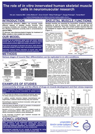
Various stages of muscle fiber differentiation can be replicated in in vitro conditions
Figure 4. Comparison of dexamethasone, agrin and
sugammadex effects on functional innervation. The number
of contracting units per contraction-positive spinal cord
explant in control (Control) co-cultures and in dexa-
methasone (Dex), agrin (Agr), sugammadex (Sug) or
dexamethasone plus sugammadex (Dex) co-cultures after 4
days treatment; n in indicates number of explants examined.
Data are means ± SD; *p < 0.05, versus control (t-test)
(Rezonja et al. Neurosci Lett. 2013, 549, 186-90).
Co-culture of rat embryonic spinal cord and human skeletal
muscle cells is one of advanced models for studying human
neuromuscular junction (NMJ) and muscle contraction in vitro.
It has all the advantages of standard cell culture, while still being
sufficiently complex to study major neuromuscular phenomena.
Innervation induces further maturation of muscle cells, which
leads to the development of functional contractile apparatus.
With co-culture of rat embryonic spinal cord and human
skeletal muscle cells we can investigate mechanisms
underlying metabolic adaptation, ion transport, myotoxicity, as
well as pharmacodynamics of various compounds.
Translational potential of our results is increased by using
contracting, functionally mature skeletal muscle cells.
Figure 1. Metabolic homeostasis of
the muscle. A schematic of energy
metabolism that occurs in skeletal
muscles and its interaction with other
organs. Skeletal muscles play an
important role in energy expenditure
and storage, but also a critical role in
metabolic homeostasis.
FA Oxidation: fatty acid oxidation,Glucose-6-
P: glucose-6-phosphate, LPL: lipoprotein
lipase, OXPHOS: oxidative phosphorylation,
TG: triglyceride, VLDL: very-low-density
lipoprotein. Adapted from Lee and Shulman.
Nat Med. 2009, Metabolic Syndrome e-Poster
http://www.nature.com/nm/e-
poster/eposter_full.html.
Glycogen
Glucose-6-P
Glucose
Brain
Sympathetic activity
(vasoconstriction)
Parasympathetic activity
(vasodilation)
Liver Gastrointestinal
tract
EXAMPLES OF STUDIES
Protein
Pyruvate
oxidation
MitochondriaTGs
Intramuscular
lipid droplets
Amino acids
Insulin Glucose
Fatty acids
Chylomicrons
White adipose
tissue
Cytokines
Adipokines
LipolysisLPL activity
Food intake =
energy needs
Glucose
production
VLDLs
Chylomicron
remnants
Synthesis Degradation
Insulin
signaling
Glucose
transport
Glucolysis
OXPHOS
FA Oxidation
Pancreas
<Figure 2. Schematic presentation of the
stages of muscle development (A) and
reproduction of these stages under in vitro
conditions (B). Myoblasts, derived from satellite
cells when placed in appropriate in vitro conditions
proliferate and at certain density start to fuse and
form multinucleated myotubes. To innervate
cultured myotubes, explants of rat embryonic
spinal cord are placed on a monolayer of muscle
cells. In those co-cultures motor neurons
functionally innervate myotubes, which then start
to contract. Adapted from Marš et al. In Handb. of
Toxicol. of Chem. Warf. Agents, Acad. Press, 2015.
>Figure 3. Phase-contrast micrograph of 11-
day-old co-culture. A representative micro-graph
of innervation area with parallely alined innervated
contracting myotubes (arrowhead) that contract
with the same contraction frequency representing
one contracting unit. Direction of explant and origin
of outgrowing axon is indicated with arrow. Scale
bar, 30 μm (Rezonja et al. Neurosci Lett. 2013,
549, 186-90).
Crossreactivity of drugs and the effects of drugs on muscle development and their stress response
Various substances can influence successful neuromuscular
transmission and functional innervation of skeletal muscle
fibers.
proliferating
mononuclear myoblast aneural
myotubes
contracting myotubes
(arrowheads)
innervated with motor
neurons (arrows)
myoblast myotube innervation of
myotube
mature myofibre
Figure 5. Secretion of IL-6 from the innervated
control and agrin-treated human muscle cell
cultures. In the human myotubes co-cultured with
rat spinal cord explants, the treatment with neural
agrin (1 nM, 3 days) increased the constitutive
secretion of IL-6. Data are means±SD, n indicates
total number of repeats analyzed. *p <0.05 vs.
control (Gros et al. J Mol Neurosci. 2014, 53, 454-
460).
In the 1st study the effects of the potential therapeutic agrin and
the widely used glucocorticoid dexamethasone were followed.
In addition, possible interaction between dexamethasone and
sugammadex, a selective relaxant binding drug, was examined.
Dexamethasone impaired functional innervation while agrin had
opposing effects (Figure 4).
This study was supported by the Slovenian Research Agency
(Research Programme # P3-0043).
In the 2nd study, the effects of agrin on secretion of interleukin-6
were studied in developing human muscle.
Secretion of IL-6 is a cytokine response in skeletal muscle to
various environmental stimuli, while autocrine and paracrine
effects of secreted IL-6 influence muscle development.
In the innervated co-cultures agrin treatment increased the
secretion of IL-6 (Figure 5).