2. 2 L.M. Fabbri et al.
chronic heart failure (CHF), coronary and peripheral vascular diseases, and the
metabolic syndrome.
Comorbidities are highly likely to affect health outcomes in COPD, and COPD•
patients are more likely to die of cardiovascular complications or cancer than of
respiratory failure.
Keywords Bronchitis • chronic diseases • chronic heart failure • emphysema • inflam-
mation • metabolic syndrome • osteoporosis
Introduction
Chronic obstructive pulmonary disease (COPD) is a major and growing cause of
morbidity and mortality [1, 2]. COPD is characterized by progressive and not fully
reversible airflow limitation, as measured by the forced expiratory volume in 1 second
(FEV1
). The airflow limitation is associated with a chronic inflammatory process in the
airways and lung parenchyma in response to noxious particles or gases, in particular,
tobacco smoking [1, 2].
The chronic inflammatory process in COPD is characterized by infiltration of
the airways by neutrophils, macrophages, and CD8+ T cells [3, 4]. Such features of
inflammation in COPD are likely driven by various cellular pathways, including pro-
inflammatory cytokines and mediators of oxidative stress released locally or systemi-
cally [5, 6]. More than airway inflammation, a systemic inflammation has also been
observed in COPD, with the detection of increased levels of cytokines and inflamma-
tory mediators, particularly from the endothelium, that can cause lung and airways
injury [5, 7–9].
The presence of systemic inflammation in COPD has been linked to a variety of
complications, including weight loss [7, 10], cachexia [11], osteoporosis [12, 13], and
cardiovascular diseases [14, 15]. Moreover, elevation of acute-phase proteins in COPD
patients suggests that individuals with increased systemic inflammatory markers, such
as fibrinogen or C-reactive protein (CRP), experience an accelerated decline in lung
function and are at increased risk of hospitalization for COPD [16, 17].
The aim of the present chapter is to discuss the pathophysiology of COPD, its
multiple components, risk factors, systemic consequences, and comorbidities based
on current knowledge.
Structural Changes
In COPD, the structural changes occurring in both the large and small airways and in
the lung parenchyma may be related to the characteristic clinical manifestations and
lung function changes of the disease, e.g., symptoms (i.e., chronic cough and sputum
production), airflow limitation, gas exchange abnormalities, pulmonary hypertension,
and cor pulmonale [18].
Inflammation of the submucosal glands and hyperplasia of goblet cells may con-
tribute to symptoms, such as chronic sputum production, although these pathological
abnormalities are not present in all patients with chronic sputum and may be present
in subjects without symptoms. The various pathological changes in the central airways
3. 1 Multiple Components of COPD 3
responsible for the symptoms of chronic cough and sputum production may continue
to be present throughout the course of the disease. Thus, these pathological changes
may be present either on their own or in combination with the changes in the peripheral
airways and lung parenchyma described below.
Fixed or poorly reversible expiratory airflow limitation is the hallmark functional
abnormality of COPD. Several pathological characteristics may contribute to airflow
limitation (Table 1.1). Airway remodeling and emphysema are most likely responsi-
ble for the fixed, poorly reversible component of airflow limitation, whereas airway
smooth muscle contraction, airway inflammation, and intraluminal accumulation of
mucus and plasma exudate may be responsible for the small part of airflow limitation
that is still reversible either spontaneously or with treatment.
The early decline in lung function in COPD is correlated with inflammatory changes
in the peripheral airways, similar to those that occur in the central airways: exudate of
fluid and cells in the airway wall and lumen, goblet and squamous cell metaplasia of
the epithelium, oedema of the airway mucosa due to inflammation, and excess mucus
in the airways due to goblet cell metaplasia. The most characteristic change in the
peripheral airways of patients with COPD is airway narrowing. Inflammation initiated
by cigarette smoking and other risk factors leads to repeated cycles of injury and repair
of the walls of the peripheral airways. Injury is caused either directly by inhaled toxic
particles and gases such as those found in cigarette smoke, or indirectly by the action
of inflammatory mediators; this injury then initiates repair processes. Although airway
repair is only partly understood, it seems likely that disordered repair processes can
lead to tissue remodeling with altered structure and function. Cigarette smoke may
impair lung repair mechanisms, thereby further contributing to altered lung structure
[19]. Even normal lung repair mechanisms can lead to airway remodeling because
tissue repair in the airways, as elsewhere in the body, may involve scar tissue forma-
tion. Inflammatory changes such as airway oedema and mucus hypersecretion also
contribute to airway narrowing in COPD. So does loss of elastic recoil, but fibrosis of
the small airways plays the largest role [18].
The relative contribution of airway remodeling and emphysema to airflow limitation
is not known. Indeed, there is still no consensus on whether the fixed airflow limitation
in COPD is mainly due to inflammation and scarring in the small airways or predomi-
nantly due to loss of support to the airways resulting from loss of alveolar walls due to
emphysema. In general, the studies assessing the lung function in relation to airway and
pulmonary structure have shown a relatively poor relationship between macroscopic
emphysema and the severity of airways obstruction as measured with spirometry.
However, the relative contribution of airway narrowing/fibrosis and emphysema to
airflow limitation may depend on the severity of COPD. Bronchiolar abnormalities
may contribute more significantly to mild–moderate chronic airflow limitation. When
Table 1.1 Causes of airflow limitation in chronic obstructive pulmonary disease
(COPD) (Adapted from [1]).
Irreversible • Fibrosis and narrowing of airways
• Loss of elastic recoil due to alveolar destruction
• Destruction of alveolar support that maintains patency of small airways
Reversible • Accumulation of inflammatory cells, mucus, and plasma exudate in bronchi
• Smooth muscle contraction in peripheral and central airways
• Dynamic hyperinflation during exercise
4. 4 L.M. Fabbri et al.
only subjects with less severe COPD are considered, several indices of bronchiolar
inflammation correlate with the degree of airflow obstruction. Indeed the most consist-
ent relationship between lung function and airway and pulmonary structure found in
subjects with severe COPD is between severe emphysema and severe airflow limitation.
The most important factor is emphysema and loss of elastic recoil. Most studies
in advanced COPD find that the best reflection of the severity of airflow limitation is
the extent of pulmonary emphysema. Thus, although both the destruction of alveolar
attachments to the outer wall of the peripheral airways and the loss of lung elastic
recoil produced by emphysema have been implicated in the pathogenesis of peripheral
airways obstruction, direct measurements of peripheral airways resistance show that
the structural changes in the airway wall are the most important cause of the increase
in peripheral airways resistance in COPD. Thus, when COPD becomes moderate or
severe, loss of elastic recoil becomes overwhelmingly important and may mask the
effects of bronchiolar disease on chronic airflow limitation [18].
Advanced COPD is also associated with gas exchange abnormalities, i.e., hypox-
emia and, later on, hypercapnia. Abnormal gas exchange may be due to several factors,
such as alveolar hypoventilation, altered gas transfer, inequalities in ventilation–
perfusion (V/Q) ratio, and right–left blood shunting. Several studies have demon-
strated a negative relationship between single breath or steady-state carbon monoxide
transfer factor (TLCO) and the degree of emphysema [20, 21]. In COPD, regardless
of the stage of disease and the presence or absence of emphysema, V/Q inequality is
generally accepted to be the major mechanism that impairs gas exchange and leads
to arterial hypoxemia. Impaired V/Q relationships may be caused by multiple patho-
logical changes in different lung structures, including the airways, parenchyma, and
pulmonary vasculature. Bronchiolar lesions are associated with V/Q mismatching, as
indicated by a significant correlation between bronchiolar inflammation and the distri-
bution of ventilation. Low V/Q units in the lungs may represent areas with a partially
blocked airway. Destruction of the lung surface area by emphysema reduces the diffus-
ing capacity and interferes with gas exchange [20]. The severity of pulmonary emphy-
sema appears to be related to the overall inefficiency of the lung as a gas exchanger.
This is reflected by the good correlation between the diffusing capacity of carbon
monoxide per liter of alveolar volume (TLCO/VA) and the severity of macroscopic
emphysema. Reduced ventilation due to loss of elastic recoil in emphysematous lung
together with loss of the capillary bed and generalized inhomogeneity of ventilation
due to the patchy nature of these changes leads to areas of V/Q mismatching which
result in arterial hypoxemia. Of the four classic mechanisms determining hypoxemia
and/or hypercapnia – alveolar hypoventilation, alveolar-end capillary diffusion limita-
tion to oxygen, increased intrapulmonary shunt, and ventilation–perfusion mismatch-
ing – the last is by far the most common intrapulmonary determinant of hypoxemia in
COPD. The role of shunt is almost negligible, even in the most life-threatening condi-
tions, and diffusion limitation is conspicuously absent. Hypercapnia can be induced
by ventilation–perfusion imbalance and/or alveolar hypoventilation, the latter being
predominant during exacerbations.
Pulmonary hypertension develops late in the natural history of patients with COPD,
is usually associated with the development of severe hypoxemia (Pa
O2 < 8 KPa or
60 mmHg), and is often hypercapnia as well. It represents the main cardiovascular
complication associated with the development of right ventricular hypertrophy (cor
pulmonale). Several factors are known to contribute to the development of pulmonary
hypertension in patients with COPD, i.e., (1) thickening of pulmonary vessel walls and
reduction of lumen; (2) hypoxia, which causes pulmonary vascular smooth muscle to
5. 1 Multiple Components of COPD 5
contract and further reduces the lumen; (3) impaired endothelium-dependent vasodilation
(reduction of nitric oxide (NO) synthesis or release in response to hypoxaemia);
(4) abnormal secretion of vasoconstrictor peptides such as endothelin-1; (5) destruc-
tion of the capillary bed, which further increases the pressure required to perfuse the
pulmonary circulation.
Cor pulmonale is defined as “hypertrophy of the right ventricle resulting from dis-
eases affecting the function and/or structure of the lungs, except when these pulmonary
alterations are the result of diseases that primarily affect the left side of the heart, as
in congenital heart disease.” This is a pathological definition and the clinical diagnosis
and assessment of right ventricular hypertrophy is difficult in life. The prevalence and
natural history of cor pulmonale in COPD are not yet clear. Pulmonary hypertension
and reduction of the vascular bed due to emphysema can lead to right ventricular
hypertrophy and right heart failure. Right heart failure is associated with venous stasis
and thrombosis that may result in pulmonary embolism and further compromise the
pulmonary circulation.
Pulmonary Inflammation
COPD is associated with inflammation of the central and peripheral airways, lung
parenchyma, and pulmonary vessels. In the central airways, the characteristics of
inflammation are (1) an increased number of mononuclear cells, particularly macro-
phages and CD8+ T lymphocytes, associated in a few cases with neutrophils, eosi-
nophils, and mast cells in the airway mucosa; (2) an increased number of neutrophils
and, in a few cases, eosinophils in bronchoalveolar lavage (BAL) fluid; (3) infiltration
of submucosal glands by neutrophils; (4) hyperplasia of goblet cells and enlarged
mucous glands; (5) metaplasia of airway epithelium that is otherwise well preserved;
and (6) no change in the structure of the lamina reticularis of the basement membrane.
The contribution of these pathological abnormalities to airflow limitation and gas
exchange abnormalities remains unclear. However, as airflow limitation progresses,
the number of T lymphocytes and macrophages increases in the submucosa, and the
number of CD8+ T lymphocytes also increases significantly [3, 4, 22, 23].
Peripheral airways show pathological abnormalities similar to those in the central
airways: (1) an increased number of mononuclear cells in the airway mucosa; (2) a sig-
nificant increase in lymphocytes and particularly in the number of CD8+ T lymphocytes
as airflow limitation progresses [3, 22, 23]; (3) an increased number of neutrophils in
the airway fluid; and (4) metaplasia of airway epithelium with hyperplasia of goblet
cells. In addition, peripheral airways show (1) increased intra-luminal mucus and exu-
date; (2) increased mass of smooth muscle; (3) fibrosis, distortion, and obliteration of
airway walls; and (4) loss of alveolar attachments to the bronchiolar walls.
In the lung parenchyma, the characteristic pathological abnormalities are
(1) panlobular emphysema and centrilobular emphysema in various combinations [24];
(2) paraseptal emphysema; and (3) loss of vascular bed linked to emphysema [25].
Patients with COPD may have a significant reversibility of airflow limitation in
response to bronchodilators and/or glucocorticosteroids [26–30]. These patients have
the same pathologic abnormalities as COPD patients, but may also have some patho-
logical features of asthma, namely, a small but significant increase of eosinophils in
BAL fluid and increased thickness of the reticular layer of the basement membrane
[27]. Moreover, in COPD with partial reversibility of airflow limitation, the bronchodilator
response is associated with increased exhaled NO and sputum eosinophilia [31].
6. 6 L.M. Fabbri et al.
Inflammatory Mediators
Migration and activation of inflammatory cells is regulated by cytokines and
chemokines, small proteins secreted by a variety of structural cells, such as epithelial
cells, endothelial cells, smooth muscle cells, and fibroblasts, as well as by inflam-
matory cells. Chemokines are a family of more than 40 small (8–11 kDa) cytokines
that have been defined primarily by their ability to mediate leukocyte chemotaxis.
Chemokines are divided into four major families on the basis of the spacing of the
most amino terminal of four conserved cysteine residues, i.e., the C, CC, CXC, and
CX3C families, where X represents any amino acid [32].
Bronchopulmonary inflammation of COPD is characterized by CD8+ T lym-
phocytes, Tc1 cells and neutrophils. Recent reports demonstrate that T cells from the
bronchial mucosa of patients with COPD predominantly express the chemokine receptor
CXCR3 [33–35] and produce gamma interferon (IFN-g), suggesting that these cells
elaborate type I cytokines [36].
CXCR3 binds three highly potent, inflammation-inducible CXC chemokine agonists:
IFN-g-inducible protein 10 (CXCL10/IP-10), a monokine induced by IFN-g, and IFN-
inducible T-cell a-chemoattractant (CXCL11/I-TAC) [36]. CD8+ cells are potent produc-
ers of IFN-g in COPD. IFN-g causes emphysema with alveolar enlargement, enhanced
lung volumes, enhanced pulmonary compliance, and macrophage- and neutrophil-rich
inflammation when inducibly targeted, in a transgenic fashion, to the adult mouse lung
[37]. Prominent protease and antiprotease alterations have also been noted in mice: the
induction and activation of matrix metalloproteinase (MMP)-12 and cathepsins B, H, D,
S, and L; the elaboration of MMP-9; and the selective inhibition of secretory leukocyte
proteinase inhibitor. Therefore, IFN-g causes emphysema and alterations in pulmonary
protease/antiprotease balance when expressed in pulmonary tissues [37].
Several chemokines involved in neutrophil chemotaxis belong to the CXC fam-
ily, of which interleukin-8 (IL-8, CXCL8) is the most prominent member relevant in
COPD. IL-8 levels are markedly elevated in the sputum and BAL fluid of patients with
COPD and correlate with the extent of neutrophilic inflammation and disease sever-
ity. The blocking of IL-8 reduces the chemotactic response of neutrophils to sputum
from COPD patients in vitro [38]. IL-8 activates human neutrophils by binding to two
G-protein-coupled receptors, designated CXCR1 and CXCR2. CXCR1 is selectively
activated by two chemokine ligands, IL-8 and granulocyte chemoattractant protein
(CXCL6/GCP2), the binding of which is coupled to activation and degranulation. CC
chemokines are also thought to be involved in COPD. Increased expression of mono-
cyte chemoattractant protein 1 (CCL2/MCP-1) and its cognate receptor CCR2 has
been demonstrated in macrophages and epithelial cells from patients with COPD [39].
CCL2/MCP-1 may play a role in recruitment of blood monocytes to the lungs of COPD
patients [40]. Elastin fragments generated at the diseased sites are potent chemoattract-
ants for monocytes in the lung in pulmonary emphysema [41]. Furthermore, an increase
in the number of dendritic cells in the airways of patients with COPD has been observed
and could be explained by the interaction of CCL20 with CCR6 on pulmonary dendritic
cells [42]; in particular, the number of dendritic cells in epithelium and adventitia of
small airways in patients with COPD increases significantly with disease severity.
Tumor necrosis factor-a (TNF-a) is also present in high concentrations in the sputum
of COPD patients and is detectable in bronchial biopsy specimens. TNF-a can induce
IL-8 expression in airway cells by activation of the transcription factor NF-kB [43].
In COPD, alveolar macrophages and neutrophils play a central role in the destruction of
lung parenchyma [44, 45]. Various proteases break down connective tissue components,
particularly elastin, in lung parenchyma to produce emphysema [46].
7. 1 Multiple Components of COPD 7
Three MMPs degrade elastin: MMP-2, MMP-9, and MMP-12. Elevated MMP-9
was found in homogenates of lung removed from patients undergoing lung volume
reduction surgery, meaning that the samples were from an advanced stage of emphy-
sema [47]. These lung homogenates revealed MMP-9-related elastolytic and gelati-
nolytic activity and significant elevations in MMP-9, with no significant increase in
neutrophil elastase by ELISA [48]. There is also an increase in the activity of MMP-9
and MMP-2 in the lung parenchyma of patients with emphysema. The role of MMP-9 in
the pathogenesis of emphysema is likely to be complex. a1
-Antitrypsin can be cleaved
by MMP-9. Increased chemotactic activity in BAL fluid is associated with the devel-
opment of smoking-related emphysema [McCrea, 1994 #188], and potentiation of IL-8
by MMP-9 could amplify the alveolar inflammation and destruction in smokers who
develop emphysema [49].
Oxidative stress may also have an important role in COPD. In smokers and in COPD
patients, there is an oxidant–antioxidant imbalance in favor of oxidants [6]. Smoking
produces a decrease in alveolar and lung glutathione (GSH) metabolism, widely recog-
nized as a central feature of COPD [50]. The severity of airway obstruction, as meas-
ured by FEV1
in smokers with COPD, correlates negatively with the concentration
of GSH in BAL fluid: the higher the concentration of GSH, the lower the FEV1
[51].
Patients with acute exacerbations of COPD show increased production of superoxide
anion by their peripheral blood neutrophils compared with stable patients. Products
of lipid peroxidation are significantly increased in plasma or BAL fluid from healthy
smokers and from patients with acute exacerbations of COPD, compared with healthy
nonsmokers [51]. Smoking also produces a fall in the plasma antioxidant capacity,
a global measure of systemic oxidative stress [50]. There are few measurements of
GSH in the lung epithelial lining fluid (ELF) from smokers and patients with COPD.
The GSH concentration in BAL fluid from patients with COPD is similar to that in
chronic smokers with no airflow obstruction. This emphasizes the effects of smoking
on GSH metabolism rather than reflecting the disease severity in the COPD patients.
The relevance of these studies is not known. It is possible that GSH concentrations in
BAL are influenced by the recent smoking of these patients.
Exposure to cigarette smoke is reported to induce goblet cell metaplasia and mucus
production [52], but the mechanism is unknown. Mucin synthesis in airways has been
reported to be regulated by the epidermal growth factor receptor (EGFR) system. In
particular, the exposure of airway epithelial cells to cigarette smoke upregulates EGFR
expression and activates EGFR tyrosine phosphorylation, causing mucin synthesis in
epithelial cells [53]. The mechanisms by which cigarette smoke induces EGFR activa-
tion are not completely defined [53]. However, it has been observed that cigarette smoke
is able to transactivate EGFR and thereby stimulate the transmembrane metalloprotei-
nase TNF-a converting enzyme, the ADAM (“A Disintegrin And Metalloproteinase”)
17. This metalloproteinase is able to cleave transmembrane amphiregulin, a ligand for
the EGFR. The binding of amphiregulin to EGFR then triggers the receptor’s activa-
tion [54]. Cigarette smoke can also cause neutrophil migration into the airways [55].
Neutrophils are also increased in the bronchial glands [56]. In vivo and in vitro stud-
ies show that these neutrophils can stimulate goblet cell degranulation [57]. Purified
neutrophil elastase has been shown to be a potent secretagogue for goblet cells in vitro
[58]. However, it is not clear whether this molecule is responsible for the effect of neu-
trophils on degranulation, since the cells themselves release little or no elastase in vitro,
even after activation with a variety of molecules (IL-8, leukotriene B4
, TNF-a). Rather,
the activation of neutrophils causes a translocation of elastase from the azurophilic
granules to the cell surface [59]. However, in a guinea pig model, the elastase inhibitor
ICI 200355 inhibited the neutrophil-dependent goblet cell degranulation seen after the
8. 8 L.M. Fabbri et al.
tracheal instillation of neutrophil chemoattractants [60].These results confirmed the
role of neutrophil elastase in degranulation, but the question of how elastase exerts
its effect still remains, since neutrophil activators and chemoattractants do not appear
to promote its release [61]. The potential role of elastase in goblet cell degranulation
implies a direct interaction between neutrophils and goblet cells. Some experimental
evidence suggests a direct interaction between neutrophils and goblet cells in the airway
epithelium, involving ICAM-1, CD11b, or CD18 cells [61]. These leukocyte adhesion
receptors play an important role in inflammation via their regulatory effects on leukocyte
adhesion, transmigration, and function [62].
Systemic Inflammation
Recent research suggests that inflammation is not confined to the lungs: inflammatory
cells and mediators generated in the lungs may enter the bloodstream and may have
systemic effects on other susceptible areas of the body. This may account for the obser-
vation that patients with COPD also present with systemic symptoms and comorbid
conditions, including muscle weakness, weight loss, cardiovascular disease, osteoporosis,
hypertension, depression, cognitive decline, sleep disorders, sexual dysfunction, and
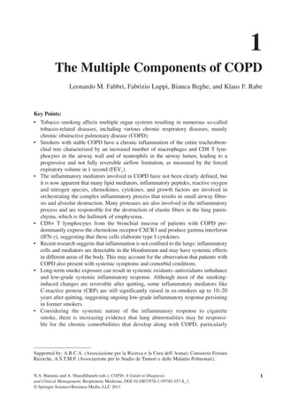
possibly diabetes [8, 13] (Fig. 1.1).
Fig. 1.1. The central role of inflammation in co-morbidity associated with chronic obstructive
pulmonary disease (COPD). Inflammation appears to play a central role in the pathogenesis of
COPD and other conditions that are increasingly being recognized as systemic inflammatory
diseases. As part of the chronic inflammatory process, TNF-a receptor polymorphisms are asso-
ciated with increased severity of disease possibly due to enhanced TNF-a effects. Also, CRP
levels can be increased directly by TNF-a and other cytokines, and elevated CRP and fibrinogen
may be crucial in the pathogenesis of cardiovascular disease. Reactive oxygen species (ROS)
released as a result of COPD may enhance the likelihood of developing cardiovascular disease,
diabetes, and osteoporosis. TNF-a tumor necrosis factor-alpha, CRP C reactive protein, ROS
reactive oxygen species
Muscle
Weakness / Wasting
Metabolic Syndrome
Type 2 diabetes
Osteoporosis
CRP
Cardiovascular
Events
Liver
+ve
?Local&Systemic
Inflammation
TNFα IL-6
CRP: C-Reactive Protein
COPD
Cigarette smoke
Pollutants
9. 1 Multiple Components of COPD 9
In recognition of the systemic component of COPD, the 2004 ATS/ERS Standards
for Diagnosis and Treatment of Patients with COPD were the first guidelines to
acknowledge that assessment of severity should ideally include systemic symptoms,
such as weight loss and muscle wasting [2]. Patients with COPD have higher baseline
levels of several circulating inflammatory markers [63]. The reasons are not clear,
and it remains unknown whether the systemic inflammation is a primary or secondary
phenomenon. Specific subsets of patients with COPD have been identified, and those
with increased resting energy expenditure and decreased fat-free mass have more
marked elevation of stable-state CRP and lipopolysaccharide binding protein [64].
Furthermore, those with higher levels of systemic inflammation lack a response to
nutritional supplementation [65], raising the possibility that this may be an associated
phenomenon rather than cause and effect. CRP levels in serum are raised in COPD
patients independent of cigarette smoking, and they are reduced in COPD patients who
use inhaled glucocorticosteroids [66]. There is also a genetic influence on CRP levels
in COPD [67], and CRP is considered a strong and independent predictor of COPD
outcomes in individuals with airflow limitation [17]. CRP is also associated with
impaired energy metabolism, impaired functional capacity, distress due to respiratory
symptoms [64], and lower quadriceps strength [68]. In the presence of exacerbated
respiratory symptoms, CRP levels in plasma may help to establish the diagnosis of a
COPD exacerbation [69].
Changes have also been noted in various inflammatory cells in peripheral blood,
including neutrophils and lymphocytes [70]. Patients with COPD have increased num-
bers of neutrophils in the lungs, increased activation of neutrophils in peripheral blood
and an increase in TNF-a and soluble TNF-R. It has been suggested that this indicates
the importance of a TNF-a/neutrophil axis in maintaining the COPD phenotype [43].
The central role of TNF-a in lung inflammation is not only supported by animal
models [43] but has also been implicated in the COPD phenotype with low body mass
index [71]. Cytokine production by macrophages is enhanced by hypoxia in vitro
[72], and thus the inverse correlation between arterial oxygen tension and circulating
TNF-a and solubleTNF-R may be the result of systemic hypoxia [72]. It is tempting
therefore to assume that TNF inhibition would be as beneficial in COPD as it has been
in other inflammatory conditions, such as rheumatoid arthritis and Crohn’s disease
[13]. However, this was also hypothesized for congestive heart failure (CHF). TNF-a
is believed to play a key role in the pathogenesis of CHF, and raised levels are associ-
ated with higher mortality in CHF [73]. However, studies using TNF-a blockade have
shown no benefit and possibly an increase in mortality for reasons that are not clear
[74], suggesting that it is not a simple cause and effect. Finally, both an increase in
CD8+ cells [75–78] and an increase in CD4+ cells [79] have been reported in patients
with COPD [75–78], highlighting the difficulties in conducting peripheral blood studies
in this disease [79].
Systemic Consequences and Comorbidities
Considering the systemic nature of the inflammatory response to irritants, particularly
cigarette smoke, there is increasing evidence that lung abnormalities may be responsible
not only for respiratory symptoms, e.g., dyspnoea, but also for the chronic comorbidi-
ties that develop along with COPD, particularly chronic heart failure (CHF), coronary
and peripheral vascular diseases, and the metabolic syndrome [8, 13]. Comorbidities are
highly likely to affect health outcomes in COPD, and COPD patients are more likely to
die of cardiovascular complications or cancer than of respiratory failure [80].
10. 10 L.M. Fabbri et al.
Progressive respiratory failure accounts for approximately one third of COPD-
related mortality; therefore, factors other than progression of lung disease must
play a substantial role. The most common comorbidities that have been described in
association with COPD are hypertension, diabetes, coronary artery disease [81, 82],
CHF [83], pulmonary infections, cancer [84], and pulmonary vascular disease [80].
The number of preexisting comorbidities in patients with COPD is associated with
increased in-hospital mortality [82]. Comorbid conditions that have been associated
with an increased mortality risk in COPD patients include chronic renal failure, cor
pulmonale [85], and pulmonary vascular disease [86]. Underlying heart diseases have
not been consistently associated with a higher mortality risk. However, since COPD is
frequently underreported, it is difficult to make an accurate estimate of how comorbid
conditions influence COPD mortality or, conversely, how COPD affects the outcome
of other diagnoses [80].
There is a growing body of evidence to indicate that persistent low-grade systemic
inflammation is present in stable COPD. Low-grade systemic inflammation has been
implicated in the pathogenesis of cardiovascular events and chronic myopathy of the
skeletal muscle. Since COPD patients suffer from excess morbidity and mortality
related to cardiovascular events, it has been suggested that systemic inflammation may be
the common link [13].
Chronic Heart Failure
CHF and COPD are two commonly encountered conditions in clinical practice. CHF
accounts for their frequent coexistence. The prevalence of COPD ranges from 20% to
32% in patients with CHF [15, 87]. FEV1
is as good a predictor of cardiovascular mor-
tality as serum cholesterol [88]. Ischemic heart disease, and not respiratory failure, is
the leading cause of death in COPD patients, with only a small fraction dying of respi-
ratory failure [87]. The relationship between COPD and cardiovascular events remains
unclear. Patients with COPD are not at increased risk for hypertension or left ventricular
hypertrophy; however, they consistently show evidence of low-grade systemic inflam-
mation that plays an increasingly recognized role in the pathogenesis of atherosclerosis
[87]. Patients with severe COPD are 2.18–2.74 times more likely to have elevated or
highly elevated circulating CRP levels than control subjects [89]. A working hypothesis
to account for the high prevalence of systolic dysfunction in patients with COPD is that
low-grade systemic inflammation accelerates the progression of coronary atheroscle-
rosis, which ultimately results in ischemic cardiomyopathy. Such a hypothesis fits the
clinical observation of a high incidence of left ventricular wall motion abnormalities
noted in patients with COPD and left ventricular dysfunction [87].
Diabetes
Type II diabetes mellitus is a disorder with an increased incidence in COPD. Systemic
inflammation may also explain why patients with COPD have an increased risk
of developing type II diabetes [90]. Some aspect of inflammation can predict the
development of diabetes and glucose disorders [91, 92], and fibrinogen, circulating
white blood cell count, and lower serum albumin predict the development of type II
diabetes [92]. Furthermore, patients with noninsulin-dependent diabetes mellitus have
increased circulating levels of TNF-a, IL-6, and CRP [93], which are also risk factors
for cardiovascular events in both sexes [94, 95].
11. 1 Multiple Components of COPD 11
The roles of circulating cytokines in the pathogenesis of diabetes and insulin resist-
ance have received increasing interest. Adipose tissue secretes numerous adipokines,
which markedly influence lipid and glucose/insulin metabolism [13]. Sonnenberg et al.
[96] proposed that TNF-a might be a mediator of the diabetic process. As described
above, this cytokine acts via its receptor to activate the nuclear transcription factor
NF-kB, leading to cytokine production, upregulation of adhesion molecules and
increasing oxidative stress. Indeed, this latter effect together with TNF-a may provide
a stimulating pathway that interferes with glucose metabolism and insulin sensitivity
[13]. This concept is supported by several clinical and experimental observations. It is
known that TNF-a expression is increased in patients with weight gain and insulin
resistance [97]. Perhaps this represents a modulating effect, as TNF-a stimulates
lipolysis [98] but TNF-a levels are associated with hyperinsulinemia and insulin resist-
ance [99]. Other studies have also confirmed that an acute-phase response (CRP) is
increased in obesity and associated with insulin resistance [100]. Furthermore,
adiponectin levels are reduced in obesity and associated with insulin resistance and
hyperinsulinemia [101]. However, the most direct supporting data for this putative axis
come from the obese, insulin-resistant mouse, in which TNF-a inhibition improves
insulin sensitivity [97]. These observations support the concept that inflammation, as
reflected in acute-phase proteins, is in some way intimately associated with the devel-
opment of glucose intolerance and insulin resistance.
Atherosclerosis
As atherosclerosis is the most common cause of coronary and peripheral artery disease
worldwide, the epidemiology and clinical consequences of peripheral arterial disease
are closely associated with classic risk factors for atherosclerosis, including cigarette
smoking.
Pai et al. [94] assessed the risk of coronary heart disease and related it to the circu-
lating levels of several inflammatory markers. They found that high levels of CRP and
IL-6 are significantly related to an increased risk in both males and females.
CRP is a type I acute-phase protein that possesses the ability to bind to bacteria, sub-
sequently facilitating the binding of complement necessary for bacterial killing and/or
phagocytosis. The protein can increase during an inflammatory process. TNF-a, IL-1,
and IL-6 stimulate CRP synthesis by inducing hepatic gene expression [102], implicat-
ing TNF-a at the core of the process. Macrophages have receptors for CRP, and CRP
can increase cytokine production [103, 104]. These features may be central to atheroma
production. CRP may deposit directly onto the arterial wall during atherogenesis, pos-
sibly via the Fcg receptor [105], facilitating monocyte adherence through the production
of the monocyte chemokine MCP-1. Further activation can result in production of other
pro-inflammatory cytokines and differentiation of the monocytes into macrophages. In
the presence of oxidized low-density lipoproteins, CRP can facilitate the production of
foam cells, which are the building blocks of atherosclerotic plaques.
A study by Smeeth et al. [106] indicated that the risk of a myocardial infarct or cer-
ebrovascular event is increased greatly within the first 3 days after an “acute systemic
respiratory tract infection,” defined by the authors as pneumonia, acute bronchitis,
“chest infections,” or influenza. These events are accompanied by a well-recognized
acute inflammatory response and cytokine production. Indeed, in patients with COPD,
not only is the baseline CRP > 3 mg/L in almost half of the patients, but the further rise
during an acute exacerbation [107] is also associated with a rise in fibrinogen [108],
12. 12 L.M. Fabbri et al.
increasing the prothrombotic risk. This may well account for the increased risk of
vascular events in COPD and particularly the increased mortality within a few months
of hospital admission for an acute exacerbation [109].
Osteoporosis
The risk of osteoporosis with steroid use is well known, but patients with COPD have
an increased risk even in the absence of steroid use. McEvoy et al. [110] observed
that vertebral fractures were present in up to 50% of steroid-naive males with COPD.
A more recent study by Bolton et al. [107] confirmed that osteopenia is a feature of
COPD and associated with an increase in circulating TNF-a. Postmenopausal oste-
oporosis is related to high serum levels of TNF-a and IL-6 [111]. It is known that
macrophages can differentiate into osteoclasts in the presence of marrow mesenchy-
mal cells. Marrow mesenchymal cells release the cytokine RANK ligand (RANKL),
a member of the TNF-a superfamily. TNF-a and IL-1 enhance this process and can
induce RANKL expression in marrow stromal cells and synergize with RANKL in
osteoclastogenesis [112]; osteoclast formation can also be induced by IL-6 independ-
ent of RANKL [113]. However, other inflammatory conditions, such as rheumatoid
arthritis [114] and periodontal disease [115], have T cells induced to produce RANKL,
and it is therefore likely that a similar process occurs in COPD.
The role of pro-inflammatory cytokines may therefore be central to the osteoporosis
associated with inflammatory disease. In support of this concept is the study reported
by Gianni et al. [111], who confirmed that raloxifene is able to decrease TNF-a tran-
scription and serum levels while increasing bone density. Again, these data support a
close association between the pro-inflammatory process and osteopenia.
Muscle Wasting
In patients with COPD, peripheral muscle dysfunction is clearly evidenced by the
significant reduction in both strength and endurance in the ambulatory muscles of
these patients in comparison with healthy subjects [116]. Other studies have shown
several morphological, structural, and biochemical abnormalities that could contribute
to the dysfunction [116].
Low body mass index, older age, and low arterial oxygen tension have been shown
to be significant independent predictors of mortality in COPD [117, 118]. More spe-
cifically, loss of fat-free mass adversely affects respiratory and peripheral muscle func-
tion, exercise capacity, and health status. Both weight loss and loss of fat-free mass
appear to be the result of a negative energy balance and are seen more commonly in
emphysema [119]. In starvation and nutritional imbalance there is an adaptive reduc-
tion in resting energy requirements [120]. In contrast, as in cachexia, increased resting
energy expenditure has been noted in many COPD patients, linked to systemic inflam-
mation [121, 122]. Furthermore, nutritional intake is also generally adequate (although
not during acute exacerbations). While there is no universally agreed upon definition
of cachexia, accelerated loss of skeletal muscle in the context of a chronic inflam-
matory response is a characteristic feature [123] not limited to COPD. Patients with
cachexia display preferential loss of fat-free mass, enhanced protein degradation [124],
and poor responsiveness to nutritional interventions [125, 126]. In addition, cachectic
patients exhibit changes in the metabolism of proteins, lipids, and carbohydrates that
are thought to be related to systemic rather than local inflammation [125, 127]. Thus,
muscle wasting in COPD displays similarities to the cachexia seen in CHF, renal failure,
13. 1 Multiple Components of COPD 13
AIDS and cancer, among others. Cachexia in these conditions is not only associated
with reduced survival [126, 128] but is also related to poor functional status and health-
related quality of life [123]. Common findings in all these conditions include increased
levels of circulating pro-inflammatory molecules, including TNF-a, IL-1, IL-6, IL-8,
and IFN-g, and reduced levels of anabolic hormones, including insulin-like growth
factors and testosterone [123].
TNF-a plays a central role in the muscle wasting and weight loss seen in COPD. It
has several direct effects (anorexia, altered levels of circulating hormones and catabolic
cytokines, and altered end organ sensitivities to them) that could promote muscle wasting
[129], predominantly via the ubiquitin pathway. This process is mediated by NF-kB, a
transcription factor that is inactive when bound to its inhibitor but that can be activated
by inflammatory cytokines, including TNF-a [130]. In muscle cells, NF-kB can interfere
with skeletal muscle differentiation and repair via inhibition of MyoD expression [131].
Conclusions
COPD can no longer be considered a disease only of the lungs. It is associated with a wide
variety of systemic consequences, most notably systemic inflammation. A better under-
standing of its origin, consequences, and potential therapy should prove to be of great
relevance and lead to better care of the patients suffering from this devastating disease.
Acknowledgements We thank M. McKenney for scientific assistance with the manuscript,
and E. Veratelli for her scientific secretarial assistance.
References
1. Rabe KF, Hurd S, Anzueto A, Barnes PJ, Buist SA, Calverley P, Fukuchi Y, Jenkins C,
Rodriguez-Roisin R, van Weel C, Zielinski J (2007) Global initiative for chronic obstruc-
tive lung disease. Global strategy for the diagnosis, management, and prevention of chronic
obstructive pulmonary disease: GOLD executive summary. Am J Respir Crit Care Med
176(6):532–555
2. Celli BR, Snider GL, Heffner J, Tiep B, Ziment I, Make B, Braman S, Olsen G, Philips Y
(1995) Standards for the diagnosis and care of patients with chronic obstructive pulmonary
disease. American Thoracic Society. Am J Respir Crit Care Med 152(5 Pt 2):S77–S121
3. Hogg JC, Chu F, Utokaparch S, Woods R, Elliott WM, Buzatu L, Cherniack RM, Rogers
RM, Sciurba FC, Coxson HO, Pare PD (2004) The nature of small-airway obstruction in
chronic obstructive pulmonary disease. N Engl J Med 350(26):2645–2653
4. Cosio MG, Saetta M, Agusti A (2009) Immunologic aspects of chronic obstructive pulmo-
nary disease. N Engl J Med 360(23):2445–2454
5. Barnes PJ, Shapiro SD, Pauwels RA (2003) Chronic obstructive pulmonary disease:
molecular and cellular mechanisms. Eur Respir J 22(4):672–688
6. MacNee W (2005) Pulmonary and systemic oxidant/antioxidant imbalance in chronic
obstructive pulmonary disease. Proc Am Thorac Soc 2(1):50–60
7. Agusti AG (2005) Systemic effects of chronic obstructive pulmonary disease. Proc Am
Thorac Soc 2(4):367–370, discussion 371–2
8. Barnes PJ, Celli BR (2009) Systemic manifestations and comorbidities of COPD. Eur
Respir J 33(5):1165–1185
9. Yanbaeva DG, Dentener MA, Creutzberg EC, Wesseling G, Wouters EF (2007) Systemic
effects of smoking. Chest 131(5):1557–1566
14. 14 L.M. Fabbri et al.
10. Vermeeren MA, Creutzberg EC, Schols AM, Postma DS, Pieters WR, Roldaan AC,
Wouters EF (2006) Prevalence of nutritional depletion in a large out-patient population of
patients with COPD. Respir Med 100(8):1349–1355
11. Rutten EP, Franssen FM, Engelen MP, Wouters EF, Deutz NE, Schols AM (2006) Greater
whole-body myofibrillar protein breakdown in cachectic patients with chronic obstructive
pulmonary disease. Am J Clin Nutr 83(4):829–834
12. Sabit R, Bolton CE, Edwards PH, Pettit RJ, Evans WD, McEniery CM, Wilkinson IB,
Cockcroft JR, Shale DJ (2007) Arterial stiffness and osteoporosis in chronic obstructive
pulmonary disease. Am J Respir Crit Care Med 175:1259–1265
13. Sevenoaks MJ, Stockley RA (2006) Chronic obstructive pulmonary disease, inflammation
and co-morbidity–a common inflammatory phenotype? Respir Res 7:70
14. Rutten FH, Cramer MJ, Zuithoff NP, Lammers JW, Verweij W, Grobbee DE, Hoes AW
(2007) Comparison of B-type natriuretic peptide assays for identifying heart failure in
stable elderly patients with a clinical diagnosis of chronic obstructive pulmonary disease.
Eur J Heart Fail 9(6–7):651–659
15. Rutten FH, Moons KG, Cramer MJ, Grobbee DE, Zuithoff NP, Lammers JW, Hoes AW
(2005) Recognising heart failure in elderly patients with stable chronic obstructive pulmo-
nary disease in primary care: cross sectional diagnostic study. BMJ 331(7529):1379
16. Dahl M, Tybjaerg-Hansen A, Vestbo J, Lange P, Nordestgaard BG (2001) Elevated plasma
fibrinogen associated with reduced pulmonary function and increased risk of chronic
obstructive pulmonary disease. Am J Respir Crit Care Med 164(6):1008–1011
17. Dahl M, Vestbo J, Lange P, Bojesen SE, Tybjaerg-Hansen A, Nordestgaard BG (2007)
C-reactive protein as a predictor of prognosis in chronic obstructive pulmonary disease.
Am J Respir Crit Care Med 175(3):250–255
18. Saetta M, Turato G, Maestrelli P, Mapp CE, Fabbri LM (2001) Cellular and structural bases
of chronic obstructive pulmonary disease. Am J Respir Crit Care Med 163(6):1304–1309
19. Global Initiative for Asthma (2002) Update of the NHLBI/WHO workshop report: global
strategy for asthma management and prevention. Issued January 1995. National Institutes
of Health, National Heart Lung and Blood Institute, Bethesda, MD, Document no.
02–3659
20. Gould GA, MacNee W, McLean A, Warren PM, Redpath A, Best JJ, Lamb D, Flenley DC
(1988) CT measurements of lung density in life can quantitate distal airspace enlargement – an
essential defining feature of human emphysema. Am Rev Respir Dis 137(2):380–392
21. McLean A, Warren PM, Gillooly M, MacNee W, Lamb D (1992) Microscopic and mac-
roscopic measurements of emphysema: relation to carbon monoxide gas transfer. Thorax
47(3):144–149
22. ÓShaughnessy TC, Ansari TW, Barnes NC, Jeffery PK (1997) Inflammation in bronchial
biopsies of subjects with chronic bronchitis: inverse relationship of CD8+ T lymphocytes
with FEV1. Am J Respir Crit Care Med 155(3):852–857
23. Saetta M, Di Stefano A, Turato G, Facchini FM, Corbino L, Mapp CE, Maestrelli P,
Ciaccia A, Fabbri LM (1998) CD8+ T-lymphocytes in peripheral airways of smokers with
chronic obstructive pulmonary disease. Am J Respir Crit Care Med 157(3 Pt 1):822–826
24. Guerassimov A, Hoshino Y, Takubo Y, Turcotte A, Yamamoto M, Ghezzo H,
Triantafillopoulos A, Whittaker K, Hoidal JR, Cosio MG (2004) The development of
emphysema in cigarette smoke-exposed mice is strain dependent. Am J Respir Crit Care
Med 170(9):974–980
25. Baraldo S, Saetta M, Cosio MG (2003) Pathophysiology of the small airways. Semin
Respir Crit Care Med 24(5):465–472
26. Anthonisen NR, Wright EC (1986) Bronchodilator response in chronic obstructive pulmonary
disease. Am Rev Respir Dis 133(5):814–819
27. Chanez P, Vignola A, ÓShaughnessy T, Enander I, Li D, Jeffery P, Bousquet J (1997)
Corticosteroid reversibility in COPD is related to features of asthma. Am J Respir Crit Care
Med 155:1529–1534
28. Enright P, Quanjer P (2007) Don’t diagnose mild COPD without confirming airway
obstruction after an inhaled bronchodilator. COPD 4(2):89–90
15. 1 Multiple Components of COPD 15
29. Gross NJ (1986) COPD: a disease of reversible air-flow obstruction. Am Rev Respir Dis
133(5):725–726
30. Perez-Padilla R, Hallal PC, Vazquez-Garcia JC, Muino A, Maquez M, Lopez MV, de Oca
MM, Talamo C, Valdivia G, Pertuze J, Jardim J, Menezes AM (2007) Impact of bronchodi-
lator use on the prevalence of COPD in population-based samples. COPD 4(2):113–120
31. Papi A, Romagnoli M, Baraldo S, Braccioni F, Guzzinati I, Saetta M, Ciaccia A, Fabbri
LM (2000) Partial reversibility of airflow limitation and increased exhaled NO and
sputum eosinophilia in chronic obstructive pulmonary disease. Am J Respir Crit Care Med
162(5):1773–1777
32. Zlotnik A, Yoshie O (2000) Chemokines: a new classification system and their role in
immunity. Immunity 12(2):121–127
33. Grumelli S, Corry DB, Song LZ, Song L, Green L, Huh J, Hacken J, Espada R, Bag R,
Lewis DE, Kheradmand F (2004) An immune basis for lung parenchymal destruction in
chronic obstructive pulmonary disease and emphysema. PLoS Med 1(1):e8
34. Panina-Bordignon P, Papi A, Mariani M, Di Lucia P, Casoni G, Bellettato C, Buonsanti C,
Miotto D, Mapp C, Villa A, Arrigoni G, Fabbri LM, Sinigaglia F (2001) The C-C chem-
okine receptors CCR4 and CCR8 identify airway T cells of allergen-challenged atopic
asthmatics. J Clin Invest 107(11):1357–1364
35. Saetta M, Mariani M, Panina-Bordignon P, Turato G, Buonsanti C, Baraldo S, Bellettato
CM, Papi A, Corbetta L, Zuin R, Sinigaglia F, Fabbri LM (2002) Increased expression of the
chemokine receptor CXCR3 and its ligand CXCL10 in peripheral airways of smokers with
chronic obstructive pulmonary disease. Am J Respir Crit Care Med 165(10):1404–1409
36. Donnelly LE, Barnes PJ (2006) Chemokine receptors as therapeutic targets in chronic
obstructive pulmonary disease. Trends Pharmacol Sci 27(10):546–553
37. Wang Z, Zheng T, Zhu Z, Homer RJ, Riese RJ, Chapman HA Jr, Shapiro SD, Elias JA
(2000) Interferon gamma induction of pulmonary emphysema in the adult murine lung.
J Exp Med 192(11):1587–1600
38. Beeh KM, Kornmann O, Buhl R, Culpitt SV, Giembycz MA, Barnes PJ (2003) Neutrophil
chemotactic activity of sputum from patients with COPD: role of interleukin 8 and leuko-
triene B4. Chest 123(4):1240–1247
39. de Boer WI, Sont JK, van Schadewijk A, Stolk J, van Krieken JH, Hiemstra PS (2000)
Monocyte chemoattractant protein 1, interleukin 8, and chronic airways inflammation in
COPD. J Pathol 190(5):619–626
40. Maus UA, Koay MA, Delbeck T, Mack M, Ermert M, Ermert L, Blackwell TS, Christman
JW, Schlondorff D, Seeger W, Lohmeyer J (2002) Role of resident alveolar macrophages
in leukocyte traffic into the alveolar air space of intact mice. Am J Physiol Lung Cell Mol
Physiol 282(6):L1245–L1252
41. Hunninghake GW, Davidson JM, Rennard S, Szapiel S, Gadek JE, Crystal RG (1981)
Elastin fragments attract macrophage precursors to diseased sites in pulmonary emphysema.
Science 212(4497):925–927
42. Demedts IK, Bracke KR, Van Pottelberge G, Testelmans D, Verleden GM, Vermassen FE,
Joos GF, Brusselle GG (2007) Accumulation of dendritic cells and increased CCL20 levels
in the airways of patients with chronic obstructive pulmonary disease. Am J Respir Crit
Care Med 175(10):998–1005
43. Mukhopadhyay S, Hoidal JR, Mukherjee TK (2006) Role of TNFalpha in pulmonary
pathophysiology. Respir Res 7:125
44. Sibille Y, Reynolds HY (1990) Macrophages and polymorphonuclear neutrophils in lung
defense and injury. Am Rev Respir Dis 141(2):471–501
45. HunninghakeGW,CrystalRG(1983)Cigarettesmokingandlungdestruction.Accumulation
of neutrophils in the lungs of cigarette smokers. Am Rev Respir Dis 128(5):833–838
46. La Rocca G, Anzalone R, Magno F, Farina F, Cappello F, Zummo G (2007) Cigarette
smoke exposure inhibits extracellular MMP-2 (gelatinase A) activity in human lung fibrob-
lasts. Respir Res 8:23
47. Atkinson JJ, Senior RM (2003) Matrix metalloproteinase-9 in lung remodeling. Am J
Respir Cell Mol Biol 28(1):12–24
16. 16 L.M. Fabbri et al.
48. Ohnishi K, Takagi M, Kurokawa Y, Satomi S, Konttinen YT (1998) Matrix metalloproteinase-
mediated extracellular matrix protein degradation in human pulmonary emphysema. Lab
Invest 78(9):1077–1087
49. Van den Steen PE, Proost P, Wuyts A, Van Damme J, Opdenakker G (2000) Neutrophil
gelatinase B potentiates interleukin-8 tenfold by aminoterminal processing, whereas it
degrades CTAP-III, PF-4, and GRO-alpha and leaves RANTES and MCP-2 intact. Blood
96(8):2673–2681
50. Peltoniemi MJ, Rytila PH, Harju TH, Soini YM, Salmenkivi KM, Ruddock LW, Kinnula
VL (2006) Modulation of glutaredoxin in the lung and sputum of cigarette smokers and
chronic obstructive pulmonary disease. Respir Res 7:133
51. MacNee W, Rahman I (2001) Is oxidative stress central to the pathogenesis of chronic
obstructive pulmonary disease? Trends Mol Med 7(2):55–62
52. Hegab AE, Sakamoto T, Nomura A, Ishii Y, Morishima Y, Iizuka T, Kiwamoto T, Matsuno
Y, Homma S, Sekizawa K (2007) Niflumic acid and AG-1478 reduce cigarette smoke-
induced mucin synthesis: the role of hCLCA1. Chest 131(4):1149–1156
53. Takeyama K, Jung B, Shim JJ, Burgel PR, Dao-Pick T, Ueki IF, Protin U, Kroschel
P, Nadel JA (2001) Activation of epidermal growth factor receptors is responsible for
mucin synthesis induced by cigarette smoke. Am J Physiol Lung Cell Mol Physiol
280(1):L165–L172
54. Lemjabbar H, Li D, Gallup M, Sidhu S, Drori E, Basbaum C (2003) Tobacco smoke-
induced lung cell proliferation mediated by tumor necrosis factor alpha-converting enzyme
and amphiregulin. J Biol Chem 278(28):26202–26207
55. Nishikawa M, Kakemizu N, Ito T, Kudo M, Kaneko T, Suzuki M, Udaka N, Ikeda H,
Okubo T (1999) Superoxide mediates cigarette smoke-induced infiltration of neutrophils
into the airways through nuclear factor-kappaB activation and IL-8 mRNA expression in
guinea pigs in vivo. Am J Respir Cell Mol Biol 20(2):189–198
56. Saetta M, Turato G, Facchini FM, Corbino L, Lucchini RE, Casoni G, Maestrelli P, Mapp
CE, Ciaccia A, Fabbri LM (1997) Inflammatory cells in the bronchial glands of smokers
with chronic bronchitis. Am J Respir Crit Care Med 156(5):1633–1639
57. Takeyama K, Agusti C, Ueki I, Lausier J, Cardell LO, Nadel JA (1998) Neutrophil-
dependent goblet cell degranulation: role of membrane-bound elastase and adhesion
molecules. Am J Physiol 275(2 Pt 1):L294–L302
58. Sommerhoff CP, Nadel JA, Basbaum CB, Caughey GH (1990) Neutrophil elastase and
cathepsin G stimulate secretion from cultured bovine airway gland serous cells. J Clin
Invest 85(3):682–689
59. Owen CA, Campbell MA, Sannes PL, Boukedes SS, Campbell EJ (1995) Cell surface-
bound elastase and cathepsin G on human neutrophils: a novel, non-oxidative mechanism
by which neutrophils focus and preserve catalytic activity of serine proteinases. J Cell Biol
131(3):775–789
60. Tokuyama K, Kuo HP, Rohde JA, Barnes PJ, Rogers DF (1990) Neural control of goblet
cell secretion in guinea pig airways. Am J Physiol 259(2 Pt 1):L108–L115
61. Nadel JA (2000) Role of neutrophil elastase in hypersecretion during COPD exacerbations,
and proposed therapies. Chest 117(5 Suppl 2):386S–389S
62. Liles WC, Dale DC, Price TH, Gaviria JM, Turner T, Saoud J, Frumkin LR (2000)
Inhibition of in vivo neutrophil transmigration by a novel humanized anti-CD11/CD18
monoclonal antibody. Cytokines Cell Mol Ther 6(3):121–126
63. Gan WQ, Man SF, Senthilselvan A, Sin DD (2004) Association between chronic obstructive
pulmonary disease and systemic inflammation: a systematic review and a meta-analysis.
Thorax 59(7):574–580
64. Broekhuizen R, Wouters EF, Creutzberg EC, Schols AM (2006) Raised CRP levels mark
metabolic and functional impairment in advanced COPD. Thorax 61(1):17–22
65. Creutzberg EC, Schols AM, Weling-Scheepers CA, Buurman WA, Wouters EF (2000)
Characterization of nonresponse to high caloric oral nutritional therapy in depleted patients with
chronic obstructive pulmonary disease. Am J Respir Crit Care Med 161(3 Pt 1):745–752
17. 1 Multiple Components of COPD 17
66. Pinto-Plata VM, Mullerova H, Toso JF, Feudjo-Tepie M, Soriano JB, Vessey RS, Celli
BR (2006) C-reactive protein in patients with COPD, control smokers and non-smokers.
Thorax 61(1):23–28
67. Hersh CP, Miller DT, Kwiatkowski DJ, Silverman EK (2006) Genetic determinants of
C-reactive protein in COPD. Eur Respir J 28(6):1156–1162
68. Yende S, Waterer GW, Tolley EA, Newman AB, Bauer DC, Taaffe DR, Jensen R,
Crapo R, Rubin S, Nevitt M, Simonsick EM, Satterfield S, Harris T, Kritchevsky SB
(2006) Inflammatory markers are associated with ventilatory limitation and muscle
dysfunction in obstructive lung disease in well functioning elderly subjects. Thorax
61(1):10–16
69. Hurst JR, Donaldson GC, Perera WR, Wilkinson TM, Bilello JA, Hagan GW, Vessey RS,
Wedzicha JA (2006) Use of plasma biomarkers at exacerbation of chronic obstructive
pulmonary disease. Am J Respir Crit Care Med 174(8):867–874
70. Sauleda J, Garcia-Palmer FJ, Gonzalez G, Palou A, Agusti AG (2000) The activity of
cytochrome oxidase is increased in circulating lymphocytes of patients with chronic
obstructive pulmonary disease, asthma, and chronic arthritis. Am J Respir Crit Care Med
161(1):32–35
71. Eid AA, Ionescu AA, Nixon LS, Lewis-Jenkins V, Matthews SB, Griffiths TL, Shale DJ
(2001) Inflammatory response and body composition in chronic obstructive pulmonary
disease. Am J Respir Crit Care Med 164(8 Pt 1):1414–1418
72. Takabatake N, Nakamura H, Abe S, Inoue S, Hino T, Saito H, Yuki H, Kato S, Tomoike H
(2000) The relationship between chronic hypoxemia and activation of the tumor necrosis
factor-alpha system in patients with chronic obstructive pulmonary disease. Am J Respir
Crit Care Med 161(4 Pt 1):1179–1184
73. Deswal A, Petersen NJ, Feldman AM, Young JB, White BG, Mann DL (2001) Cytokines
and cytokine receptors in advanced heart failure: an analysis of the cytokine database from
the Vesnarinone trial (VEST). Circulation 103(16):2055–2059
74. Coletta AP, Clark AL, Banarjee P, Cleland JG (2002) Clinical trials update: renewal (ren-
aissance and recover) and attach. Eur J Heart Fail 4(4):559–561
75. de Jong JW, van der Belt-Gritter B, Koeter GH, Postma DS (1997) Peripheral blood lym-
phocyte cell subsets in subjects with chronic obstructive pulmonary disease: association
with smoking, IgE and lung function. Respir Med 91(2):67–76
76. Domagala-Kulawik J, Hoser G, Dabrowska M, Chazan R (2007) Increased proportion
of Fas positive CD8+ cells in peripheral blood of patients with COPD. Respir Med
101(6):1338–1343
77. Glader P, von Wachenfeldt K, Lofdahl CG (2006) Systemic CD4+ T-cell activation is
correlated with FEV1 in smokers. Respir Med 100(6):1088–1093
78. Kim WD, Kim WS, Koh Y, Lee SD, Lim CM, Kim DS, Cho YJ (2002) Abnormal peripheral
blood T-lymphocyte subsets in a subgroup of patients with COPD. Chest 122(2):437–444
79. Koch A, Gaczkowski M, Sturton G, Staib P, Schinkothe T, Klein E, Rubbert A, Bacon
K, Wassermann K, Erdmann E (2007) Modification of surface antigens in blood CD8+
T-lymphocytes in COPD: effects of smoking. Eur Respir J 29(1):42–50
80. Mannino DM, Watt G, Hole D, Gillis C, Hart C, McConnachie A, Davey Smith G, Upton
M, Hawthorne V, Sin DD, Man SF, Van Eeden S, Mapel DW, Vestbo J (2006) The natural
history of chronic obstructive pulmonary disease. Eur Respir J 27(3):627–643
81. Holguin F, Folch E, Redd SC, Mannino DM (2005) Comorbidity and mortality in COPD-
related hospitalizations in the United States, 1979 to 2001. Chest 128(4):2005–2011
82. Sidney S, Sorel M, Quesenberry CP Jr, DeLuise C, Lanes S, Eisner MD (2005) COPD and
incident cardiovascular disease hospitalizations and mortality: Kaiser Permanent Medical
Care Program. Chest 128(4):2068–2075
83. Sin DD, Wu L, Anderson JA, Anthonisen NR, Buist AS, Burge PS, Calverley PM, Connett
JE, Lindmark B, Pauwels RA, Postma DS, Soriano JB, Szafranski W, Vestbo J (2005)
Inhaled corticosteroids and mortality in chronic obstructive pulmonary disease. Thorax
60(12):992–997, Epub 2005 Oct 14
18. 18 L.M. Fabbri et al.
84. Balkwill F, Mantovani A (2001) Inflammation and cancer: back to Virchow? Lancet
357(9255):539–545
85. Naeije R (2005) Pulmonary hypertension and right heart failure in chronic obstructive
pulmonary disease. Proc Am Thorac Soc 2(1):20–22
86. Naeije R, Vizza D (2005) Current perspectives modern hemodynamic evaluation of the
pulmonary circulation. Application to pulmonary arterial hypertension and embolic pul-
monary hypertension. Ital Heart J 6(10):784–788
87. Le Jemtel TH, Padeletti M, Jelic S (2007) Diagnostic and therapeutic challenges in patients
with coexistent chronic obstructive pulmonary disease and chronic heart failure. J Am Coll
Cardiol 49(2):171–180
88. Hole DJ, Watt GC, Davey-Smith G, Hart CL, Gillis CR, Hawthorne VM (1996) Impaired
lung function and mortality risk in men and women: findings from the Renfrew and Paisley
prospective population study. Bmj 313(7059):711–715, discussion 715–6
89. Sin DD, Man SF (2003) Why are patients with chronic obstructive pulmonary disease at
increased risk of cardiovascular diseases? The potential role of systemic inflammation in
chronic obstructive pulmonary disease. Circulation 107(11):1514–1519
90. Rana JS, Mittleman MA, Sheikh J, Hu FB, Manson JE, Colditz GA, Speizer FE, Barr RG,
Camargo CA Jr (2004) Chronic obstructive pulmonary disease, asthma, and risk of type 2
diabetes in women. Diab Care 27(10):2478–2484
91. Barzilay JI, Abraham L, Heckbert SR, Cushman M, Kuller LH, Resnick HE, Tracy RP
(2001) The relation of markers of inflammation to the development of glucose disorders in
the elderly: the cardiovascular health study. Diabetes 50(10):2384–2389
92. Schmidt MI, Duncan BB, Sharrett AR, Lindberg G, Savage PJ, Offenbacher S, Azambuja
MI, Tracy RP, Heiss G (1999) Markers of inflammation and prediction of diabetes
mellitus in adults (atherosclerosis risk in communities study): a cohort study. Lancet
353(9165):1649–1652
93. Pickup JC, Mattock MB, Chusney GD, Burt D (1997) NIDDM as a disease of the innate
immune system: association of acute-phase reactants and interleukin-6 with metabolic
syndrome X. Diabetologia 40(11):1286–1292
94. Pai JK, Pischon T, Ma J, Manson JE, Hankinson SE, Joshipura K, Curhan GC, Rifai N,
Cannuscio CC, Stampfer MJ, Rimm EB (2004) Inflammatory markers and the risk of
coronary heart disease in men and women. N Engl J Med 351(25):2599–2610
95. Ridker PM, Rifai N, Rose L, Buring JE, Cook NR (2002) Comparison of C-reactive protein
and low-density lipoprotein cholesterol levels in the prediction of first cardiovascular
events. N Engl J Med 347(20):1557–1565
96. Sonnenberg GE, Krakower GR, Kissebah AH (2004) A novel pathway to the manifestations
of metabolic syndrome. Obes Res 12(2):180–186
97. Hotamisligil GS, Peraldi P, Budavari A, Ellis R, White MF, Spiegelman BM (1996) IRS-1-
mediated inhibition of insulin receptor tyrosine kinase activity in TNF-alpha- and obesity-
induced insulin resistance. Science 271(5249):665–668
98. Porter MH, Cutchins A, Fine JB, Bai Y, DiGirolamo M (2002) Effects of TNF-alpha on
glucose metabolism and lipolysis in adipose tissue and isolated fat-cell preparations. J Lab
Clin Med 139(3):140–146
99. Zinman B, Hanley AJ, Harris SB, Kwan J, Fantus IG (1999) Circulating tumor necrosis
factor-alpha concentrations in a native Canadian population with high rates of type 2 diabetes
mellitus. J Clin Endocrinol Metab 84(1):272–278
100. Tamakoshi K, Yatsuya H, Kondo T, Hori Y, Ishikawa M, Zhang H, Murata C, Otsuka R,
Zhu S, Toyoshima H (2003) The metabolic syndrome is associated with elevated circulating
C-reactive protein in healthy reference range, a systemic low-grade inflammatory state.
Int J Obes Relat Metab Disord 27(4):443–449
101. Weyer C, Funahashi T, Tanaka S, Hotta K, Matsuzawa Y, Pratley RE, Tataranni PA (2001)
Hypoadiponectinemia in obesity and type 2 diabetes: close association with insulin resist-
ance and hyperinsulinemia. J Clin Endocrinol Metab 86(5):1930–1935
102. Albert MA (2000) The role of C-reactive protein in cardiovascular disease risk. Curr
Cardiol Rep 2(4):274–279
19. 1 Multiple Components of COPD 19
103. Ballou SP, Lozanski G (1992) Induction of inflammatory cytokine release from cultured
human monocytes by C-reactive protein. Cytokine 4(5):361–368
104. Zahedi K, Tebo JM, Siripont J, Klimo GF, Mortensen RF (1989) Binding of human
C-reactive protein to mouse macrophages is mediated by distinct receptors. J Immunol
142(7):2384–2392
105. Devaraj S, Du Clos TW, Jialal I (2005) Binding and internalization of C-reactive protein
by Fcgamma receptors on human aortic endothelial cells mediates biological effects.
Arterioscler Thromb Vasc Biol 25(7):1359–1363
106. Smeeth L, Thomas SL, Hall AJ, Hubbard R, Farrington P, Vallance P (2004) Risk of
myocardial infarction and stroke after acute infection or vaccination. N Engl J Med
351(25):2611–2618
107. Stockley RA, ÓBrien C, Pye A, Hill SL (2000) Relationship of sputum color to nature and
outpatient management of acute exacerbations of COPD. Chest 117(6):1638–1645
108. Wedzicha JA, Seemungal TA, MacCallum PK, Paul EA, Donaldson GC, Bhowmik A,
Jeffries DJ, Meade TW (2000) Acute exacerbations of chronic obstructive pulmonary
disease are accompanied by elevations of plasma fibrinogen and serum IL-6 levels.
Thromb Haemost 84(2):210–215
109. Almagro P, Calbo E, Ochoa de Echaguen A, Barreiro B, Quintana S, Heredia JL, Garau J
(2002) Mortality after hospitalization for COPD. Chest 121(5):1441–1448
110. McEvoy CE, Ensrud KE, Bender E, Genant HK, Yu W, Griffith JM, Niewoehner DE
(1998) Association between corticosteroid use and vertebral fractures in older men
with chronic obstructive pulmonary disease. Am J Respir Crit Care Med 157(3 Pt 1):
704–709
111. Gianni W, Ricci A, Gazzaniga P, Brama M, Pietropaolo M, Votano S, Patane F, Agliano
AM, Spera G, Marigliano V, Ammendola S, Agnusdei D, Migliaccio S, Scandurra R
(2004) Raloxifene modulates interleukin-6 and tumor necrosis factor-alpha synthesis
in vivo: results from a pilot clinical study. J Clin Endocrinol Metab 89(12):6097–6099
112. Cenci S, Weitzmann MN, Roggia C, Namba N, Novack D, Woodring J, Pacifici R (2000)
Estrogen deficiency induces bone loss by enhancing T-cell production of TNF-alpha. J Clin
Invest 106(10):1229–1237
113. Kudo O, Sabokbar A, Pocock A, Itonaga I, Fujikawa Y, Athanasou NA (2003) Interleukin-6
and interleukin-11 support human osteoclast formation by a RANKL-independent mecha-
nism. Bone 32(1):1–7
114. Kong YY, Feige U, Sarosi I, Bolon B, Tafuri A, Morony S, Capparelli C, Li J, Elliott R,
McCabe S, Wong T, Campagnuolo G, Moran E, Bogoch ER, Van G, Nguyen LT, Ohashi
PS, Lacey DL, Fish E, Boyle WJ, Penninger JM (1999) Activated T cells regulate bone
loss and joint destruction in adjuvant arthritis through osteoprotegerin ligand. Nature
402(6759):304–309
115. Teng YT, Nguyen H, Gao X, Kong YY, Gorczynski RM, Singh B, Ellen RP, Penninger JM
(2000) Functional human T-cell immunity and osteoprotegerin ligand control alveolar bone
destruction in periodontal infection. J Clin Invest 106(6):R59–R67
116. Couillard A, Prefaut C (2005) From muscle disuse to myopathy in COPD: potential
contribution of oxidative stress. Eur Respir J 26(4):703–719
117. Celli BR, Cote CG, Marin JM, Casanova C, Montes de Oca M, Mendez RA, Pinto Plata V,
Cabral HJ (2004) The body-mass index, airflow obstruction, dyspnea, and exercise
capacity index in chronic obstructive pulmonary disease. N Engl J Med 350(10):
1005–1012
118. Schols AM, Slangen J, Volovics L, Wouters EF (1998) Weight loss is a reversible factor in
the prognosis of chronic obstructive pulmonary disease. Am J Respir Crit Care Med 157(6
Pt 1):1791–1797
119. Engelen MP, Schols AM, Lamers RJ, Wouters EF (1999) Different patterns of chronic
tissue wasting among patients with chronic obstructive pulmonary disease. Clin Nutr
18(5):275–280
120. Schols AM (2003) Nutritional and metabolic modulation in chronic obstructive pulmonary
disease management. Eur Respir J Suppl 46:81s–86s
20. 20 L.M. Fabbri et al.
121. Baarends EM, Schols AM, Westerterp KR, Wouters EF (1997) Total daily energy expendi-
ture relative to resting energy expenditure in clinically stable patients with COPD. Thorax
52(9):780–785
122. Schols AM, Buurman WA, Staal van den Brekel AJ, Dentener MA, Wouters EF (1996)
Evidence for a relation between metabolic derangements and increased levels of inflam-
matory mediators in a subgroup of patients with chronic obstructive pulmonary disease.
Thorax 51(8):819–824
123. Kotler DP (2000) Cachexia. Ann Intern Med 133(8):622–634
124. Morrison WL, Gibson JN, Scrimgeour C, Rennie MJ (1988) Muscle wasting in emphy-
sema. Clin Sci (Lond) 75(4):415–420
125. Schols AM, Soeters PB, Mostert R, Pluymers RJ, Wouters EF (1995) Physiologic effects
of nutritional support and anabolic steroids in patients with chronic obstructive pulmonary
disease. A placebo-controlled randomized trial. Am J Respir Crit Care Med 152(4 Pt 1):
1268–1274
126. Tisdale MJ (1997) Biology of cachexia. J Natl Cancer Inst 89(23):1763–1773
127. Debigare R, Cote CH, Maltais F (2001) Peripheral muscle wasting in chronic obstructive
pulmonary disease. Clinical relevance and mechanisms. Am J Respir Crit Care Med
164(9):1712–1717
128. Anker SD, Ponikowski P, Varney S, Chua TP, Clark AL, Webb-Peploe KM, Harrington D,
Kox WJ, Poole-Wilson PA, Coats AJ (1997) Wasting as independent risk factor for mortality
in chronic heart failure. Lancet 349(9058):1050–1053
129. Stewart CE, Newcomb PV, Holly JM (2004) Multifaceted roles of TNF-alpha in myoblast
destruction: a multitude of signal transduction pathways. J Cell Physiol 198(2):237–247
130. Reid MB, Li YP (2001) Tumor necrosis factor-alpha and muscle wasting: a cellular
perspective. Respir Res 2(5):269–272
131. Gordon JN, Green SR, Goggin PM (2005) Cancer cachexia. Qjm 98(11):779–788

![2 L.M. Fabbri et al.
chronic heart failure (CHF), coronary and peripheral vascular diseases, and the
metabolic syndrome.
Comorbidities are highly likely to affect health outcomes in COPD, and COPD•
patients are more likely to die of cardiovascular complications or cancer than of
respiratory failure.
Keywords Bronchitis • chronic diseases • chronic heart failure • emphysema • inflam-
mation • metabolic syndrome • osteoporosis
Introduction
Chronic obstructive pulmonary disease (COPD) is a major and growing cause of
morbidity and mortality [1, 2]. COPD is characterized by progressive and not fully
reversible airflow limitation, as measured by the forced expiratory volume in 1 second
(FEV1
). The airflow limitation is associated with a chronic inflammatory process in the
airways and lung parenchyma in response to noxious particles or gases, in particular,
tobacco smoking [1, 2].
The chronic inflammatory process in COPD is characterized by infiltration of
the airways by neutrophils, macrophages, and CD8+ T cells [3, 4]. Such features of
inflammation in COPD are likely driven by various cellular pathways, including pro-
inflammatory cytokines and mediators of oxidative stress released locally or systemi-
cally [5, 6]. More than airway inflammation, a systemic inflammation has also been
observed in COPD, with the detection of increased levels of cytokines and inflamma-
tory mediators, particularly from the endothelium, that can cause lung and airways
injury [5, 7–9].
The presence of systemic inflammation in COPD has been linked to a variety of
complications, including weight loss [7, 10], cachexia [11], osteoporosis [12, 13], and
cardiovascular diseases [14, 15]. Moreover, elevation of acute-phase proteins in COPD
patients suggests that individuals with increased systemic inflammatory markers, such
as fibrinogen or C-reactive protein (CRP), experience an accelerated decline in lung
function and are at increased risk of hospitalization for COPD [16, 17].
The aim of the present chapter is to discuss the pathophysiology of COPD, its
multiple components, risk factors, systemic consequences, and comorbidities based
on current knowledge.
Structural Changes
In COPD, the structural changes occurring in both the large and small airways and in
the lung parenchyma may be related to the characteristic clinical manifestations and
lung function changes of the disease, e.g., symptoms (i.e., chronic cough and sputum
production), airflow limitation, gas exchange abnormalities, pulmonary hypertension,
and cor pulmonale [18].
Inflammation of the submucosal glands and hyperplasia of goblet cells may con-
tribute to symptoms, such as chronic sputum production, although these pathological
abnormalities are not present in all patients with chronic sputum and may be present
in subjects without symptoms. The various pathological changes in the central airways](data:image/gif;base64,R0lGODlhAQABAIAAAAAAAP///yH5BAEAAAAALAAAAAABAAEAAAIBRAA7)