VIP Service Call Girls Sindhi Colony 📳 7877925207 For 18+ VIP Call Girl At Th...
Antibiotic allergy
1. Review
www.thelancet.com Vol 393 January 12, 2019 183
Antibiotic allergy
Kimberly G Blumenthal, Jonny G Peter, Jason ATrubiano, Elizabeth J Phillips
Antibiotics are the commonest cause of life-threatening immune-mediated drug reactions that are considered off-
target, including anaphylaxis, and organ-specific and severe cutaneous adverse reactions. However, many antibiotic
reactions documented as allergies were unknown or not remembered by the patient, cutaneous reactions unrelated to
drug hypersensitivity, drug-infection interactions, or drug intolerances. Although such reactions pose negligible risk
to patients, they currently represent a global threat to public health. Antibiotic allergy labels result in displacement of
first-line therapies for antibiotic prophylaxis and treatment. A penicillin allergy label, in particular, is associated with
increased use of broad-spectrum and non-β-lactam antibiotics, which results in increased adverse events and
antibiotic resistance. Most patients labelled as allergic to penicillins are not allergic when appropriately stratified for
risk, tested, and re-challenged. Given the public health importance of penicillin allergy, this Review provides a global
update on antibiotic allergy epidemiology, classification, mechanisms, and management.
Introduction
Antibiotics can result in adverse drug reactions (ADRs)
and hypersensitivity reactions (HSRs) through a variety of
mechanisms. Antibiotic allergies are frequently docu
mented in the electronic health record, which results in
changes to the care of future infectious diseases.
Inaccurately determined allergies might result in the use
of unnecessarily broad-spectrum or inferior antibiotics,
posing a threat to patient safety and public health. Despite
these threats, the histories associated with documented
allergies are rarely reconciled, or acted on, by the health-
care team. Ideally, patients at low risk for allergy would
have their allergy evaluated without specialist intervention,
and high-risk patients would be referred for allergy
diagnostic testing and have potential reaction mech
anism(s) implicated. Although some allergy investigations
are validated diagnostic tests approved by governing bodies
globally, many tests for immunologically mediated drug
hypersensitivity remain under investigation.
In this Review, we provide a global perspective on
antibiotic allergies, with a focus on updated classification,
epidemiology, effect on public health, diagnosis, and
management. We also advise on the crucial steps
required to appropriately combat unverified penicillin
allergy labels as an emergent threat for individuals and
public health.
Classification, presentation, and mechanism
ADRs include any untoward medication effect experienced
at normal therapeutic doses of the drug, and HSRs are
ADRs that are immunologically mediated. As our
mechanistic understanding of ADRs improves, limitations
of previous ADR classifications have become apparent.
Consequently, a high-level classification of on-target and
off-target reactions, with further categorisation of off-
target immune and non-immune reactions has been
proposed (figure 1).2,3
Both on-target and off-target effects
can show concentration-exposure relationships that can
differ between individuals, due to acquired or genetic host
factors. The type and intensity of interaction between the
drug and target may relate to both the dose and duration
of treatment. This classification recognises that in many
ADRs, for example drug-induced liver injury, the
aetiopathogenesis is not due to a singular mechanism.
The targeting of eukaryotic and prokaryotic cellular
differences by antibiotics means that there are few, true
on-target antibiotic ADRs, which are due to an
augmentation of the known therapeutic and pharma
cological action of a drug. The negative consequences of
disrupting commensal microbial communities, such as
antibiotic-associated diarrhoea or Clostridioides difficile
infection, are perhaps an exception. Some off-target ADRs
are both directly immune-mediated and associated with
immunological memory of varied duration (drug
hypersensitivity), whereas others without immunological
memory might have an immunological phenotype, such
as non-IgE-mediated mast-cell activation seen with the
use of fluoroquinolones. In this scheme, immunologically
mediated drug hypersensitivity comprises the antibody-
mediated and T-cell-mediated off-target ADRs.
Lancet 2019; 393: 183–98
Published Online
December 14, 2018
http://dx.doi.org/10.1016/
S0140-6736(18)32218-9
Division of Rheumatology,
Allergy and Immunology,
Department of Medicine,
Massachusetts General
Hospital, Boston, MA, USA
(K G Blumenthal MD); Harvard
Medical School, Boston, MA,
USA (K G Blumenthal); Division
of Allergy and Clinical
Immunology, Department of
Medicine, University of
CapeTown, CapeTown,
South Africa (J G Peter PhD);
Allergy and Immunology Unit,
University of CapeTown Lung
Institute, CapeTown,
South Africa (J G Peter);
Department of Infectious
Diseases, Austin Health,
Melbourne,VIC, Australia
(J ATrubiano MBBS);
Department of Medicine,
University of Melbourne,
Melbourne,VIC, Australia
(J ATrubiano);The National
Centre for Infections in Cancer,
Peter McCallum Cancer Centre,
Melbourne,VIC, Australia
(J ATrubiano); Institute for
Immunology and Infectious
Search strategy and selection criteria
We searched MEDLINE from Jan 1, 2005, to April 30, 2018, but did not exclude commonly
referenced landmark articles published before 2005. Primary search terms included:
“drug“, “antibiotics”, “drug-induced”, “penicillin”, “beta-lactam”, “sulfonamide”,
“nevirapine”, “abacavir”, “antiretroviral”, “rifampin”, “rifamycin”, “vancomycin”,
“fluoroquinolone”, “anesthesia”, “itch”, “erythema”, “pruritus”, “rhinitis”, “wheezing”,
“urticaria”, “hive”, “angioedema”, “edema”, “swelling”, “anaphylaxis”, “serum sickness”,
“fever”, “rash”, “eczema”, “contact”, “dermatitis”, “maculopapular”, “interstitial”,
“nephritis”, “erythema multiforme”, “exfoliative dermatitis”, “drug reaction with
eosinophilia and systemic symptoms”, “drug-induced hypersensitivity syndrome”, “severe
cutaneous adverse reaction”, “Stevens-Johnson syndrome”, “toxic epidermal necrolysis”,
“liver”, “DILI”, “hepatotoxicity”, “acute generalized exanthematous pustulosis”, “fixed
drug reaction”, “linear IgA exanthem”, “allergy”, “hypersensitivity”, “cross-reactivity.”
Priority review was given to studies with more rigorous study designs, those with more
numbers or diversity of patients, and those published in higher-quality journals within the
last 5 years.We excluded clinical drug trials, case reports, and animal model studies.
Additional articles were identified through review of all reference lists from relevant
articles identified by this search strategy. Detailed sources on primary severe cutaneous
adverse reaction (SCAR) data were excluded given a recent Seminar.1
Review articles,
practice parameters, and position statements were included to narrow references and
provide readers with key topic resources.
2. Review
184 www.thelancet.com Vol 393 January 12, 2019
Diseases, Murdoch University,
Murdoch,WA, Australia
(Prof E J Phillips MD); and
Department of Medicine,
Vanderbilt University Medical
Center, Nashville,TN, USA
(Prof E J Phillips)
Correspondence to:
Prof Elizabeth J Phillips,
Department of Medicine,
Vanderbilt University Medical
Center, Nashville,
TN 37232–2582, USA
elizabeth.j.phillips@vanderbilt.
edu
Other HSR frameworks, including Gell and Coombs
hypersensitivity mechanisms (eg, types I through IV
HSRs) and reaction chronology and onset (immediate vs
delayed), remain clinically important. Immediate anti
biotic HSRs may either be mediated by IgE or by other
factors (figure 1, table). The pathways of IgE-mediated
reactions are well described.6
Recently a mechanism for
some reactions not driven by IgE, previously called
pseudoallergic or anaphylactoid, has been further
elucidated. A receptor on murine mast cells, Mrgprb2, the
orthologue of the human G-protein-coupled receptor
MRGPRX2, was necessary for certain non-IgE-mediated
drug reactions.5
Vancomycin and fluoroquinolones are
the most commonly recognised mast-cell activators that
cause non-IgE-mediated reactions to antibiotics,8,9
thus
producing a reaction with an immunological phenotype,
but without im
muno
logical memory. Typically, non-IgE
reactions have less cardiovascular symptomatology and
hypotension, but are otherwise not easily distinguished
from IgE-mediated allergy (table).10
Delayed HSRs are mediated by T cells or antibodies
other than IgE (table). Antibody-mediated cytopenias,
such as haemolytic anaemia, neutropenia, and thrombo
cytopenia (Gell and Coombs Type II), and serum sickness
(Gell and Coombs Type III), are uncommon. Organ-
specific HSRs to antibiotics often involve the liver
(eg, drug-induced liver injury), kidney, or both (eg, acute
interstitial nephritis).3
The commonest T-cell-mediated
reaction to antibiotics is maculopapular rash, considered
to be a type IVb HSR (table).11
This is the mechanism
of the drug rash observed with aminopenicillin use.
Other specific cutaneous HSRs from antibiotics include
fixed drug eruptions, reported from tetracyclines, sulph
onamides, β-lactams, vancomycin, and fluconazole.2
Generalised fixed drug eruptions can have bullae and
mimic Stevens-Johnson syndrome or toxic epidermal
necrolysis.7
Vancomycin is the most common antibiotic
cause of linear IgA bullous disease, a blistering cutaneous
adverse reaction that can also mimic Stevens-Johnson
syndrome and toxic epidermal necrolysis.12
The major
severe cutaneous adverse reaction (SCAR) pheno
types
include Stevens-Johnson syndrome, toxic epidermal
necrolysis, drug reaction eosinophilia and systemic
symptoms (DRESS) syndrome, and acute generalised
Figure 1: Classification of on-target and off-target ADRs
Pink panel illustrates an example of an on-target ADR. Blue panel (left) illustrates non-immunologically-mediated off-target effects: direct cellular toxicity or disruption of normal physiology,
interaction with non-immune receptors, and interaction with immune receptors (eg, non-IgE-mediated mast-cell activation via G-protein coupled receptors). Blue panel (right) shows
immunologically mediated adaptive immune responses (antibody-mediated [eg, IgE] immediate reactions orT-cell-mediated delayed reactions). Predisposition to both on-target and off-target
reactions is driven by genetic variation, but also ecological factors that can vary over the course of an individual’s lifetime. ADR=adverse drug reaction. Bid=BH3 interacting-domain death.
C difficile=Clostridioides difficile. ER=endoplasmic reticulum. FcεR1=high-affinity IgE receptor. HSR=hypersensitivity reaction. MRGPRX2=MAS-related G-protein coupled receptor member X2.
PKC=protein kinase C. PLCβ=phospholipase C β. ROS=reactive oxygen species.TCR=T cell receptor. UPR=unfolded protein response. *Dose-dependent. Reproduced from Peter et al.2
ADR
mechanisms
Abacavir hypersensitivity
Penicillin angioedema
Fluoroquinolone urticaria
Aminoglycosides acute tubular necrosis
C difficile associated
pseudomembranous colitis
ADR
phenotype/example
Antibiotic associated diarrhoea
eg, C difficile
Antibiotics
Microbiome
Predictable based on drug
action*
Non-immunologically-mediated HSRs
Cellular toxicity and disrupted physiology*
Non-immune cell receptor interaction*
Immune receptor interaction* Antibody-mediated PureT-cell-mediated*
Immunologically-mediated HSRs
On-target ADRs Off-target ADRs
Antibiotics
Megalin
Cubilin
Endosomes,
lysosomes
Golgi
ER
Blocks protein folding
Apoptosis
Cell death
Inflammation
Ca2+
Caspase 12/4
Calpain
Bid
↓ATP
UPR
Cytochrome C
Executioner
caspases
↑ROS
ER
stress
Ca2+
Ca2+
Ca2+
Ca2+
Ca2+
Ca2+
Quinolone
Non-IgE-mediated
mast cell activation
Degranulation
PLCβ
PKC
αi/o
MRGPRX2
Mast cell
Degranulation
Antigen
IgE
FcεRI
T-cell receptor membrane
Antigen-presenting
cell membrane
TCR
Peptide
Abacavir
3. Review
www.thelancet.com Vol 393 January 12, 2019 185
exanthematous pustulosis, which are detailed in the
table.1,2
In addition to causing HSRs through immunologic
mechanisms, drugs can also be implicated as the
cause through coincidential association with a viral
exanthem or through drug-infection interactions.13
A
notable example of a drug-infection interaction is the
rash observed with Epstein-Barr virus and amino
penicillin treatment, present in at least 30% of such
patients.14
Bacterial (eg, rash and mucositis associated
with Mycoplasma pneumoniae) and viral (eg, herpes
simplex virus) infections are directly linked to the onset
of erythema multiforme mimicking Stevens-Johnson
syndrome.13
A more traditional illness that resembles
Stevens-Johnson syndrome and toxic epidermal necro
lysis has also been associated with viruses such as
Coxsackie A6.15
Viral reactivation to human herpesvirus
(HHV) 6 and 7, cytomegalovirus, and Epstein-Barr
virus has been described and thought to occur as
a consequence of regulatory T-cell expansion and the
immune dys
regulation associated with DRESS, rather
than as a trigger of DRESS syndrome.13
Epidemiology
Adverse drug reactions and hypersensitivity reactions
ADRs account for more than 3% of hospital admissions16
and complicate the inpatient care of 10–20% of
hospitalised patients.17,18
Drug HSRs comprise up
to 20% of ADRs and are reported in approximately
8% of general populations.19,20
Cutaneous reactions,
including rash and hives, are the most commonly
reported HSRs.21,22
Although most patients are labelled
Mechanism Presentation Chronology or
onset
Antibiotic
examples
Diagnosis Genetic (HLA)
association4
Treatment Antibiotic
recommendations
Non-IgE-mediated*
Flushing, itching,
urticaria, and
angio-oedema;
occasionally
presents like
anaphylaxis
Direct mast-cell
stimulationor
basophil activation;
MRGPRX2
implicated for
certaindirect
mast-cell
degranulators5
Cutaneous
symptoms (most
common), then
respiratory
symptoms (eg,
wheezing), then
cardiovascular
symptoms (eg,
hypotension)
Minutes to <1 h
(typically during
infusion)
Vancomycinor
fluoroquinolones
History and physical
exam; serumtryptase
within 30 minto 1·5 h
after reaction usually
normal; drug
challengetypically
negative with lower
dose (dose-dependent
reaction)
·· Antihistamines
alone typically
suffice; epinephrine
for those meeting
anaphylaxis criteria;
adjunctive treatment
with corticosteroids
and inhaled beta
agonists as needed
Slow infusion or
premedication with
antihistamines or
corticosteroids; use
fewer associated
drugs with similar
mast-cell effects
(eg, opioids)
Antibody-mediated
IgE-mediated (type I HSR)
Urticaria,
angio-oedema,
bronchospasm,
and
anaphylaxis
Mast-cell and
basophil
degranulation via
IgE-crosslinking
bound to the
high-affinity IgE
receptor (FceR1)6
Itching, palmar
erythema, rhinitis,
wheezing, urticaria,
angio-oedema, or
anaphylaxis
<1 h typical, but
can be considered
within 6 h of
exposure
Penicillins or
cephalosporins
History, physical
exam, elevated serum
tryptase (measured
within 30 min to 1·5 h
after reaction), skin
testing, and drug
challenge
·· Antihistamines;
epinephrine for
those meeting
anaphylaxis criteria;
adjunctive treatment
with corticosteroids
and inhaled beta
agonists as needed
Desensitisation
protocol for
implicated drug(s);
caution with use of
drugs in the same
class and and
structurally related
drugs which are
potentially
cross-reactive
IgG-mediated (type II HSR)
Cytopenias Antigen-antibody
interactions; IgG
and complement-
mediated
phagocytosis or
cytotoxicity
Haemolytic
anaemia,
thrombocytopenia,
or vasculitis
Often <72 h, but
can be up to
15 days
Penicillins,
cephalosporins,
sulphonamides,
dapsone, or
rifampicin
History, physical
exam, targeted
laboratory evaluation,
and biopsy as
indicated
·· Corticosteroids,other
immunosuppressants
or
immunomodulators
Avoidance of
implicated drug(s);
caution with use of
same class and and
structurally related
drugs which are
potentially
cross-reactive
Serum sickness or serum sickness-like reaction (type III HSR)
Serum
sickness
High antibody
titres and
circulating
immune-
complexes; IgM or
IgG and
complement†
Fever, rash, or
arthralgia;
uncommon in
adults
Days to weeks
(typically
1–3 weeks)
Penicillin,
amoxicillin, cefaclor,
ortrimethoprim-
sulfamethoxazole
History, physical
exam, and laboratory
evaluation including
differential blood
count, sedimentation
rate, C-reactive
protein, total
complement, C3, C4,
urinalysis to assess for
proteinuria, and skin
biopsy
·· Antihistamines and
corticosteroids
(systemic for severe
cases only)
Avoidance of
implicated drug(s);
caution with use of
same class and and
structurally related
drugs which are
potentially
cross-reactive
(Table continues on next page)
4. Review
186 www.thelancet.com Vol 393 January 12, 2019
Mechanism Presentation Chronology or
onset
Antibiotic
examples
Diagnosis Genetic (HLA)
association4
Treatment Antibiotic
recommendations
(Continued from previous page)
Cell-mediated
Primary single organ disease
Acute
interstitial
nephritis‡
CD4 or monocyte
immune injury to
the renal
tubulointerstitium
Rash, acute kidney
injury, white cell
casts in urinary
sediment,
peripheral blood
eosinophilia, or
eosinophiluria
3 days to 4 weeks Semi-synthetic
anti-staphylococcal
penicillins
(eg, nafcillin and
oxacillin)
fluoroquinolones,
or rifampicin
History, physical
exam, laboratory,
urinalysis, and renal
biopsy (severe cases)
·· Antihistamines,
topical or systemic
corticosteroids, and
mycophenolate
mofetil or
cyclophosphamide
(for renal failure not
responsive to
systemic
corticosteroids)
Avoidanceof
implicateddrug(s) and
drugs inthe same class
advisable; limiteddata
to supportor negate
cross-reactivitywithin
same family
(eg, cephalosporins
oftentoleratedwith
semi-synthetic
penicillin acute
interstitial nephritis)
Drug-induced
liver injury
CD4 then CD8
T-cell activation
and FasL;TNF
alpha and perforin
to hepatocyte cell
death
Transaminitis
(cholestatic or
mixed picture);
hepatitis is the
main presentation,
but some cases are
accompanied by
rash, fever, or
eosinophilia
From 5 days to
12 weeks (typically
more than
4 weeks)
Amoxicillin–
clavulanate,
flucloxacillin,
rifampicin,
co-trimoxazole,
nevirapine,
efavirenz,
nitrofurantoin,‡ or
minocycline‡
History, physical
exam, laboratory,§
and liver biopsy
(severe cases)
HLA-B*57:01
(flucloxacillin)
HLA-A*02:01;
HLA-
DRB1*15:01;
HLA-
DQB1*06:02
(amoxicillin–
clavulanate)
HLA-
DRB1*01:01
and 01:02
(nevirapine)
Corticosteroids (after
toxic or viral etiology
excluded);
antihistamines and
topical
corticosteroids
(if concurrent rash)
Avoidance of
implicated drug(s),
drugs in same class,
and structurally
related drugs which
are potentially
cross-reactive
Isolated cutaneous disease¶
Maculopapular
rash
Eosinophilic
inflammation
(CD4 andTh2) via
IL-4, IL-5, IL-13,or
eotaxin (type IVb
HSR)
Morbilliform rash,
often with
peripheral blood
eosinophilia
Days to weeks
(typically in second
week of therapy)
Amoxicillin or
sulphonamide
antibiotics
History, physical exam,
laboratory evaluation
(eosinophilia, no
organ involvement),
and biopsy (severe
casesonly)with
eosinophilic infiltrate
inthedermisor
variable non-specific
picture
·· Antihistamines,
topical
corticosteroids, or
systemic
corticosteroids
(severe cases only)
Repeat exposureto
implicateddrug(s)
may not result in same
reaction, especially
after a periodof
unexposedtime;
cross-reactivity is less
defined;data existson
atreat-through
approach for patients
requiringtherapywho
developthis hyper
sensitivity reaction
with monitoring for
signsof SCAR
Fixed drug
eruption||
Activated
intraepidermal
CD8T cells release
IFN gamma and
cytotoxic granules
Erythematous or
oedematous
plaques of a round
shape with gray or
dusky center at
same sites (often
lip, tongue, face, or
genitalia) with
each exposure;
burning and pain
at involved sites
Days to weeks
(within minutes on
re-challenge)
Sulphonamide
antibiotics or
vancomycin
History, physical exam,
biopsywith basal cell
degeneration,
pigmentary
incontinence,dermal
melanophages, patch
testing (topical
provocation), and
drug challenge
(systemic
provocation)
·· Antihistamines,
topical
corticosteroids, or
systemic
corticosteroids
(severe cases only)
Avoidance of
implicated drug(s)
advisable
Contact
dermatitis or
eczema**
Monocytic
inflammation
(Th1 and IFN
gamma)
Erythema and
oedema with
vesicles or bullae**
Days to weeks Bacitracin or
ampicillin**
History, physical
exam, biopsy (mixed
superficial
perivascular
inflammation), patch
testing, and drug
challenge
·· Treatment similar to
that for atopic
dermatitis (mild
cleansers, emollients,
topical
corticosteroids, and
antihistamines) or
systemic
corticosteroids
(severe cases only)
Avoidance of
implicated drug(s)
advisable
(Table continues on next page)
5. Review
www.thelancet.com Vol 393 January 12, 2019 187
with an antibiotic allergy at the time of hospital admission,
new onset cutaneous HSRs were found to affect
approximately 2% of inpatients.11
Severe, immediate
allergies are infrequent; however, anaphylaxis comprised
3% of reactions documented in a US electronic health
record repository of allergy.21
Mechanism Presentation Chronology or
onset
Antibiotic
examples
Diagnosis Genetic (HLA)
association4
Treatment Antibiotic
recommendations
(Continued from previous page)
Systemic or multisystem disease1,7,9
Drug reaction
eosinophilia
and systemic
symptoms
syndrome
CD4 (IL-4, IL-5,
IL-13) and CD8
T cells implicated
(release ofTNF
alpha and IFN
gamma); primary
dermal
lymphocytic
infiltrate
Fever, rash,
peripheral blood
eosinophilia,
lymphadenopathy,
or organ
involvement (often
liver or kidney)
2–6 weeks Vancomycin,
rifamycin,
sulphonamide
antibiotics,
dapsone, or all
β-lactam
antibiotics
History, physical
exam, laboratory
(assessment of
absolute eosinophil
count and organ
involvement), biopsy,
clinical scoring
RegiSCAR,†† causality
assessment
Naranjo,‡‡ and patch
testing (may identify
culprit)
HLA-B*13:01
(dapsone in
southeast
Asians);
HLA-B*35:05
(nevirapine in
southeast
Asians);
HLA-B*53:01
(raltegravir in
African
ancestry)
Immediate removal
of drug;
antihistamines or
corticosteroids
(severe cases only)
Avoidance of
implicated drug(s),
drugs in the same
class, and structurally
related drugs which
are potentially
cross-reactive
Abacavir
hyper
sensitivity
syndrome
CD8T cells;
non-covalent
binding to floor of
antigen blinding
cleft with altered
peptide repertoire
of endogenous
peptides bound to
HLA-B*57:01
Fever, malaise,
gastrointestinal or
respiratory
symptoms; rash is
mild to moderate,
present in 70% of
patients, and
occurs late
From days to
3 weeks (typically
1 week)
Abacavir (no other
drugs to date cause
identical
syndrome)
History, physical
exam, and patch test
(to confirm culprit)
HLA-B*57:01
(screening is
guideline-
based therapy
in developed
world)
Immediate removal
of drug
Avoidance of abacavir
only
Stevens-
Johnson
syndrome and
toxicepidermal
necrolysis
CD8 cytotoxic
T cells via perforin,
granulysin,
granzyme B, or FasL
(keratinocyte
death,
type IVc HSR)
Rash with
desquamation,
mucosal lesions
(mouth, eyes,
genitals) with
mucositis, or fever
SJS: <10%
BSA SJS–TEN
overlap: 10–30%
BSATEN:
>30% BSA
4 days to 4 weeks
(for many
antimicrobials
shorter latency is
typical)
Sulphonamide
antimicrobials,
nevirapine,
antimycobacterials,
macrolides,or
quinolones
History (blistering
rash with skin
sloughing), physical
exam (Nikolsky and
Asboe-Hansen signs),
skin biopsy with
keratinocyte necrosis
(from partial to full
thickness) of the
epidermis, and clinical
scoring (SCORETEN,§§
ALDEN,¶¶
Naranjo‡‡)
HLA-C*04:01
(nevirapine in
Africans)
Immediate removal
of drug; aggressive
supportive care in
intensive care unit or
burn unit setting;
pulse corticosteroids,
etanercept, or
cyclosporine
Avoidance of
implicated drug(s),
drugs in the same
class, and structurally
related drugs which
are potentially
cross-reactive
Acute
generalised
exanthe
matous
pustulosis
T cells via IL-8 and
granulocyte-
macrophage
colony-stimulating
factor (neutrophilic
inflammation,
type IVd HSR)
Acute pustular
eruption
characterised by
widespread
non-follicular
sterile pustules
with fever, facial
oedema, or
neutrophilia;
25% of patients
have oral
involvement
<48 h (typically
within 24 h); longer
latency for
pristinamycin and
hydroxychloroquine
Aminopenicillins,
clindamycin,other
β-lactams,
fluoroquinolones,
sulphonamides,
pristinamycin,
terbinafine,or
hydroxychloroquine
(anti-malarial)
History, physical
exam, fever,
laboratory evaluation
showing neutrophilic
leukocytosis with
mild eosinophilia;
skin biopsy
(subcorneal pustules
or intraepidermal
pustules filled with
neutrophils), and
patch testing (to help
identify culprit)
·· Immediate removal
of drug, topical
corticosteroids, or
systemic
corticosteroids
(severe cases and
widespread
involvement)
Avoidance of
implicated drug(s),
drugs in the same
class, and structurally
related drugs which
are potentially
cross-reactive; drugs
reintroduced may be
guided by patch
testing
BSA=body surface area.C3=complementC3.C4=complementC4. FasL=Fas ligand (CD95). HSR=hypersensitivity reaction. IFN=interferon. IL=interleukin. MRGPRX2=MAS-relatedG-protein coupled receptor member
X2. SCAR=severe cutaneous adverse reaction. SJS=Stevens-Johnson syndrome.TEN=toxic epidermal necrolysis.Th=T-helper cell.TNF=tumour necrosis factor. *Previously called pseudoallergicor anaphylactoid
reactions. †Serum sickness reaction largely relatesto interactionsof large molecules (non-human protein)with antibodies and immune complex formation. Serum sickness-like reaction, associatedwith cefaclor and
likelyother small molecule antibiotics,does not involve immune complexes, soC3 andC4 are normal and nephritis is notobserved.Thedrugs associatedwith serum sickness-like reaction fromdrugor reactive
metabolites have an alternative, potentiallydirectlytoxicorT-cell-mediated mechanism. ‡Autoimmunedrug-induced hepatitis. §Most autoimmune hepatitis istype 1 (96%). Drug-induced autoimmune hepatitis is
often associatedwith antineutrophil antibody, anti-liver-kidney microsomal antibody, and anti-smooth muscle antibody (>1:80); however,thesewilloftenonly be present acutely and not afterdrugwithdrawalor
clinical resolution. Drug-induced autoimmune hepatitis patients also have a polyclonal gammopathy, making IgG levels auseful laboratory evaluationwith IgG >1·5timestheupper limitof normal. ¶All phenotypes
presentwith itching and rash. ||Generalised bullous fixeddrug eruption can be severe and associatedwith systemic features. **Canoccasionally be more extensive (symmetricaldrug-related intertriginous and flexural
exanthem, formerlytermed baboon syndrome), presentingwith sharplydemarcated erythemaof buttock and innerthighs (in aV-shape). ††Fromthe European Registryof SevereCutaneousAdverse Reactionsto
Drugs andCollectionof Biological Samples group. An adversedrug reaction probability scalethat can beused for any adversedrug reactionto assess causality. §§A score for severityof illness fortoxic epidermal
necrolysis. ¶¶An algorithm for assessmentofdrug causality in Stevens-Johnson syndrome andtoxic epidermal necrolysis.
Table: Hypersensitivity reactions and clinical phenotypes
6. Review
188 www.thelancet.com Vol 393 January 12, 2019
Early studies identified antibiotics, particularly
β-lactams, as the most common HSR culprits.11
However,
antibiotic HSRs are easily misdiagnosed because
alternative explanations for rashes exist (eg, infections
from viruses such as Herpesviridae, or bacteria such as
Streptococcuspyogenes,anddrug-infectioninteractions).14,22,23
Antibiotic allergy labels, which are those documented in
health records but unverified, might also be recorded
incorrectly in patients’ charts after a non-immunological
reaction, such as gastrointestinal upset, headache, or
fatigue.21
β-Lactams, which include penicillins, cephalosporins,
carbapenems, and monobactams (figure 2), are the most
common antibiotic classes reported to cause HSRs.24,25
β-Lactam ADRs are documented in 5–15% of patients’
charts.25,26
Sulphonamide antibiotics are another commonly
reported antibiotic allergy, with ADRs documented in
2–10% of cases.24–26
Patients labelled as sulfa allergic could
have had a reaction previously to sulphonamide antibiotics
or a non-antibiotic sulphonamide, and notably there is no
cross-reactivity between sulphonamide antibiotics and
non-antibiotic sulphonamides.27
Sulphonamide antibiotics
are implicated in benign T-cell-mediated rashes and
SCARs.1,28
A third of reported cases of Stevens-Johnson
syndrome and toxic epidermal necrolysis documented in
electronic health records is attributed to sulphonamide
antibiotics.25,29
Other notable antibiotic allergies reported to cause
HSRs are fluoroquinolones, macrolides, tetracyclines,
and glycopeptides.25,30
Although these antibiotic classes
generally cause cutaneous reactions, the glycopeptide
vancomycin is also the commonest antibiotic implicated
in non-IgE-mediated reactions and up to 40% of DRESS
syndrome cases.12,29,31–34
β-Lactam antibiotics
Penicillin was first widely used in the 1940s, with reports
of immediate drug hypersensitivity surfacing soon
thereafter.34
Early reported allergies to penicillins
included injection reactions, serum sickness-like
reactions, and delayed T-cell-mediated cutaneous
eruptions. Studies confirm an approximate penicillin
reaction rate from 0·5% to 5·0% of administrations.8,11
Today, from 5% to 15% of patients in developed countries
carry a penicillin allergy label.24–26,35
Aminopenicillins,
largely administered orally, have been used since the
1970s. Although they are recognised as the most common
cause of drug-induced delayed rashes and drug viral
interactions,14
they infrequently cause true IgE-mediated
reactions.
In the USA, cephalosporin ADRs are documented in
1–2% of patients’ charts,36
with rash being the most
commonly reported reaction. The use of carbapenems is
uncommongloballyandisoftenrestrictedbyantimicrobial
Figure 2: β-Lactam structure and cross-reactivity
β-Lactam antibiotics include penicillins, cephalosporins, carbapenems, and monobactams. Cross-reactivity is possible through the core β-lactam ring, adjacent
thiazolidine (penicillin) or dihydrothiazine (cephalosporin) ring, and also from a side chain, R1, or R2 group (left panel). Cephalosporins have both an R1 and R2 group
and penicillins only an R1. Despite varied mechanisms, true cross-reactivity is largely based on R1 side chains. Identical side chains in patients with IgE-mediated allergy
pose the highest risk. However, cross-reactivity from side chains that are similar, but not identical, and from R2 group similarity is possible and reported.The centre
panel demonstrates the structure and rates of cross-reactivity between penicillins, cephalosporins, carbapenems, and monobactams.The right panel details the most
clinically important cross-reactivity considerations. *Except for shared group aminopenicillins and cephalosporins. †Monobactams have no shared cross-reactivity
with other β-lactams, with the exception for aztreonam and ceftazidime, which share an identical R1. ‡Amoxicillin and ampicillin are structurally similar
aminopenicillins and should be considered clinically cross-reactive with each other and the respective cephalosporins with shared R1 groups listed in the figure. Similar
considerations exist for the aminocephalosporins.
O
O
C N
N
S
H
R1
R2
Basic structures β-Lactam structures and rates of cross-reactivity Clinically relevant cross-reactivity
Similar side-chains penicillins (R1):
Shared side-chains, penicillins, and
cephalosporins (R1):
Shared side-chains cephalosporins (R1):
No shared side-chains, penicillins, and
cephalosporins (R1):
• PenicillinVK and penicillin G
• Amoxicillin‡
and cefadoxil, cefprozil, cefatrizine
• Ampicillin‡
and cefaclor, cephalexin, cephradine,
cephaloglycin
• Cefadroxil, cefprozil, cefatrizine
• Cefaclor, cephalexin, cephradine, cephaloglycin
• Cefepime, ceftriaxone, cefotaxime, cefpodoxime,
ceftizoxime
• Ceftazidime and aztreonam
• Cefazolin
O
O O
C N
N
S
H
HO
CH3
CH3
R1
O
O
O
OH
N
S
O
C N
H
R1
O O
N
HO
R1
R2
R3
O
O
C N
N
S
H
R1
R2
Penicillins
O
O O
C N
N
S
H
HO
CH3
CH3
R1
Penicillin structure
β-Lactam ring
β-Lactam ring
β-Lactam ring
Acyl side chain
Acyl side chain
Thiazolidine
ring
Dihydrothiazine
ring
Cephalosporins
Cephalosporin structure
Monobactams† Carbapenems
O
NH
<1% <1%
<2%*
None
None
7. Review
www.thelancet.com Vol 393 January 12, 2019 189
stewardship programmes, because of the drugs’ broad-
spectrum activity and formulation as parenteral and
intramuscular antibiotics. As such, ADRs and HSRs
reported from carbapenems are substantially lower than
those reported from penicillins and cephalosporins.24,25
β-Lactam IgE-mediated HSRs
Although IgE-mediated reactions are not uncommon in
patients treated with penicillin, anaphylaxis is rare
(approximately 0·001% for parenteral exposures and
0·0005% for oral exposures).37,38
IgE-mediated penicillin
HSRs are less frequent today than described previously,
and the prevalence of penicillin anaphylaxis has also
declined over time.39
There was one fatal amoxicillin
reaction in the UK during the period from 1972 to 2007.37
The changing epidemiology of IgE-mediated penicillin
allergy might be attributed to newer, less allergenic
formulations and changes in administration route.40,41
Penicillin antibiotics commonly prescribed today are
used orally, such as for bacterial pharyngitis, sinusitis,
lower respiratory tract infections, or skin and soft tissue
infections. In addition to oral administration, cephalo
sporins are vital intra
muscular (eg, ceftriaxone) and
parenteral (eg, cefazolin, cefepime, ceftriaxone) anti
biotics. The cephalosporin cefazolin is identified as a
common causative agent in perioperative anaphylaxis in
countries where it is available and frequently used (USA,
Canada, UK, France, Australia, South Africa, and parts of
Southeast Asia and South America).8
Other β-lactam HSRs
The most common β-lactam reaction is a delayed-type
rash, often a T-cell-mediated eruption. β-Lactams are also
key culprits in serum sickness-like reactions observed that
are due to cephalosporins, often cefaclor, and penicillins,
typically with high-dose parenteral penicillin therapy.8
SCARs are the most severe non-immediate HSRs
and can be attributed to antibiotics in a quarter to half
of cases.33,42
A recent USA-based study calculated an annual
incidence per million inhabitants of 8·61–9·69 cases for
Stevens-Johnson syndrome, 1·46–1·84 cases for Stevens-
Johnson syndrome and toxic epidermal necrolysis overlap,
and 1·58–2·26 cases for toxic epidermal necrolysis.43
Antibiotics, including penicillins, are reported as SCAR
culprits but are also common drugs started at the first sign
of the Stevens-Johnson syndrome and toxic epidermal
necrolysis prodrome that mimics an infection.32
Illnesses
similar to Stevens-Johnson syndrome and not induced
by drugs, such as erythema multiforme, are often mis
classified as Stevens-Johnson syndrome, and the anti
biotics and non-steroidal anti-inflammatory drugs
intro
duced during the prodromal stage of illness may be
implicated as causative.7
Patients with antibiotic-associated
SCAR are often treated with more than one antimicrobial
at the time of diagnosis. Aminopenicillins and cephalo
sporins uncommonly cause SCARs but may be implicated
when drug causality is unclear.28
In a US study of over
800 000 patients exposed to over 1 million cephalosporins,
there were three cases of cephalosporin-associated SCARs
documented, but patients were on other drugs that could
also have caused the SCAR.44
Special patient groups
The frequency of documented drug allergy is higher in
women, those of self-reported European ancestry,
adults, and in inpatients.20,24
Female predominance has
been stable across multiple studies for reported
allergies, especially for antibiotic allergies, but no sex
effect has been demonstrated in children.24,25,45
Patients
whose self-determined ancestry is European report
more IgE-mediated HSRs.39
Genetic associations for
SCAR risk to specific drugs are more relevant in certain
populations in which allele frequencies are higher, for
example self-reporting Han Chinese or Black African
(table).1
Adults have more self-reported drug allergy
because of more cumulative drug exposures (ie, the
strongest drug allergy risk factor). Almost a quarter of
patients admitted to hospital have an antibiotic ADR
documented in the allergy section of their electronic
health records.46
Both penicillin and cephalosporin
allergy labels are more common among inpatients and
those linked to ongoing ambulatory care, compared
with single-visit outpatients.24,44
Inter
nationally, a
penicillin allergy label among patients admitted to
hospital ranges from 6% (Netherlands) to 19% (Canada),
although data from low-income and middle-income
countries are scarce.35,46–48
Patients with documented allergies to multiple unrelated
drugs or antibiotics are considered to have multiple drug
allergy syndrome, which affects 1–5% of patients seeking
health care.22,49
Such patients might have more depression,
anxiety, and somatic illnesses, but this syndrome could
have a biological basis in differential histamine-releasing
factors, tolerances of small chemicals, drug-induced
interferon gamma release, or pre-activated CD4 T cells.49
Patients with multiple drug allergy syndrome have allergy
labels that interfere with optimal medical care and they
often have subjective symptoms when drug allergies are
formally evaluated.24,49
A high prevalence (23–35%) of reported antibiotic allergy
is observed in patients with cancer.50,51
Patients with HIV/
AIDS also have a high frequency of reported drug allergy
(up to one in four); these patients have 10–100 times more
cutaneous reactions caused by drugs (including SCARs)
than individuals without HIV/AIDS, especially from
sulphonamide antibiotics.52,53
Over 10% of patients with
HIV have a reported sulphonamide antibiotic allergy or
intolerance,54
although data from endemic populations
are insufficient. Compared with patients without cystic
fibrosis, patients with cystic fibrosis have a threefold
higher incidence of antibiotic allergy, with approximately a
third of patients reporting an antibiotic allergy.55
Although
this high frequency might be related to high drug-infection
interactions or a need for high-dose parenteral antibiotic
8. Review
190 www.thelancet.com Vol 393 January 12, 2019
treatment, most reactions in patients with cystic fibrosis
are not IgE-mediated.56
Unverified antibiotic allergy labels
Most patients labelled with a β-lactam allergy are not
allergic (ie, they tolerate penicillin and related drugs).57
This mislabel occurs for a variety of reasons. First, the
original reaction might not have been an allergy (there
could be intolerance, a viral exanthem, or a drug-infection
interaction). Even if the original reaction were immuno
logical, it might not recur with re-challenge. IgE-mediated
reactions to β-lactams can wane over time; approximately
80% of patients who are positive for a penicillin skin test
and 60% of those positive for a cephalosporin skin test
are no longer sensitive, as measured by skin testing after
a period of 10 and 5 years, respectively.58,59
Mild delayed
reactions that in many cases were T-cell-mediated do
not reliably occur with re-challenge;60–62
such reactions,
therefore, either did not represent adaptive immune
responses or were immune responses that were lost in
the absence of ongoing drug exposure.
Among patients admitted to hospital with a documented
penicillin allergy who were skin tested and challenged,
95% were not allergic and were de-labelled.63
Outpatients
with documented penicillin allergies have also been
largely (>98%) tolerant to penicillin.64,65
However, notable
global variation in the frequency of confirmed IgE-
mediated penicillin allergy exists. Although some
international variation might be tied to differential
antibiotic prescribing patterns, other variations could
be explained by differences in patient selection or
demographic and genetic differences. For example,
European studies confirm penicillin allergy in 18%–30% of
evaluated patients, although confirmed allergy could
include diagnostics in vitro.66,67
Children might have an even lower incidence of true
β-lactam allergy because the observed allergy could have
been confused with a viral exanthem. Most children with
documented β-lactam allergies presenting to a US
emergency department (76%) were determined to have
low-risk allergy histories, unlikely to represent true
allergy.68,69
Protocols in children have recently included
one-step amoxicillin challenge without preceding skin
testing and more than 90% had no immediate reactions.61,70
Although validated skin tests do not exist for non-
penicillin antibiotics, skin testing with non-irritating
concentrations and challenge procedures have identified
that 11% of US patients in one study71
and less than 1% in
another72
were allergic to the drug reported to cause an
allergy that prompted specialist evaluation. In European
studies, less than 20% of patients with reported reactions
have their allergy confirmed.73
Therefore, more than
80% of patients seen by allergy specialists for evaluation of
non-penicillin antibiotic allergies are likely tolerant.
Although such patients could also benefit from drug
allergy evaluations to confirm them or rule them out, to
date, there are no direct data supportive of the need for
such evaluations for improved quality, safety, and public
health.
Effect of antibiotic allergy labels
Precise assessment and subsequent documentation of
antibiotic allergies is a key mechanism to ensure patients
do not receive a medication to which they are allergic.
However, most allergy labels are untrue and less
than 1% of reported antibiotic allergies globally are
interrogated through allergy evaluation methods, despite
known negative consequences of allergy mislabels for
patients, health-care systems, and communities.
Effect on patients
Patients with only a penicillin allergy documented receive
alternative antibiotics that are more broad-spectrum
and have lower efficacy or increased side-effects, such
as vancomycin, clindamycin, gentamicin, and fluoro
quinolones.47
Alternatives are used even when β-lactams
are indicated.74,75
Canadian inpatients with a β-lactam
allergy label had a three-fold increased risk of adverse
events, compared with patients without a documented
β-lactam allergy.48
Effect on health-care associated infections
Antibiotic allergies have a strong impact on the
development of health-care associated infections, which
are globally and uniformly important to patients, hospitals,
and health-care systems. These infections are monitored
for quality, safety, and public health purposes.76
The US Centers for Disease Control and Prevention
consider C difficile infections an urgent threat to public
health with over half a million cases annually.77
Prevalence
of this infection type was increased by 23% in US patients
admitted to hospital with penicillin allergy labels compared
with those without a penicillin allergy label.78
Patients with
penicillin allergy in a UK cohort had a 26% increased
incidence of C difficile infection, compared to matched
comparators after adjustment for other known C difficile
risk factors.79
Over a third of the heightened C difficile risk
in patients with penicil
lin allergy was attributable to
subsequent β-lactam alternative antibiotic use, with
subsequent fluoro
quinolone use alone responsible for
more than 10% of the increased risk.79
Infections that occur postoperatively, termed sur
gical site infections, represent almost half of health-care
associated infections80
and result in substantial patient
morbidity.81
When patients with penicillin allergy labels
get surgical site infections, inferior perioperative
prophylactic antibiotic choice may be the cause.82
For
most surgical procedures, the β-lactams cefazolin or
cefoxitin are the preferred perioperative antibiotics.83
For
patients who report a previous penicillin allergy, the
non-β-lactam antibiotics clindamycin, vancomycin, or
teicoplanin are often administered, even though there is
very limited and unproven cross-reactivity between
penicillins and cefazolin in patients with a documented
9. Review
www.thelancet.com Vol 393 January 12, 2019 191
IgE-mediated allergy to penicillin (figure 2).84
Among
8385 perioperative patients in the USA, penicillin allergy
labels resulted in 50% increased odds of surgical site
infections attributed to perioperative antibiotic choice or
timing, compared with patients without a penicillin
allergy label.82
Alternative non-β-lactam antibiotics such
as clindamycin and vancomycin can also confer
additional negative sequelae, including postoperative
C difficile infections85
and non-IgE-mediated reactions
respectively, even when used sparingly in the peri
operative setting.86,87
Effect on antibiotic resistance
Each year in the USA, at least 2 million people become
infected with bacteria that are resistant to antibiotics, with
at least 50 000 Americans and Europeans dying annually
as a direct result of these infections.77,88
A UK report
predicted that 10 million people globally could die from
antimicrobial resistance per year by 2050.88
Some of the
most common resistant pathogens include methicillin-
resistant Staphylococcus aureus (MRSA) and vancomycin-
resistant Enterococci (VRE). One previous study
document
ed a 14% increased prevalence of MRSA and
30% increased prevalence of VRE in hospital inpatients
with a penicillin allergy matched to those without a
penicillin allergy label.78
A UK study identified that a
penicillin allergy label conferred a 69% increased incidence
of MRSA79
and 55% of the increased risk was attributable
to administration of β-lactam alternative antibiotics.
One of the core actions recommended to prevent
antibiotic resistance is improving antibiotic prescribing
and stewardship,89
which includes penicillin allergy
evaluations as a method to reclaim narrow-spectrum
β-lactams.90
International guidelines have begun to
recommend penicillin allergy assessments as part of
antibiotic stewardship interventions.91
Diagnosis and management of suspected
hypersensitivity
The evaluation of patients with antibiotic allergies begins
with an allergy history that includes symptom details,
timing of reaction, timing since reaction, treatment of the
reaction, and relevant ingestions concurrent with, and
since, the reaction. When relevant, review of historical
details, such as: rash description, photos, and biopsy;
concomitant medication list; concomitant diagnoses;
laboratory; and imaging details should be obtained.
Although allergy specialists widely agree on these
important history components, limited drug allergy
history tools have been developed, endorsed, and
validated.92
Further, tools have largely been for specialist
use, although a practical history risk tool that uses low-
risk and high-risk signals from the salient history is
needed (figure 3). Drug allergy history tools for general
use have included clinical decision support for inpatient
providers93,94
and a history tool for perioperative patients
implemented by pharmacists.95
Potentially IgE-mediated reactions
Patients with reactions that are, by history, immediate
and potentially IgE-mediated can undergo further
evaluation (figure 4). Although it is appropriate for this
initial evaluation and risk-stratification to be performed
by non-specialists, patients with severe immediate or
delayed reactions should be evaluated by the relevant
specialist, such as an allergist or dermatologist.
For reactions that could be IgE-mediated, skin testing can
be considered (figure 4, appendix). Antibiotic skin testing
for immediate reactions uses both epicutaneous (ie, prick,
puncture, or scratch) testing and intradermal skin testing
(if the epicutaneous step is negative). For penicillin skin
testing, the major antigenic determinant penicilloyl-
polylysine (also known as PPL) injection is used8,96,99
and is
available as the PRE-PEN97
or Diater DAP-kit.98
Skin tests
for drugs and drug antigens are performed and compared
with a positive control (histamine phosphate) and a
negative control (normal saline). Penicillin skin testing has
been successfully implemented by internists,100
infectious
diseases physicians,101
and pharma
cists,102
largely in patients
with non-severe allergy phenotypes.
To skin-test patients for immediate reactions to anti
biotics other than penicillin, non-irritating concen
trations
Figure 3: Patient-reported history for risk stratification
When limited allergy details are available, patient-reported historical details can
be used to distinguish patients at high and low risk. In the case of penicillin
allergy, patients with low risk histories are unlikely to be allergic and could be
referred on large scales for allergy evaluations.When details are available about
the purported reaction, the following questions are important components of
the drug allergy history. (1)What were the symptoms? (raised, red, itchy spots
with each lesion lasting less than 24 h [hives or urticaria]; swelling of the mouth,
eyes, lips, or tongue [angioedema]; blisters or ulcers involving the lips, mouth,
eyes, urethra, vagina, or peeling skin [severe type IV HSRs, SCARs]; respiratory or
haemodynamic changes [anaphylaxis]; joint pains [serum sickness and
serum-sickness like reaction]; organs involvement such as kidneys, lungs, or liver
[severe type IV HSRs]). (2)What was the timing of the reaction after taking
penicillin [minutes, hours, or days later]?Was it after the first dose or after
multiple doses? (3) How long ago did the reaction happen? (4) How was the
reaction treated?Was there a need for urgent care or was epinephrine
administered? (5) Has the patient tolerated similar medications, such as
ampicillin, amoxicillin, or cephalexin since the penicillin reaction?
HSR=hypersensitivity reaction. SCAR=severe cutaneous adverse reaction.
SJS=Stevens-Johnson syndrome.
Mucosal involvement (eg, SJS) or profound skin desquamation
Organ (eg, kidney) involvement or vital sign change
Emergency visit or hospital admission
Epinephrine use
Rash with high or protracted fever
Parenteral steroid use
Skin biopsy performed
Oral steroid use
Cutaneous symptoms only
Antihistamines used
Remote reactions (>10 years ago)
No therapy was needed
Itching only
Tested positive without exposure
Non-allergic symptoms (eg, headache)
Family history only
High
Low
Risk
factors
See Online for appendix
10. Review
192 www.thelancet.com Vol 393 January 12, 2019
are used.103
Antibiotics that typically cause non-IgE-
mediated reactions, such as fluoroquinolones and
vancomycin, have measurable non-specific mast-cell
activation that renders immediate hypersensitivity skin
testing challenging to interpret.8
Drug challenge procedures, whereby a therapeutic dose
of the culprit drug is administered under medical
observation, are the current standard for excluding
IgE-mediated allergy. Challenge procedures are often
performed using escalating drug doses in one, two, or
three steps, and 30–60 min of observation in between
steps. A common challenge to disprove IgE-mediated
penicillin allergy is a two-step amoxicillin challenge, for
example administering 50 mg of amoxicillin orally with
an observation period of 30–60 min. If there is no reaction,
then 500 mg is administered orally, followed by another
period of observation of 60–90 min. A common one-step
amoxicillin challenge for patients at low risk of allergy is
simply the administration of 250–500 mg of amoxicillin to
a patient and observing them for 60–120 min. In patients
at high risk for IgE-mediated allergy, skin testing should
precede drug challenge, when available. The skin test and
challenge together have more than 99% negative predictive
value for excluding IgE-mediated penicillin allergy.8
Drug
challenge procedures for patients labelled with penicillin
allergy have been implemented in pediatric outpatients,61,104
military recruits,65
hospitalised patients,93
and allergy
outpatients.60,71,72
Cross-reactivity between β-lactam antibiotics has been
described for IgE-mediated HSRs (figure 2).84
Early
cephalosporin formulations were likely to be contaminated
with penicillin, leading to high estimates of β-lactam
cross-reactivity (10%).105
Although the cross-reactivity rate
is currently calculated to be lower than these initial
estimates (2%),8
European allergy referral populations
have documented high rates of β-lactam cross-reactivity in
skin tests, predicted by shared side-chain structures.106,107
Testing can often be able to distinguish a non-IgE-
mediated reaction from an IgE-mediated reaction. If a
serum mast-cell tryptase was drawn at the time of a
Figure 4: Diagnostic approach to antibiotic allergy
Immediate reactions commonlyoccurwithin 1 h but canoccurupto 6 h afterdrug
administration. Serumtryptasedrawn 30–90 min after reactiononset is auseful
biomarkerto helpdifferentiate anaphylaxis from non-IgE-mediated mast-cell
activation. Drug-specificdiagnostictests for immediate reactions include
(A) epicutaneous skintesting (ie, prick, puncture,or scratch) and (B) intradermal
skintesting.Thedefinitionof a positive penicillin skintest varies globally.96–98
Delayed reactionstypicallyoccur in morethan 6 h andupto 8weeks afterdrug
exposure and canoccur afterdrugdiscontinuation.Testing fordelayed reactions
varies geographically and is not standardised. In-vivotesting fordelayed reactions
can include (C) patchtesting, inwhich non-irritantdrug concentrations in a base
vehicle are applied by a Finn chamber and adhesivetape for 48 h and are read at
96 h and 1week,or (D)delayed intradermaltesting, inwhich results are read 24 h
and 48 h afterthedrug solution is injected. Drug challenge,when safeto perform,
isoftenthe final stepto confirmor exclude adrug allergy, after negative
epicutaneous, immediateordelayed intradermal,or patchtesting. In immediate
reactions,drug challenges can feature a single fulldoseor be graded,with 2to
3dosing increments. Indelayed reactions,dosing can be continued for multiple
days but might be consideredto be anunnecessary exposureto antibiotics. Drug
challenge is contraindicated for SCAR and single-organdisease. Several additional
ex-vivo and in-vitrodiagnosticoptions are available in some subspecialty centres
but are currently atthe levelof researchtoolsthat require further validation. See
appendix.ALDEN=an algorithm for assessmentofdrug causality in
Stevens-Johnson syndrome andtoxic epidermal necrolysis. ELISpot=enzyme-linked
immunosorbent assay. Naranjo=an adversedrug reaction probability scalethat can
beusedto assess causality for any adversedrug reaction.
PPL=penicilloyl-polylysine. RegiSCAR=the European Registryof SevereCutaneous
Adverse Reactionsto Drugs andCollectionof Biological Samples group.
SCAR=severe cutaneous adverse reaction.
Acute
disease
In-vivo
allergy
testing
Research
or
limited
use
• History
• Physical examination
• Phenotyping score (eg, RegiSCAR)
• Drug causality assessment (eg, ALDEN, Naranjo)
• Histopathology with direct
immunofluorescence, when relevant
IgE-mediated; mast cells and basophils T-cell-mediated
• Lymphocyte transformation testing
• ELISpot assay for drug-specificT cells
• HLA typing or other pharmacogenomic risk
allele testing
• Drug challenge (positive skin test excluded)
- Single or full dose
- Graded
• Drug challenge (severe rash and single organ
disease excluded)
- Single or full dose
- Multiple day
Immediate (min to 1 h, up to 6 h) Delayed (> 6 h)
A Epicutaneous C Patch
B Intradermal D Intradermal
PPL
PPL
Histamine
Histamine
Saline
Saline
PPL
Histamine
Saline
Ampicillin 25 mg/mL
15 min
15 min
15 min 24 h
In place for 48 h
Positive
Amoxicillin 10%
Ampicillin 10%
Cephalexin 10%
Negative
Petrolatum
control
• History
• Physical examination
• Mast-cell tryptase
• Basophil activation testing
• Serum-specific IgE
11. Review
www.thelancet.com Vol 393 January 12, 2019 193
reaction and elevated, an IgE (rather than non-IgE)
mechanism is likely.8
For clear non-IgE-mediated mast-
cell activation, future administrations require pre-medi
cations, slowed infusions, or altering drug choice (table).
When an IgE mechanism is excluded, future antibiotic
use is considered safe; however, few longer-term studies
exist. We know that patients with previous penicillin
allergy who had negative penicillin allergy evaluation
received a subsequent series of parenteral courses of
penicillin without difficulty.108
However, despite a negative
IgE allergy evaluation, approximately 3% of adult patients
and up to 10% in pediatric patients61
could have a benign,
delayed, possibly T-cell-mediated eruption to the drug.109
These reactions are nevertheless considered to be close to
their baseline incidence in the general population.11
Although some allergists advocate for prolonged multiple-
day oral challenges of 3, 5, or 7 days to ensure there is no
evidence of delayed hypersensitivity,110
general antibiotic
stewardship principles caution against unnecessary
antibiotic usage. Therefore, prolonged multiple drug
challenges need to only be employed in carefully selected
patients.62
When IgE-mediated allergy is confirmed by skin testing
or drug challenge, patients can only receive the drug in
question by an induction of tolerance or desensitisation
procedure (appendix).111
For patients whose clinical history
alone is high-risk for true, IgE-mediated allergy (eg, severe
or recurrent immediate reactions), or in situations in which
anaphylaxis would pose an unacceptable risk (eg, those
with unstable coronary or respiratory status or pregnancy),
desensitisation procedures can be used without skin testing
to safely administer a first-line antibiotic therapy despite
the allergy.112
Desensitisations are particularly beneficial to
facilitate use of β-lactam antibiotics when alternatives have
inferior efficacy (eg, methicillin-sensitive Staphylococcus
aureus endocarditis or bacteraemia, strepto
coccal or
enterococcal endocarditis and syphilis in pregnancy).8,74
Non-immediate reactions
For non-immediate reactions, delayed intradermal testing
or patch testing can be used (figure 4). Delayed intradermal
testing is more convenient for patients than patch testing,
as multiple reads are not required and positives can be
identified within 24 h. It also appears more sensitive
than patch testing for DRESS and acute generalised
exanthematous pustulosis. For Stevens-Johnson syndrome
and toxic epidermal necrolysis, in which delayed intra
dermal testing is contraindicated despite a low risk of
provoking a systemic reaction, the sensitivity of patch
testing is less than 30% and is therefore not recommended
unless the benefit outweighs any risk. Patch testing is
generally avoided when the culprit drug can be identified
with high likelihood on the basis of clinical history
alone.113–115
Patch testing is performed by applying a drug in
a soluble base (usually petroleum), with subsequent patch
removal after 48 h and taking readings for erythema,
induration, and vesiculopapular eruption at 48 h, 96 h, and
7 days to maximise sensitivity. Patch testing has proved
clinically useful for specific drug hypersensitivity pheno
types (eg, acute generalised exan
thematous pustulosis,
intra-lesional fixed drug eruptions) and culprit drugs
(eg, abacavir hypersensitivity syndrome).116,117
For non-SCAR T-cell-mediated hypersensitivity, re-
challenge is safe and cross-reactivity is less defined.8
Administration of small, escalating doses over hours,
days, and weeks have also been successfully used
in patients with reported non-SCAR T-cell-mediated
hypersensitivity, typically for delayed rashes from a
sulphonamide antibiotic.112,118
For severe T-cell-mediated reactions, such as Stevens-
Johnson syndrome and toxic epidermal necrolysis,
DRESS, and organ-specific reactions, there are few long-
term antibiotic re-challenge or cross-reactivity data to
guide future therapy.32
However, since ex-vivo and in-
vitro studies have demonstrated long-lived immune
responses,119
patients with severe T-cell-mediated
allergies associated with antibiotics should refrain from
re-exposure to the same drug and, ideally, all potentially
cross-reactive drugs. The exception to this is when SCAR
occurs in the setting of multiple drug therapy for
tuberculosis, in which the benefit of selective drug re-
challenges might outweigh the risk of death from an
inadequately treated infection.53
The SCAR should
remain a permanent part of the patients’ allergy history.3
New and investigational allergy tools
Advancing diagnostic testing for drug HSRs requires
distinguishing patients who are reportedly allergic from
those who are truly allergic with subsequent phenotyping
and translational studies. To date, this research has been
hampered by the disproportionate labelling of allergy,
lack of standard time from HSR to clinical presentation
to allergy specialists, and lack of known antigens for
most drug allergens. Despite this, new investigational
tools are being evaluated for both immediate and non-
immediate HSRs (appendix, figure 4).
A global call for action
Although penicillin allergy evaluations are recognised as
important by a variety of government bodies, foundations,
and professional organisations,91,120–122
there is no standard
approachtopenicillinallergyevaluationordocumentation.
However, a systematic approach to remove the penicillin
allergy label is now warranted.
Global implementation of penicillin allergy evaluations
must be supported on an international scale to improve
the quality and safety of health care delivered to patients
with documented penicillin allergies. The simplest
intervention might be a universal drug allergy history tool
aimed at improving allergy documentation and identifying
patients with penicillin allergy histories that should
undergo further investigation. Even when there are
limited allergy details, most patients will describe low-risk
history elements (figure 3). Patients at low risk are most
12. Review
194 www.thelancet.com Vol 393 January 12, 2019
appropriate for de-labelling with direct re-challenge
procedures. For patients at moderate risk for IgE-mediated
allergy by history, de-labelling can be accomplished by
first using penicillin skin testing, followed by a drug
challenge for those with negative skin test results.
Although penicillin skin testing was developed in the
1960s, and the primary reagent penicilloyl-polylysine is
commercially available, no clear guidance for its use exists
on a global scale. Non-allergists need instruction and
training on how to perform and interpret skin tests.
Given the large numbers of patients with documented
penicillin allergy, evaluation programmes must prioritise
immunocompromised, preoperative, or actively infected
patients first and use different methods to remove the
penicillin allergy label in low-risk patients, such as history
alone, direct re-challenge, and skin testing. Variation by
treatment setting must also be encouraged, since there
are limitations in the inpatient setting that could make
skin testing less desirable than drug challenges,93,123
whereas in preoperative settings skin testing might be
preferable to direct challenges.124–126
There are existing
treatment algorithms, questionnaires, and electronic
clinical decision support systems for patients with
β-lactam allergies, some of which consider direct
cephalosporin use in patients reporting penicillin allergy
(figure 5).61,94,123,127,128
Similar treatment algorithms have
increased first-line antibiotic therapy and increased use of
β-lactam antibiotics overall.57,74,127
Although allergists have unique expertise that make
them suited to evaluate patients with suspected drug
allergy, there is an inadequate supply of allergy specialists
to address this problem alone.129,130
A quarter of US
infectious diseases specialists describe not having any
local options for antibiotic allergy testing,131
with similar
deficiencies noted in Australia and New Zealand.132
In
the UK, wait time to see an allergist exceeds 3 months.87
When straightforward, investigations for low-risk
penicillin allergy can be accomplished by generalists
throughout the world, then the complex cases can be
appropriately triaged to allergists and specialist centres.
There are many examples of penicillin allergy
evaluations led by trained non-allergists with various
specialist medical backgrounds.95,100,101,133,134
The most
impactful multidisciplinary antibiotic allergy testing
Figure 5:Treatment algorithm for patients with penicillin allergy histories
This algorithm, adapted from expert opinion, published studies, and guidelines,93,127,128
can be used to identify how to optimally prescribe β-lactam antibiotics acutely to patients with prior penicillin
allergies. Reactions are divided into those with immediate and delayed onset, with reactions subsequently grouped as severe and non-severe. ADR=adverse drug reaction. AGEP=acute generalised
exanthematous pustulosis. AIN=acute interstitial nephritis. DRESS=drug reaction with eosinophilia and systemic symptoms. SJS/TEN=Stevens-Johnson syndrome and toxic epidermal necrolysis.
*Non-immune mediated ADRs are typically pharmacologically predictable side effects which do not preclude penicillin usage. †SCARS include DRESS, SJS/TEN, and AGEP (table 2).
Treatment options
Safe to administer a non-β-lactam
antibiotic or aztreonam
Non-cross-reactive cephalosporins
and carbapenems can be
considered in monitored settings,
consider drug challenge
Avoid penicillins and cross-reactive
cephalosporins
If these are indicated, consider:
(1) penicillin allergy evaluation
with skin prick and intradermal
testing followed by drug challenge
(if skin test negative) or (2)
desensitisation
Treatment options
Safe to administer a non-β-lactam
antibiotic or aztreonam
Avoid penicillins, cephalosporins,
and carbapenems
Avoid desensitisation or drug
challenge
If penicillin, cephalosporin or
carbapenem indicated, specialty
consultation is advised
Treatment options
Safe to administer cephalosporins,
carbapenems, and aztreonam
Penicillins can be considered in
monitored settings, consider
drug challenge
Treatment options
Safe to administer a non-β-lactam
antibiotic or aztreonam or
carbapenem
Safe to use non-cross-reactive
cephalosporins
Avoid penicillins and
cross-reactive cephalosporins
If these are indicated, consider:
(1) penicillin allergy evaluation
with skin prick and intradermal
testing followed by drug challenge
(if skin test negative) or (2) drug
challenge in closely monitored
hospital settings
Severe
Airway involvement,
bronchospasm, wheezing
Anaphylaxis
Angioedema
Extensive urticaria
Arrhythmia, cardiovascular
collapse
Hypotension
Non-severe
Isolated urticaria or mild rash
Severe
Severe cutaneous adverse
reactions (SCAR),† mucosal lesions
Serum sickness like reaction
Exfoliative dermatitis, extensive
skin desquamation
Cytopenia
Organ involvement (eg, AIN)
Non-severe
Itching
Isolated exanthem
(maculopapular eruption,
unknown childhood history)
Immediate (potential IgE)
Penicillin allergy history
Delayed (potentialT cell)
Treatment options
Safe to use all β-lactams
Not allergic*
Intolerances (eg, nausea, headache)
Family history only
13. Review
www.thelancet.com Vol 393 January 12, 2019 195
programmes have been those embedded in antimicrobial
stewardship services.57,102,127,128
However, barriers to
engaging non-allergists in penicillin allergy evaluations
remain, as drug allergy is not universally taught in
medical school, and Allergy and Immunology rotations
for postgraduate trainees are not required in most centres
outside of Europe.135
Most survey studies of general
provider knowledge identified substantial educational
gaps in the knowledge of drug allergy.136,137
Thus, any
intervention must include appropriate, multi-dimensional
education for health-care team members on the
importance of penicillin allergy verification, drug allergy
history taking, testing indication and methods, electronic
health record documentation, and the implications of
negative testing.
The education of patients and the general public
is similarly crucial to advance penicillin allergy evaluations.
Patients often use the term allergy interchangeably with
side-effects, and most electronic health records contain
missing, erroneous reactions that are inconsistently
and incompletely documented138,139
or entirely discrepant
with the patient report.140
Educating and empowering
patients to define and manage their drug allergies and
intolerances might improve allergy documentation
generally and lead to more penicillin allergy evaluations
and de-labelling. Patient education must also focus on the
harms of unverified penicillin allergies, since patients can
be resistant to allergy testing. Previous studies have shown
that more than 15% of patients decline penicillin
evaluation with skin testing when offered.127,141
Multimedia
educational materials might also serve to assuage fear of
future reactions in those found not allergic. For example,
18% of parents refused penicillins for their child because
of continued fear of a penicillin reaction, despite a negative
penicillin allergy evaluation.142
Patient education has the
potential to affect the uptake and effectiveness of any
penicillin allergy evaluation programme.
Conclusions
Although antibiotic ADRs are commonly reported,
immuno
logically mediated hypersensitivity is un
common
and true IgE-mediated antibiotic allergy is verified in only
a small minority. For those with true antibiotic HSRs,
appropriate specialty assessment is indicated to prevent
future ADR-related morbidity and mortality. This assess
ment includes defining the most likely drug implicated
in the allergic reaction, the probable mechanism(s), and
the potential cross-reactive drugs that should be avoided in
the future. Despite the threat associated with true antibiotic
allergy, the highest burden lies with those reporting a
penicillin allergy who do not have one. These patients have
multiple lifelong negative sequelae that begin with inferior
and unnecessarily broad-spectrum infection prophylaxis
and treatment. Given the associ
ations between unverified
penicillin allergy and health-care associated infections and
multidrug-resistant organisms, the capacity to appro
priately address penicillin and other antibiotic allergy
labels must increase globally across health-care settings.
To address this threat, international efforts might better
define risk groups, determine optimal use and method of
penicillin allergy evaluations, and identify a workforce to
spread evaluations to environments in which the epidemi
ology of antibiotic allergy differs. Along with a strategic
implementation plan, health-care provider and patient
education materials and support are required. Despite
the anticipated barriers to penicillin allergy de-labeling
programmes, the benefits of reclaiming β-lactams are to
improve infectious disease care and antibiotic stewardship,
while unmasking drug hypersensitivity phenotypes to
advance drug hypersensitivity discovery, outcomes that are
desirable, feasible, and imminently necessary.
Contributors
KGB performed the literature search and drafted the first report of this
Review. All authors contributed equally to the development of visuals
included in this Review and participated in the critical revision of the
manuscript.
Declaration of interests
The authors declare no competing interests. KGB receives research
funding and career development support from the National Institutes
of Health, USA and the American Academy of Allergy, Asthma and
Immunology Foundation, USA; has authored copy-written material
and an electronic decision support tool and app used institutionally at
Partners HealthCare System for hospitalised patients with historical
β-lactam allergies; has received royalties from UpToDate (Waltham,
MA, USA) and honorarium from the New England Society of Allergy,
USA. JGP is supported by a grant from the National Institutes of
Health, USA (K43 TW011178-01). JAT is supported by a National
Health and Medical Research Council (NHMRC) of Australia Early
Careers Fellowship and a post-doctoral scholarship from the National
Centre for Infections in Cancer (NCIC, Australia). EJP reports grants
from NHMRC of Australia; grants from the National Institute of
Health, USA (1P50GM115305–01, 1P30AI110527–01A1, R21AI139021,
and R34AI136815); grants from the Australian Centre for HIV and
Hepatitis Virology Research (all paid to the institution); personal fees
from UpToDate and Biocryst; consulting fees not directly received
from Aicuris, outside the submitted work; and is co-director of IIID
Pty Ltd that holds patent for HLA-B*57:01 testing for abacavir
hypersensitivity, without any financial remuneration and not directly
related to the submitted work. Funders played no role in any aspect of
this Review.
Acknowledgments
We thank Yu Li for her research assistance and Karen Adamson for her
design expertise.
References
1 Duong TA, Valeyrie-Allanore L, Wolkenstein P, Chosidow O.
Severe cutaneous adverse reactions to drugs. Lancet 2017;
390: 1996–2011.
2 Peter JG, Lehloenya R, Dlamini S, et al. Severe delayed cutaneous and
systemic reactions to drugs: a global perspective on the science and
art of current practice. J Allergy Clin Immunol Pract 2017; 5: 547–63.
3 White KD, Chung WH, Hung SI, Mallal S, Phillips EJ.
Evolving models of the immunopathogenesis of T cell-mediated
drug allergy: the role of host, pathogens, and drug response.
J Allergy Clin Immunol 2015; 136: 219–34.
4 Garon SL, Pavlos RK, White KD, Brown NJ, Stone CA Jr, Phillips EJ.
Pharmacogenomics of off-target adverse drug reactions.
Br J Clin Pharmacol 2017; 83: 1896–911.
5 McNeil BD, Pundir P, Meeker S, et al. Identification of a
mast-cell-specific receptor crucial for pseudo-allergic drug reactions.
Nature 2015; 519: 237–41.
6 Oettgen HC. Chapter 23: immunobiology of IgE and IgE receptors.
In: Adkinson NF Jr, Bochner BS, Burks AW, et al. Middleton’s
allergy principles and practice, 8th edn. Philadelphia, PA: Elsevier
Saunders, 2014: 364–75.
14. Review
196 www.thelancet.com Vol 393 January 12, 2019
7 White KD, Abe R, Ardern-Jones M, et al. SJS/TEN 2017: building
multidisciplinary networks to drive science and translation.
J Allergy Clin Immunol Pract 2018; 6: 38–69.
8 Joint Task Force on Practice Parameter, American Academy of
Allergy Asthma and Immunology and American College of Allergy
Asthma and Immunology. Drug allergy: an updated practice
parameter. Ann Allergy Asthma Immunol 2010; 105: 259–73.
9 Dona I, Moreno E, Perez-Sanchez N, Andreu I,
Hernandez Fernandez de Rojas D, Torres MJ. Update on quinolone
allergy. Curr Allergy Rep 2017; 17: 56.
10 Mertes PM, Lambert M, Gueant-Rodriguez RM, et al. Perioperative
anaphylaxis. Immunol Allergy Clin North Am 2009; 29: 429–51.
11 Bigby M, Jick S, Jick H, Arndt K. Drug-induced cutaneous reactions.
A report from the Boston Collaborative Drug Surveillance Program on
15,438 consecutive inpatients, 1975 to 1982. JAMA 1986; 256: 3358–63.
12 Minhas JS, Wickner PG, Long AA, Banerji A, Blumenthal KG.
Immune-mediated reactions to vancomycin: a systematic case
review and analysis. Ann Allergy Asthma Immunol 2016; 116: 544–53.
13 Pavlos R, White KD, Wanjalla C, Mallal SA, Phillips EJ.
Severe delayed drug reactions: role of genetics and viral infections.
Immunol Allergy Clin North Am 2017; 37: 785–815.
14 Chovel-Sella A, Ben Tov A, Lahav E, et al. Incidence of rash after
amoxicillin treatment in children with infectious mononucleosis.
Pediatrics 2013; 131: e1424–27.
15 Chung WH, Shih SR, Chang CF, et al. Clinicopathologic analysis of
coxsackievirus a6 new variant induced widespread mucocutaneous
bullous reactions mimicking severe cutaneous adverse reactions.
J Infect Dis 2013; 208: 1968–78.
16 Senst BL, Achusim LE, Genest RP, et al. Practical approach to
determining costs and frequency of adverse drug events in a health
care network. Am J Health Syst Pharm 2001; 58: 1126–32.
17 Hug BL, Keohane C, Seger DL, Yoon C, Bates DW. The costs of
adverse drug events in community hospitals.
Jt Comm J Qual Patient Saf 2012; 38: 120–26.
18 Miguel A, Azevedo LF, Araujo M, Pereira AC. Frequency of adverse
drug reactions in hospitalized patients: a systematic review and
meta-analysis. Pharmacoepidemiol Drug Saf 2012; 21: 1139–54.
19 Blumenthal KG, Lai KH, Huang M, Wallace ZS, Wickner PG,
Zhou L. Adverse and hypersensitivity reactions to prescription
nonsteroidal anti-inflammatory agents in a large health care system.
J Allergy Clin Immunol Pract 2017; 5: 737–43.e3.
20 Sousa-Pinto B, Fonseca JA, Gomes ER. Frequency of self-reported
drug allergy: a systematic review and meta-analysis with
meta-regression. Ann Allergy Asthma Immunol 2017;
119: 362–73.e2.
21 Goss FR, Lai KH, Topaz M, et al. A value set for documenting
adverse reactions in electronic health records.
J Am Med Inform Assoc 2018; 25: 661–69.
22 Thompson DF, Ramos CL. Antibiotic-induced rash in patients
with infectious mononucleosis. Ann Pharmacother 2017;
51: 154–62.
23 Hocqueloux L, Guinard J, Buret J, Causse X, Guigon A. Do penicillins
really increase the frequency of a rash when given during
Epstein-Barr virus primary infection? Clin Infect Dis 2013; 57: 1661–62.
24 Macy E, Ho NJ. Multiple drug intolerance syndrome: prevalence,
clinical characteristics, and management.
Ann Allergy Asthma Immunol 2012; 108: 88–93.
25 Zhou L, Dhopeshwarkar N, Blumenthal KG, et al. Drug allergies
documented in electronic health records of a large healthcare
system. Allergy 2016; 71: 1305–13.
26 Gomes E, Cardoso MF, Praca F, Gomes L, Marino E, Demoly P.
Self-reported drug allergy in a general adult Portuguese population.
Clin Exp Allergy 2004; 34: 1597–601.
27 Strom BL, Schinnar R, Apter AJ, et al. Absence of cross-reactivity
between sulfonamide antibiotics and sulfonamide nonantibiotics.
N Engl J Med 2003; 349: 1628–35.
28 Mockenhaupt M, Viboud C, Dunant A, et al. Stevens-Johnson
syndrome and toxic epidermal necrolysis: assessment of medication
risks with emphasis on recently marketed drugs.
The EuroSCAR-study. J Invest Dermatol 2008; 128: 35–44.
29 Blumenthal KG, Wickner PG, Lau JJ, Zhou L. Stevens-Johnson
syndrome and toxic epidermal necrolysis: a cross-sectional analysis
of patients in an integrated allergy repository of a large health care
system. J Allergy Clin Immunol Pract 2015; 3: 277–80.e1.
30 Demoly P, Adkinson NF, Brockow K, et al. International Consensus
on drug allergy. Allergy 2014; 69: 420–37.
31 Kardaun SH, Sekula P, Valeyrie-Allanore L, et al. Drug reaction
with eosinophilia and systemic symptoms (DRESS): an original
multisystem adverse drug reaction. Results from the prospective
RegiSCAR study. Br J Dermatol 2013; 169: 1071–80.
32 Lin YF, Yang CH, Sindy H, et al. Severe cutaneous adverse reactions
related to systemic antibiotics. Clin Infect Dis 2014; 58: 1377–85.
33 Trubiano JA, Aung AK, Nguyen M, et al. A comparative analysis
between antibiotic- and nonantibiotic-associated delayed cutaneous
adverse drug reactions. J Allergy Clin Immunol Pract 2016; 4: 1187–93.
34 Waldbott GL. Anaphylactic death from penicillin. JAMA 1949;
139: 526–27.
35 van Dijk SM, Gardarsdottir H, Wassenberg MW, Oosterheert JJ,
de Groot MC, Rockmann H. The high impact of penicillin allergy
registration in hospitalized patients. J Allergy Clin Immunol Pract 2016;
4: 926–31.
36 Macy E, Poon KYT. Self-reported antibiotic allergy incidence and
prevalence: age and sex effects. Am J Med 2009; 122: 778.e1–7.
37 Lee P, Shanson D. Results of a UK survey of fatal anaphylaxis after
oral amoxicillin. J Antimicrob Chemother 2007; 60: 1172–73.
38 Thornhill MH, Dayer MJ, Prendergast B, Baddour LM, Jones S,
Lockhart PB. Incidence and nature of adverse reactions to
antibiotics used as endocarditis prophylaxis.
J Antimicrob Chemother 2015; 70: 2382–88.
39 Dhopeshwarkar N, Sheikh A, Doan R, et al. Drug-induced
anaphylaxis documented in electronic health records.
J Allergy Clin Immunol Pract 2018; published online June 30.
DOI:10.1016/j.jaip.2018·06·010 (preprint).
40 Sogn DD. Prevention of allergic reactions to penicillin.
J Allergy Clin Immunol 1986; 78: 1051–52.
41 Adkinson NF Jr. Risk factors for drug allergy.
J Allergy Clin Immunol 1984; 74: 567–72.
42 Chaby G, Valeyrie-Allanore L, Duong TA, et al. Severe cutaneous
adverse reactions due to inappropriate medication use.
Br J Dermatol 2018; 179: 329–36.
43 Hsu DY, Brieva J, Silverberg NB, Silverberg JI. Morbidity and
mortality of Stevens-Johnson syndrome and toxic epidermal
necrolysis in United States adults. J Invest Dermatol 2016; 136: 1387–97.
44 Macy E, Contreras R. Adverse reactions associated with oral and
parenteral use of cephalosporins: a retrospective population-based
analysis. J Allergy Clin Immunol 2015; 135: 745–52.e5.
45 Barranco P, Lopez-Serrano MC. General and epidemiological
aspects of allergic drug reactions. Clin Exper Allergy 1998;
28 (suppl 4): 61–62.
46 Trubiano JA, Pai Mangalore R, Baey YW, et al. Old but not
forgotten: antibiotic allergies in general medicine (the AGM Study).
Med J Aust 2016; 204: 273.
47 Lee CE, Zembower TR, Fotis MA, et al. The incidence of
antimicrobial allergies in hospitalized patients: implications
regarding prescribing patterns and emerging bacterial resistance.
Arch Intern Med 2000; 160: 2819–22.
48 MacFadden DR, LaDelfa A, Leen J, et al. Impact of reported
β-lactam allergy on inpatient outcomes: a multicenter prospective
cohort study. Clin Infect Dis 2016; 63: 904–10.
49 Blumenthal KG, Li Y, Acker WW, et al. Multiple drug intolerance
syndrome and multiple drug allergy syndrome: epidemiology and
associations with anxiety and depression. Allergy 2018; published
online March 25. DOI:10.1111/all.13440 (preprint).
50 Huang KG, Cluzet V, Hamilton K, Fadugba O. The impact of
reported β-lactam allergy in hospitalized patients with hematologic
malignancies requiring antibiotics. Clin Infect Dis 2018; 67: 27–33.
51 Trubiano JA, Leung VK, Chu MY, Worth LJ, Slavin MA, Thursky KA.
The impact of antimicrobial allergy labels on antimicrobial usage in
cancer patients. Antimicrob Resist Infect Control 2015; 4: 23.
52 Coopman SA, Johnson RA, Platt R, Stern RS. Cutaneous disease and
drug reactions in HIV infection. N Engl J Med 1993; 328: 1670–74.
53 Lehloenya RJ, Dheda K. Cutaneous adverse drug reactions to
anti-tuberculosis drugs: state of the art and into the future.
Expert Rev Anti Infect Ther 2012; 10: 475–86.
54 May SM, Motosue MS, Park MA. Dapsone is often tolerated in
HIV-infected patients with history of sulfonamide antibiotic
intolerance. J Allergy Clin Immunol Pract 2017; 5: 831–33.
15. Review
www.thelancet.com Vol 393 January 12, 2019 197
55 Petroni DH, Aitken ML, Ham E, et al. Approach to the evaluation of
adverse antibiotic reactions in patients with cystic fibrosis.
Ann Allergy Asthma Immunol 2016; 117: 378–81.
56 Jenkins RE, Yaseen FS, Monshi MM, et al. β-Lactam antibiotics
form distinct haptenic structures on albumin and activate
drug-specific T-lymphocyte responses in multiallergic patients with
cystic fibrosis. Chem Res Toxicol 2013; 26: 963–75.
57 Trubiano JA, Adkinson NF, Phillips EJ. Penicillin allergy is not
necessarily forever. JAMA 2017; 318: 82–83.
58 Romano A, Gaeta F, Valluzzi RL, Zaffiro A, Caruso C, Quaratino D.
Natural evolution of skin-test sensitivity in patients with IgE-mediated
hypersensitivity to cephalosporins. Allergy 2014; 69: 806–09.
59 Blanca M, Romano A, Torres MJ, et al. Update on the evaluation of
hypersensitivity reactions to betalactams. Allergy 2009; 64: 183–93.
60 Bourke J, Pavlos R, James I, Phillips E. Improving the effectiveness
of penicillin allergy de-labeling. J Allergy Clin Immunol Pract 2015;
3: 365–74.e1.
61 Mill C, Primeau MN, Medoff E, et al. Assessing the diagnostic
properties of a graded oral provocation challenge for the diagnosis
of immediate and nonimmediate reactions to amoxicillin in
children. JAMA Pediatr 2016; 170: e160033.
62 Mori F, Cianferoni A, Barni S, Pucci N, Rossi ME, Novembre E.
Amoxicillin allergy in children: five-day drug provocation test in the
diagnosis of nonimmediate reactions. J Allergy Clin Immunol Pract
2015; 3: 375–80.e1.
63 Sacco KA, Bates A, Brigham TJ, Imam JS, Burton MC.
Clinical outcomes following inpatient penicillin allergy testing:
a systematic review and meta-analysis. Allergy 2017; 72: 1288–96.
64 Macy E, Ngor EW. Safely diagnosing clinically significant penicillin
allergy using only penicilloyl-poly-lysine, penicillin, and oral
amoxicillin. J Allergy Clin Immunol Pract 2013; 1: 258–63.
65 Tucker MH, Lomas CM, Ramchandar N, Waldram JD. Amoxicillin
challenge without penicillin skin testing in evaluation of penicillin
allergy in a cohort of Marine recruits. J Allergy Clin Immunol Pract
2017; 5: 813–15.
66 Moreno E, Laffond E, Munoz-Bellido F, et al. Performance in real
life of the European Network on Drug Allergy algorithm in
immediate reactions to β-lactam antibiotics. Allergy 2016;
71: 1787–90.
67 Mota I, Gaspar A, Chambel M, Piedade S, Morais-Almeida M.
Hypersensitivity to β-lactam antibiotics: a three-year study.
Eur Ann Allergy Clin Immunol 2016; 48: 212–19.
68 Vyles D, Adams J, Chiu A, Simpson P, Nimmer M, Brousseau DC.
Allergy testing in children with low-risk penicillin allergy
symptoms. Pediatrics 2017; 140: e20170471.
69 Vyles D, Chiu A, Simpson P, Nimmer M, Adams J, Brousseau DC.
Parent-reported penicillin allergy symptoms in the pediatric
emergency department. Acad Pediatr 2017; 17: 251–55.
70 Norton AE, Konvinse K, Phillips EJ, Broyles AD. Antibiotic allergy
in pediatrics. Pediatrics 2018; 141: e20172497.
71 Iammatteo M, Blumenthal KG, Saff R, Long AA, Banerji A. Safety
and outcomes of test doses for the evaluation of adverse drug
reactions: a 5-year retrospective review. J Allergy Clin Immunol Pract
2014; 2: 768–74.
72 Kao L, Rajan J, Roy L, Kavosh E, Khan DA. Adverse reactions during
drug challenges: a single US institution’s experience.
Ann Allergy Asthma Immunol 2013; 110: 86–91.e1.
73 Messaad D, Sahla H, Benahmed S, Godard P, Bousquet J,
Demoly P. Drug provocation tests in patients with a history
suggesting an immediate drug hypersensitivity reaction.
Ann Intern Med 2004; 140: 1001–06.
74 Blumenthal KG, Shenoy ES, Huang M, et al. The impact of reporting
a prior penicillin allergy on the treatment of methicillin-sensitive
Staphylococcus aureus bacteremia. PLoS One 2016; 11: e0159406.
75 Jeffres MN, Narayanan PP, Shuster JE, Schramm GE.
Consequences of avoiding β-lactams in patients with β-lactam
allergies. J Allergy Clin Immunol 2016; 137: 1148–53.
76 US Centers for Disease Control and Prevention. Surveillance for
surgical site infection (SSI) events. https://www.cdc.gov/nhsn/
acute-care-hospital/ssi/index.html (accessed May 7, 2018).
77 US Centers for Disease Control and Prevention. Antibiotic/
antimicrobial resistance (AR/AMR): biggest threats and data.
https://www.cdc.gov/drugresistance/biggest_threats.html (accessed
May 7, 2018).
78 Macy E, Contreras R. Health care use and serious infection
prevalence associated with penicillin “allergy” in hospitalized
patients: a cohort study. J Allergy Clin Immunol 2014; 133: 790–96.
79 Blumenthal K, Lu N, Zhang Y, Li Y, Walensky RP, Choi HK. Risk of
methicillin-resistant Staphylococcus aureus and Clostridium difficile in
patients with a documented penicillin allergy: a population-based
matched cohort study. BMJ 2018; 361: k2400.
80 Zimlichman E, Henderson D, Tamir O, et al. Health care-associated
infections: a meta-analysis of costs and financial impact on the US
health care system. JAMA Intern Med 2013; 173: 2039–46.
81 Awad SS. Adherence to surgical care improvement project
measures and post-operative surgical site infections.
Surg Infect 2012; 13: 234–37.
82 Blumenthal KG, Ryan EE, Li Y, Lee H, Kuhlen JL, Shenoy ES.
The impact of a reported penicillin allergy on surgical site infection
risk. Clin Infect Dis 2018; 66: 329–36.
83 Bratzler DW, Dellinger EP, Olsen KM, et al. Clinical practice
guidelines for antimicrobial prophylaxis in surgery.
Am J Health Syst Pharm 2013; 70: 195–283.
84 Macy E, Blumenthal KG. Are cephalosporins safe for use in
penicillin allergy without prior allergy evaluation?
J Allergy Clin Immunol Pract 2018; 6: 82–89.
85 Balch A, Wendelboe AM, Vesely SK, Bratzler DW. Antibiotic
prophylaxis for surgical site infections as a risk factor for infection
with Clostridium difficile. PLoS One 2017; 12: e0179117.
86 Romanelli VA, Howie MB, Myerowitz PD, et al. Intraoperative and
postoperative effects of vancomycin administration in cardiac
surgery patients: a prospective, double-blind, randomized trial.
Crit Care Med 1993; 21: 1124–31.
87 Cook T, Harper N. Anaesthesia, surgery and life-threatening
allergic reactions: report and findings of the royal college of
anaesthetists’ 6th national audit project. 2018. http://www.
nationalauditprojects.org.uk/NAP6Report (accessed
Aug 25, 2018).
88 O’Neill J. Antimicrobial resistance: tackling a crisis for the health
and wealth of nations. 2014. https://amr-review.org/sites/default/
files/AMR%20Review%20Paper%20-%20Tackling%20a%20
crisis%20for%20the%20health%20and%20wealth%20of %20
nations_1.pdf (accessed Nov 21, 2018).
89 Pollack LA, Srinivasan A. Core elements of hospital antibiotic
stewardship programs from the Centers for Disease Control and
Prevention. Clin Infect Dis 2014; 59 (suppl 3): S97–100.
90 Ressner RA, Gada SM, Banks TA. Antimicrobial stewardship and
the allergist: reclaiming our antibiotic armamentarium.
Clin Infect Dis 2016; 62: 400–01.
91 Barlam TF, Cosgrove SE, Abbo LM, et al. Implementing an
antibiotic stewardship program: guidelines by the Infectious
Diseases Society of America and the Society for Healthcare
Epidemiology of America. Clin Infect Dis 2016; 62: e51–77.
92 Bousquet PJ, Demoly P, Romano A, et al. Pharmacovigilance of drug
allergy and hypersensitivity using the ENDA-DAHD database and
the GALEN platform. The Galenda project. Allergy 2009; 64: 194–203.
93 Blumenthal KG, Shenoy ES, Wolfson AR, et al. Addressing
inpatient β-lactam allergies: a multihospital implementation.
J Allergy Clin Immunol Pract 2017; 5: 616–25.e7.
94 Krishna MT, Huissoon AP, Li M, et al. Enhancing antibiotic
stewardship by tackling “spurious” penicillin allergy.
Clin Exp Allerg 2017; 47: 1362–73.
95 Vaisman A, McCready J, Hicks S, Powis J. Optimizing preoperative
prophylaxis in patients with reported β-lactam allergy: a novel
extension of antimicrobial stewardship. J Antimicrob Chemother
2017; 72: 2657–60.
96 Sogn DD, Evans R 3rd, Shepherd GM, et al. Results of the National
Institute of Allergy and Infectious Diseases Collaborative Clinical
Trial to test the predictive value of skin testing with major and
minor penicillin derivatives in hospitalized adults.
Arch Intern Med 1992; 152: 1025–32.
97 AllerQuest, ALK-Abelló. PRE-PEN benzylpenicilloyl polylysine
injection, solution [package insert]. 2013. https://www.
penallergytest.com/wp-content/uploads/PRE-PEN-Package-Insert.
pdf (accessed Aug 24, 2018).
98 Diater Terapéuticas. DAP-kit [package insert]. 2009.
http://www.medecapharma.com/dap/dap-amoxicillin-en.pdf
(accessed Aug 24, 2018).
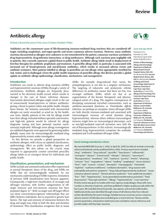
![Review
184 www.thelancet.com Vol 393 January 12, 2019
Diseases, Murdoch University,
Murdoch,WA, Australia
(Prof E J Phillips MD); and
Department of Medicine,
Vanderbilt University Medical
Center, Nashville,TN, USA
(Prof E J Phillips)
Correspondence to:
Prof Elizabeth J Phillips,
Department of Medicine,
Vanderbilt University Medical
Center, Nashville,
TN 37232–2582, USA
elizabeth.j.phillips@vanderbilt.
edu
Other HSR frameworks, including Gell and Coombs
hypersensitivity mechanisms (eg, types I through IV
HSRs) and reaction chronology and onset (immediate vs
delayed), remain clinically important. Immediate anti
biotic HSRs may either be mediated by IgE or by other
factors (figure 1, table). The pathways of IgE-mediated
reactions are well described.6
Recently a mechanism for
some reactions not driven by IgE, previously called
pseudoallergic or anaphylactoid, has been further
elucidated. A receptor on murine mast cells, Mrgprb2, the
orthologue of the human G-protein-coupled receptor
MRGPRX2, was necessary for certain non-IgE-mediated
drug reactions.5
Vancomycin and fluoroquinolones are
the most commonly recognised mast-cell activators that
cause non-IgE-mediated reactions to antibiotics,8,9
thus
producing a reaction with an immunological phenotype,
but without im
muno
logical memory. Typically, non-IgE
reactions have less cardiovascular symptomatology and
hypotension, but are otherwise not easily distinguished
from IgE-mediated allergy (table).10
Delayed HSRs are mediated by T cells or antibodies
other than IgE (table). Antibody-mediated cytopenias,
such as haemolytic anaemia, neutropenia, and thrombo
cytopenia (Gell and Coombs Type II), and serum sickness
(Gell and Coombs Type III), are uncommon. Organ-
specific HSRs to antibiotics often involve the liver
(eg, drug-induced liver injury), kidney, or both (eg, acute
interstitial nephritis).3
The commonest T-cell-mediated
reaction to antibiotics is maculopapular rash, considered
to be a type IVb HSR (table).11
This is the mechanism
of the drug rash observed with aminopenicillin use.
Other specific cutaneous HSRs from antibiotics include
fixed drug eruptions, reported from tetracyclines, sulph
onamides, β-lactams, vancomycin, and fluconazole.2
Generalised fixed drug eruptions can have bullae and
mimic Stevens-Johnson syndrome or toxic epidermal
necrolysis.7
Vancomycin is the most common antibiotic
cause of linear IgA bullous disease, a blistering cutaneous
adverse reaction that can also mimic Stevens-Johnson
syndrome and toxic epidermal necrolysis.12
The major
severe cutaneous adverse reaction (SCAR) pheno
types
include Stevens-Johnson syndrome, toxic epidermal
necrolysis, drug reaction eosinophilia and systemic
symptoms (DRESS) syndrome, and acute generalised
Figure 1: Classification of on-target and off-target ADRs
Pink panel illustrates an example of an on-target ADR. Blue panel (left) illustrates non-immunologically-mediated off-target effects: direct cellular toxicity or disruption of normal physiology,
interaction with non-immune receptors, and interaction with immune receptors (eg, non-IgE-mediated mast-cell activation via G-protein coupled receptors). Blue panel (right) shows
immunologically mediated adaptive immune responses (antibody-mediated [eg, IgE] immediate reactions orT-cell-mediated delayed reactions). Predisposition to both on-target and off-target
reactions is driven by genetic variation, but also ecological factors that can vary over the course of an individual’s lifetime. ADR=adverse drug reaction. Bid=BH3 interacting-domain death.
C difficile=Clostridioides difficile. ER=endoplasmic reticulum. FcεR1=high-affinity IgE receptor. HSR=hypersensitivity reaction. MRGPRX2=MAS-related G-protein coupled receptor member X2.
PKC=protein kinase C. PLCβ=phospholipase C β. ROS=reactive oxygen species.TCR=T cell receptor. UPR=unfolded protein response. *Dose-dependent. Reproduced from Peter et al.2
ADR
mechanisms
Abacavir hypersensitivity
Penicillin angioedema
Fluoroquinolone urticaria
Aminoglycosides acute tubular necrosis
C difficile associated
pseudomembranous colitis
ADR
phenotype/example
Antibiotic associated diarrhoea
eg, C difficile
Antibiotics
Microbiome
Predictable based on drug
action*
Non-immunologically-mediated HSRs
Cellular toxicity and disrupted physiology*
Non-immune cell receptor interaction*
Immune receptor interaction* Antibody-mediated PureT-cell-mediated*
Immunologically-mediated HSRs
On-target ADRs Off-target ADRs
Antibiotics
Megalin
Cubilin
Endosomes,
lysosomes
Golgi
ER
Blocks protein folding
Apoptosis
Cell death
Inflammation
Ca2+
Caspase 12/4
Calpain
Bid
↓ATP
UPR
Cytochrome C
Executioner
caspases
↑ROS
ER
stress
Ca2+
Ca2+
Ca2+
Ca2+
Ca2+
Ca2+
Quinolone
Non-IgE-mediated
mast cell activation
Degranulation
PLCβ
PKC
αi/o
MRGPRX2
Mast cell
Degranulation
Antigen
IgE
FcεRI
T-cell receptor membrane
Antigen-presenting
cell membrane
TCR
Peptide
Abacavir](data:image/gif;base64,R0lGODlhAQABAIAAAAAAAP///yH5BAEAAAAALAAAAAABAAEAAAIBRAA7)