1. Novel Supercomplex Organization of Photosystem I in
Anabaena and Cyanophora paradoxa
Mai Watanabe, Hisako Kubota, Hajime Wada, Rei Narikawa and Masahiko Ikeuchi*
Department of Life Sciences (Biology), Graduate School of Arts and Science, University of Tokyo, Komaba, Meguro,
Tokyo, 153-8902, Japan
Regular Paper
*Corresponding author: E-mail, mikeuchi@bio.c.u-tokyo.ac.jp; Fax, +81-3-5454-4337
(Received September 2, 2010; Accepted November 17, 2010)
The supercomplex organization of photosystem complexes 2003a, Guskov et al. 2009). It is generally accepted that the
was studied in various cyanobacteria, a glaucocystophyte PSII complex can be isolated as a monomer or dimer in organ-
and a primitive rhodophyte by blue-native PAGE with a isms from cyanobacteria to higher plants (Rogner et al. 1987,
¨
wide range of detergent concentrations. In contrast to Bald et al. 1996, Hankamer et al. 1997, Adachi et al. 2009,
known cyanobacteria that produced the PSI trimer, a fila- Watanabe et al. 2009). On the other hand, the PSI complex
mentous N2-fixing cyanobacterium Anabaena sp. PCC 7120 can be isolated as a monomer or a trimer from cyanobacteria
Downloaded from pcp.oxfordjournals.org at Rutgers University on April 4, 2011
and a glaucocystophyte Cyanophora paradoxa NIES 547 had (Takahashi et al. 1982, Rogner et al. 1990), while it is isolated
¨
a PSI tetramer and dimer but no trimer at all. This was as a monomer or a supercomplex of a monomer and
confirmed by sucrose density gradient centrifugation. A light-harvesting Chl complex of PSI (LHCI) from algae and
primitive rhodophyte Cyanidioschyzon merolae had two higher plants (Bengis and Nelson 1975, Ben-Shem et al.
species of PSI monomeric complex with a light-harvesting 2003b). The trimeric PSI complex was predominantly recovered
Chl complex of a different composition. These results from a thermophilic cyanobacterium Thermosynechococcus
are discussed with regard to the evolution of the PSI elongatus, and its atomic structure has been determined
supercomplex. (Jordan et al. 2001). Both monomeric and trimeric PSI
Keywords: Anabaena Blue-native PAGE Cyanophora para- complexes were yielded by a mesophilic cyanobacterium
doxa PSI Supercomplex. Synechocystis sp. PCC 6803 (Kruip et al. 1997). Gene targeting
in Synechocystis revealed that the PsaL subunit is essential
Abbreviations: BN, blue-native; CBB, Coomassie Brilliant for trimerization (Chitnis and Chitnis 1993, Xu et al. 1995). In
Blue; DM, n-dodecyl-b-D-maltopyranoside; LHCI, light- accordance with this mutagenesis, direct contact of the PsaL
harvesting Chl complex of PSI. subunits of three monomeric complexes was found at the
center of the trimeric crystal structure (Fromme et al. 2001,
Jordan et al. 2001). The trimerization of PSI may be affected
Introduction under variable environmental conditions (Karapetyan et al.
In oxygenic photosynthesis, PSII oxidizes water at the lumenal 1999). The trimerization may also be involved in energy distri-
side and reduces plastoquinone at the cytoplasmic (stromal) bution among the PSI monomers (Grotjohann and Fromme
side of the thylakoid membrane, whereas PSI oxidizes plasto- 2005). On the other hand, the interaction of the PSI monomer
cyanin or cytochrome c6 on the lumenal side and reduces with LHC appears to be important in green algae and higher
ferredoxin on the cytoplasmic side. PSI and PSII have evolved plants. The binding of the plant-specific PsaH subunit to PsaL
from a common ancestor that drives electron transfer across prevents the trimer formation (Ben-Shem et al. 2003a,
the plasma membrane from the extracytoplasmic side to the Ben-Shem et al. 2004) and enables regulation of state transition
cytoplasmic side to generate energized electrons and mem- (Lunde et al. 2000).
brane potential by using light energy. The basic framework of Previously, we studied photosystem complexes of the
PSI and PSII is similar, but they have been differentiated into thermophilic T. elongatus by blue-native PAGE (BN-PAGE)
their own systems in evolution and combined to drive the and found that the ratio of the PSII monomer to the dimer
coordinated electron transfer from water to ferredoxin to gen- varied depending on the concentrations of n-dodecyl-b-D-mal-
erate reducing equivalents and ATP for CO2 fixation. topyranoside (DM) applied for solubilization (Watanabe et al.
Functional PSI and PSII have been isolated as multisubunit 2009). In contrast, the PSI complex was almost exclusively
membrane supercomplexes from cyanobacteria, algae and land recovered as a trimer, which hardly depended on the DM
plants, and some of their detailed structures have been revealed concentration. In the mesophilic Synechocystis, it has been
by X ray crystallography (Jordan et al. 2001, Ben-Shem et al. reported that the PSI complexes were separated into trimer
Plant Cell Physiol. 52(1): 162–168 (2011) doi:10.1093/pcp/pcq183, available online at www.pcp.oxfordjournals.org
! The Author 2010. Published by Oxford University Press on behalf of Japanese Society of Plant Physiologists.
All rights reserved. For permissions, please email: journals.permissions@oup.com
162 Plant Cell Physiol. 52(1): 162–168 (2011) doi:10.1093/pcp/pcq183 ! The Author 2010.
2. Novel PSI supercomplex
and monomer fractions (Chitnis and Chitnis 1993, C. paradoxa at a high molecular mass region, which migrated
Herranen et al. 2004, Kubota et al. 2009). Recent genome more slowly than the trimeric PSI of T. elongatus. Instead, the
studies of cyanobacteria and algae have provided vast se- trimeric PSI band was not detected in those species. PSI of
quence information about photosystem genes (Vanselow C. merolae was resolved as two closely migrated bands near
et al. 2009). However, comparative biochemical studies of the dimeric PSII band.
the photosystem complexes have not been performed The recovery of the high molecular mass PSI band was
extensively. In this study, we investigated the organization of examined under a wide range of DM concentrations. In
the PSI and PSII complexes in mesophilic cyanobacteria Fig. 2A, the high molecular mass PSI band of Anabaena was
(Synechocystis and Anabaena sp. PCC 7120), a glaucocysto- abundantly recovered at 0.6–2% DM, less abundantly at 3% and
phyte (Cyanophora paradoxa) and a primitive rhodophyte detected as a faint band at 5%. Conversely, a putative PSI dimer
(Cyanidioschyzon merolae) by BN-PAGE. We found unexpected band was abundantly detected near the PSII dimer bands at 3–
variations in the organization of the PSI complexes depending 5% DM and slightly detected at 1–2% DM. Another faint PSI
on the species. band at a far larger position was often recovered at 2–5% DM.
On the other hand, recovery of the PSI monomer band
was rather independent of the DM concentration. However,
Results and Discussion the PSI trimer band of T. elongatus and Synechocystis was not
Downloaded from pcp.oxfordjournals.org at Rutgers University on April 4, 2011
The thylakoid membranes from cyanobacteria, a glaucocysto- detected at all in Anabaena under any solubilization conditions.
phyte and a primitive rhodophyte were solubilized with 1% DM Two-dimensional PAGE confirmed that all the PSI bands con-
and subjected to BN-PAGE (Fig. 1). In agreement with previous sisted of at least six identical spots (Fig. 2B). By N-terminal
reports (Herranen et al. 2004, Aro et al. 2005, Watanabe et al. sequencing of these subunits, we identified PsaD, PsaL, PsaF,
2009), four green bands (PSI trimer, PSII dimer, PSI monomer PsaK and PsaJ as described previously (Nyhus et al. 1992).
and PSII monomer) were detected in Synechocystis sp. PCC We also detected PsaC, PsaE, PsaX and PsaI as minor spots
6803, while three green bands but no PSI monomer band (not shown in Fig. 2B). Apparently, there were no differences
were detected in T. elongatus perhaps due to heat stability in the subunit composition between these PSI bands. It is sug-
(Fig. 1). In contrast, the separation patterns of Anabaena and gested that the high molecular mass PSI complex was disso-
algae were distinctively different from those of T. elongatus and ciated into the dimeric complex but not the monomeric
Synechocystis. A PSI green band was found in Anabaena and complex.
tis
tis
s
s
a
a
ys
ys
tu
tu
ox
ae
ox
ae
na
na
oc
oc
ga
ga
ad
ad
ol
ol
ae
ae
ch
ch
r
on
on
er
er
ke
ar
ar
ab
ab
ne
ne
.m
.m
el
el
.p
.p
ar
An
An
Sy
Sy
T.
T.
M
C
C
C
A B C
High molecular PSI
1. PSI trimer kDa
669
2. PSII dimer
440
3. PSI monomer
4. PSII monomer 232
158
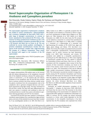
Fig. 1 BN-PAGE of several cyanobacteria, a glaucocystophyte and a primitive rhodophyte. Thylakoid membranes were solubilized with 1.0% DM.
The gel is shown before (A) and after staining with CBB R-250 (B). The PSI trimer, PSII dimer, PSI monomer and PSII monomer of T. elongatus and
Synechocystis are shown on the left (bands 1–4). The green bands of Anabaena, C. paradoxa and C. merolae are indicated with arrowheads. Red
arrowheads indicate high molecular mass PSI bands. The size of soluble molecular markers is given on the left.
Plant Cell Physiol. 52(1): 162–168 (2011) doi:10.1093/pcp/pcq183 ! The Author 2010. 163
3. M. Watanabe et al.
A 0.6 0.8 1.0 2.0 3.0 5.0 % DM A 0.8 1.0 2.0 2.5 3.0 3.5 4.0 5.0 %DM
PSI
High molecular PSI
High molecular PSI
kDa kDa
669 669
PSI dimer PSI dimer
PSII dimer PSII dimer
440
PSI monomer 440
232 PSII monomer PSI monomer
232
PSII monomer
158
158
Downloaded from pcp.oxfordjournals.org at Rutgers University on April 4, 2011
B SI
la rP PS
I B PS
I
om
e r
PS
I
m
er
cu er me
r lar er e
r
0.8% DM lar er e
r on 5.0% DM lar r er ono
0.8% DM ole om no 3.0% DM cu r er nom nom cu m om m cu er er ome om s m
m on o ole ime im o o ole er no on Iess ole m n n s
h m II m m d II d I m II m m Id
im o m
3- m I dim II di mo I mo 3-Ie
I I h I I m II
hig
h h I I
PS PS PS hig PS PS PS PS hig PS PS PS CP4 hig PS PS PS PS CP
4
PsaA/B
PsaD
PsaL
PsaF
PsaK
PsaJ
Fig. 3 BN-PAGE of C. paradoxa. (A) BN-PAGE. Thylakoid membranes
Fig. 2 BN-PAGE of Anabaena. (A) BN-PAGE. Thylakoid membranes were solubilized with 0.8, 1.0, 2.0, 2.5, 3.0, 3.5, 4.0 and 5.0% DM. (B)
were solubilized with 0.6, 0.8, 1.0, 2.0, 3.0 and 5.0% DM. (B) Two-dimensional PAGE. Arrowheads indicate the PSI subunit poly-
Two-dimensional PAGE. Arrowheads indicate the PSI subunit poly- peptides. The gel was silver stained.
peptides. The gel was silver stained.
the absence of the trimeric band is very similar to the situation
The Anabaena PSII complexes were separated into the for Anabaena. However, higher concentrations of DM (1%)
monomer and the dimer and their ratio was also dependent gave a more intense band of the high molecular mass PSI band
on the concentrations of DM. At higher concentrations of DM, than lower concentrations (0.8%) (Fig. 3A), which contrasts
the recovery of the PSII dimer was greater than that of the with Anabaena. We also did not detect any differences in the
monomer. These features of the Anabaena PSII were very simi- subunit composition between the high molecular mass, dimeric
lar to those of T. elongatus PSII (Watanabe et al. 2009). It is of and monomeric PSI bands in C. paradoxa (Fig. 3B). Notably,
note that the PSII to PSI ratio was much smaller in Anabaena recovery of the dimer and monomer bands of PSII was also
than in T. elongatus, as shown in two-dimensional PAGE dependent on the detergent concentration, as in Anabaena
(Fig. 2B; and Watanabe et al. 2009). and T. elongatus.
The high molecular mass PSI band was also observed in the We attempted to determine the molecular size of the high
glaucocystophyte C. paradoxa, in addition to the dimer and the molecular mass PSI band of Anabaena and C. paradoxa by
monomer (Fig. 1, lane 4; Fig. 3). The mobility of the high mo- extrapolating the relationship to the mobility (Fig. 4). We
lecular mass PSI band was very close to that of Anabaena, and plotted known proteins of photosystems and soluble enzymes
164 Plant Cell Physiol. 52(1): 162–168 (2011) doi:10.1093/pcp/pcq183 ! The Author 2010.
4. Novel PSI supercomplex
2000 tis
soluble marker ys
PSI tetramer T. elongatus
ho
c na
Anabaena
ec bae
PSI triamer C. paradoxa
Syn A na
Molecular mass (kDa)
1000
800 PSI dimer
600 PSII dimer
400 PSI monomer
PSII monomer
200
PSI/PSII monomer PSI/PSII monomer
100
2 3 4 5 6 7 8 9
Mobility (cm) (PSI dimer)
Fig. 4 Mobility/molecular mass relationship of photosystems and PSI trimer
Downloaded from pcp.oxfordjournals.org at Rutgers University on April 4, 2011
soluble molecular markers. The mobilities in Fig. 1B were used for PSI tetramer
the plot.
as a marker and found that the consensus line of the photo- Fig. 5 Sucrose density gradient centrifugation of Anabaena and
systems was more reliable than that of the soluble proteins Synechocystis. Thylakoid was solubilized with 1% DM.
for estimation of the green bands. The PSI dimer bands of
Anabaena and C. paradoxa fit very well with the line of the
known photosystems. By extrapolation, we obtained a molecu- from C. merolae at 0.8 and 5.0% DM (Fig. 6B). This is in contrast
lar mass of approximately 1,387 kDa for the high molecular to previous report by Takahashi et al. (2009). They claimed
mass PSI bands of Anabaena and C. paradoxa. This value cor- that only PSII monomer was detected at 0.6–1.2% DM by
responds to the 3.9-mer of the monomer of 356 kDa (Jordan one-dimensional BN-PAGE. Perhaps the one-dimensional
et al. 2001). These results suggest that the high molecular mass BN-PAGE may not be sensitive enough to detect small amounts
PSI complex is the tetramer. We fractionated photosystem of the PSII dimer.
complexes of Anabaena and Synechocystis by sucrose density To date, the PSI trimer and monomer have been isolated
gradient centrifugation after solubilization with 1% DM (Fig. 5). from many cyanobacteria. The crystal structure of the PSI
Obviously, the high molecular mass PSI band of Anabaena trimer was reported in T. elongatus (Jordan et al. 2001). Single
migrated faster than the Synechocystis trimer band, while the particle analysis revealed a similar trimeric organization of PSI
position of the PSI monomer band was practically identical. In prepared from Synechocystis sp. PCC 6803, Synechococcus sp.
Fig. 5, almost no PSI dimer was recovered, but treatment with PCC 7002, Synechococcus sp. PCC 7942, Acaryochloris marina
higher concentrations of DM produced a dimer band (data not and Gloeobacter violaceus (Tsiotis et al. 1995, Kruip et al. 1997,
shown). These results are consistent with those of BN-PAGE. Boekema et al. 2001, Mangels et al. 2002, Chen et al. 2005a). In
Single particle analysis of the Anabaena PSI complexes is now in the filamentous cyanobacteria Phormidium laminosum and
progress. Spirulina platensis, the PSI trimer was also reported based on
Cyanidioschyzon merolae produced two green bands of PSI electron microscopy and gel filtration chromatography (Ford
between the typical monomer and dimer regions (Fig. 6). 2D and Holzenburg 1988, Rakhimberdieva et al. 2001). On the
PAGE revealed that both green bands consisted of identical PSI other hand, a PSI complex of Nostoc punctiforme was assigned
subunits and two LHCI bands near 20 kDa. Based on the mo- to the trimer according to BN-PAGE (Cardona et al. 2007,
lecular size relationship of the photosystems, these PSI-LHCI Cardona et al. 2009) but it migrated near the PSII dimer,
bands were estimated as 613 and 539 kDa, indicative of both which resembles the Anabaena PSI dimer in our study.
monomeric PSI complexes. Although it is difficult to estimate Further, we can see another high molecular mass spot of PSI
the band intensity of silver staining, we can see the difference in (see Fig. 4 of Cardona et al. 2007), which again resembles the
the band intensity of Coomassie Brilliant Blue (CBB) R-250 tetramer of Anabaena PSI. These features seem to suggest that
staining between the two monomeric complexes. Fig. 6C PSI of N. punctiforme can be fractionated as the dimer and
shows that PSI monomer 1 contained more LHCI subunits tetramer but not as the trimer. Since N. punctiforme is close
than PSI monomer 2 relative to the upper PSI band. in phylogeny to Anabaena sp. PCC 7120, heterocyst differenti-
It is of note that our 2D PAGE clearly demonstrated that ation or some common features may be related to the unique
both dimeric and monomeric PSII complexes were resolved PSI organization of the dimer and tetramer. In the literature, the
Plant Cell Physiol. 52(1): 162–168 (2011) doi:10.1093/pcp/pcq183 ! The Author 2010. 165
5. M. Watanabe et al.
A 0.6 0.8 1.0 2.0 3.0 5.0 % DM
has been reported in cyanobacteria as well as in higher plants
(Guskov et al. 2009). Tetrameric PSII is not so common, but has
been reported in Acaryochloris (Chen et al. 2005b). A similar
organization of the PSI dimer may give the tetrameric or higher
kDa oligomeric structure.
669 In the case of the trimeric organization of T. elongatus,
PSII dimer the PsaL subunit of one monomer directly interacts with the
PSII monomer 1
440 PSI monomer 2 PsaL subunits of the other two monomers (Jordan et al. 2001).
232 PSII monomer The phylogenetic tree (Supplementary Fig. S1A) revealed
1
er er
om om
2
that PsaL proteins of Anabaena and related heterocyst-forming
C on on
158 Im Im cyanobacteria were clustered together into a unique clade
PS PS
distinct from the other cyanobacteria. Similar clustering was
PSI also found in the tree of the small membrane protein PsaI
LHCI (Supplementary Fig. S1B) that is located next to PsaL in
LHCI
the 3D structure. On the other hand, phylogeny of the
other PSI subunits such as PsaF and PsaA showed that the
Downloaded from pcp.oxfordjournals.org at Rutgers University on April 4, 2011
B 1 2 er 1 2 er heterocyst-forming cyanobacteria are positioned within
er er er er
0.8% DM er nom om nom 5.0% DM er nom om nom
II
m
I I
n
di mo mo I mo
I II
m
I I
n
di mo mo I mo
I
the other cyanobacteria (Supplementary Fig. S1C, D). These
PS PS PS PS PS PS PS PS
results clearly illustrated the unique evolutionary position of
PsaL and PsaI of the heterocyst-forming cyanobacteria.
Interestingly, PsaL of the heterocyst-forming cyanobacteria,
algae and plants has a short deletion in the C-terminal
region, which directly interacts with PsaL of the other PSI
monomers in the trimeric complex. Gene targeting of the
PsaL or PsaI subunit prevented assembly of the trimer in
Synechocystis (Chitnis and Chitnis 1993, Xu et al. 1995). These
facts may suggest that the trimeric or tetrameric organization
could be determined by the set of PsaL and PsaI in the PSI
supercomplex. Since C. paradoxa seems to be the only eukary-
ote that does not have LHCI (Reyes-Prieto and Bhattacharya
2007), the tetrameric organization of the other algae including
C. merolae might have been canceled when LHCI was intro-
duced in evolution. Mutagenesis of psaL and psaI in
Anabaena may shed light on the structure/function and evo-
lution of the PSI supercomplex organization.
Fig. 6 BN-PAGE of C. merolae. (A) BN-PAGE. Thylakoid membranes
were solubilized with 0.6, 0.8, 1.0, 2.0, 3.0 and 5.0% DM. (B) Materials and Methods
Two-dimensional PAGE. White arrowheads and open arrowheads in-
dicate the PSI subunit polypeptides and LHCI subunits, respectively. Growth conditions
The gel was silver stained. (C) CBB R-250-stained profile of the sub- Anabaena sp. PCC 7120 and Synechocystis sp. PCC 6803 cells
units of PSI and LHCI that correspond to the silver-stained profile were grown in a liquid BG-11 medium at 31 C (Midorikawa
boxed in B. et al. 2009). Thermosynechococcus elongatus BP-1 cells were
grown in BG-11 at 45 C (Watanabe et al. 2009). Cyanophora
PSI monomer was predominantly isolated from C. paradoxa by paradoxa strain NIES 547 was obtained from the National
solubilization with DM, sucrose density gradient centrifugation Institute for Environmental Studies, Tsukuba, Japan and
and gel filtration chromatography (Koike et al. 2000). This grown as in Koike et al. (2000). Cyanidioschyzon merolae 10D
result may fit with our observation of the relatively large recov- was obtained from Dr. Naoki Sato, University of Tokyo and
ery of the PSI monomer (Fig. 3). grown in a liquid A medium at 45 C as reported previously
Since recovery of the PSI dimer and tetramer was affected (Moriyama et al. 2008). Cultures were grown with bubbling
by DM concentrations, the assembly of the tetramer may with 1% CO2-containing air under white light.
depend on the hydrophobic interaction within the thylakoid
membrane but not on the interaction between the hydrophilic Isolation of thylakoid membranes
surfaces of the complex. The PSI dimer may be formed in an Thylakoid membranes of Anabaena, Synechocystis, T. elongatus
arrangement similar to that of the PSII dimer, whose structure and C. merolae were isolated as described in Watanabe et al.
166 Plant Cell Physiol. 52(1): 162–168 (2011) doi:10.1093/pcp/pcq183 ! The Author 2010.
6. Novel PSI supercomplex
(2009). Thylakoid membrane from C. paradoxa was isolated as Bald, D., Kruip, J. and Rogner, M. (1996) Supramolecular archi-
¨
described in Koike et al. (2000) with modifications as described tecture of cyanobacterial thylakoid membranes: how is the phyco-
below. Cyanelles were isolated by osmotic shock and then dis- bilisome connected with the photosystems?. Photosynth. Res. 49:
rupted with zirconia beads by a Bead-beater (Biospec). 103–118.
Bengis, C. and Nelson, N. (1975) Purification and properties of the
Agitation was performed for 10 s and they were then cooled
photosystem I reaction center from chloroplasts. J. Biol. Chem. 250:
on ice for 2 min. This cycle was repeated 20 times. After removal
2783–2788.
of the unbroken cyanelles, the resulting supernatant was cen- Ben-Shem, A., Frolow, F. and Nelson, N. (2003a) Crystal structure of
trifuged at 300,000 Â g for 30 min at 4 C to precipitate the plant photosystem I. Nature 426: 630–635.
thylakoid membranes. The thylakoid was resuspended with Ben-Shem, A., Frolow, F. and Nelson, N. (2004) Evolution of photosys-
50 mM HEPES-NaOH (pH 7.5), 2 mM ethylene glycol tem I—from symmetry through pseudo-symmetry to asymmetry.
tetra-acetic acid, 1 mM MgCl2 and 0.5 M sucrose, and stored FEBS Lett. 564: 274–280.
at À80 C. Ben-Shem, A., Nelson, N. and Frolow, F. (2003b) Crystallization and
initial X-ray diffraction studies of higher plant photosystem I.
BN-PAGE and two-dimensional PAGE Acta Crystallogr. D Biol. Crystallogr. 59: 1824–1827.
Boekema, E.J., Hifney, A., Yakushevska, A.E., Piotrowski, M.,
BN-PAGE and two-dimensional PAGE were performed as
Keegstra, W., Berry, S. et al. (2001) A giant chlorophyll–protein
described in Watanabe et al. (2009). Thylakoid membranes complex induced by iron deficiency in cyanobacteria. Nature 412:
Downloaded from pcp.oxfordjournals.org at Rutgers University on April 4, 2011
{1 [(mg Chl)] mlÀ1} were solubilized with DM on ice for 745–748.
30 min, followed by centrifugation at 300,000 Â g for 30 min Cardona, T., Battchikova, N., Agervald, A., Zhang, P., Nagel, E., Aro, E.M.
at 4 C. The solubilized supernatant was subjected to et al. (2007) Isolation and characterization of thylakoid membranes
BN-PAGE using CBB G-250 (Serva) as described in Schagger from the filamentous cyanobacterium Nostoc punctiforme.
and von Jagow (1991). After electrophoresis, the BN-PAGE Physiol. Plant. 131: 622–634.
gel was stained with CBB R-250 (Bio-Rad) and the two- Cardona, T., Battchikova, N., Zhang, P., Stensjo, K., Aro, E.M.,
dimensional gel was stained with silver as described in Aro Lindblad, P. et al. (2009) Electron transfer protein complexes
et al. (2005). in the thylakoid membranes of heterocysts from the cyano-
bacterium Nostoc punctiforme. Biochim. Biophys. Acta 1787:
Sucrose density gradient centrifugation 252–263.
Chen, M., Bibby, T.S., Nield, J., Larkum, A. and Barber, J. (2005a) Iron
Sucrose density gradient centrifugation was performed as deficiency induces a chlorophyll D-binding Pcb antenna system
described in Ikeda et al. (2008). Thylakoid membranes were around photosystem I in Acaryochloris marina. Biochim. Biophys.
solubilized with 1% DM and centrifuged at 300,000 Â g for Acta 1708: 367–374.
30 min at 4 C. The solubilized material was loaded on a 10– Chen, M., Bibby, T.S., Nield, J., Larkum, A.W. and Barber, J. (2005b)
30% linear sucrose density gradient and centrifuged at Structure of a large photosystem II supercomplex from
130,000 Â g for 16 h at 4 C. Acaryochloris marina. FEBS Lett. 579: 1306–1310.
Chitnis, V.P. and Chitnis, P.R. (1993) PsaL subunit is required for
the formation of photosystem I trimers in the cyanobacterium
Supplementary data Synechocystis sp. PCC 6803. FEBS Lett. 336: 330–334.
Ford, R.C. and Holzenburg, A. (1988) Investigation of the structure of
Supplementary data are available at PCP online. trimeric and monomeric photosystem I reaction centre complexes.
EMBO J. 7: 2287–2293.
Fromme, P., Jordan, P. and Krauss, N. (2001) Structure of photosystem
Funding I. Biochim. Biophys. Acta 1507: 5–31.
Grotjohann, I. and Fromme, P. (2005) Structure of cyanobacterial
This work was supported by the Ministry of Education and photosystem I. Photosynth. Res. 85: 51–72.
Science [Grants-in-Aid for Young Scientists (to R.N.), Guskov, A., Kern, J., Gabdulkhakov, A., Broser, M., Zouni, A. and
Scientific Research and the GCOE program (to M.I.)]. Saenger, W. (2009) Cyanobacterial photosystem II at 2.9-A
resolution and the role of quinones, lipids, channels and chloride.
Nat. Struct. Mol. Biol. 16: 334–342.
References Hankamer, B., Barber, J. and Boekema, E.J. (1997) Structure and mem-
brane organization of photosystem II in green plants. Annu. Rev.
Adachi, H., Umena, Y., Enami, I., Henmi, T., Kamiya, N. and Shen, J.R. Plant Physiol. Plant Mol. Biol. 48: 641–671.
(2009) Towards structural elucidation of eukaryotic photosystem II: Herranen, M., Battchikova, N., Zhang, P., Graf, A., Sirpio, S.,
purification, crystallization and preliminary X-ray diffraction Paakkarinen, V. et al. (2004) Towards functional proteomics of
analysis of photosystem II from a red alga. Biochim. Biophys. Acta membrane protein complexes in Synechocystis sp. PCC 6803.
1787: 121–128. Plant Physiol. 134: 470–481.
Aro, E.M., Suorsa, M., Rokka, A., Allahverdiyeva, Y., Paakkarinen, V., Ikeda, Y., Komura, M., Watanabe, M., Minami, C., Koike, H., Itoh, S.
Saleem, A. et al. (2005) Dynamics of photosystem II: a prote- et al. (2008) Photosystem I complexes associated with fucoxanthin–
omic approach to thylakoid protein complexes. J. Exp. Bot. 56: chlorophyll-binding proteins from a marine centric diatom,
347–356. Chaetoceros gracilis. Biochim. Biophys. Acta 1777: 351–361.
Plant Cell Physiol. 52(1): 162–168 (2011) doi:10.1093/pcp/pcq183 ! The Author 2010. 167
7. M. Watanabe et al.
Jordan, P., Fromme, P., Witt, H.T., Klukas, O., Saenger, W. and Reyes-Prieto, A. and Bhattacharya, D. (2007) Phylogeny of
Krauss, N. (2001) Three-dimensional structure of cyanobacterial nuclear-encoded plastid-targeted proteins supports an early
photosystem I at 2.5 A resolution. Nature 411: 909–917. divergence of glaucophytes within Plantae. Mol. Biol. Evol. 24:
Karapetyan, N.V., Holzwarth, A.R. and Rogner, M. (1999) The photo-
¨ 2358–2361.
system I trimer of cyanobacteria: molecular organization, excitation Rogner, M., Dekker, J.P., Boekema, E.J. and Witt, H.T. (1987) Size, shape
¨
dynamics and physiological significance. FEBS Lett. 460: 395–400. and mass of the oxygen-evolving photosystem II complex from the
Koike, H., Shibata, M., Yasutomi, K., Kashino, Y. and Satoh, K. (2000) thermophilic cyanobacterium Synechococcus sp. FEBS Lett. 219:
Identification of photosystem I components from a glaucocysto- 207–211.
phyte, Cyanophora paradoxa: the PsaD protein has an N-terminal Rogner, M., Muhlenhoff, U., Boekema, E.J. and Witt, H.T. (1990) Mono-,
¨
stretch homologous to higher plants. Photosynth. Res. 65: 207–217. di- and trimeric PSI reaction center complexes isolated from the
Kruip, J., Chitnis, P.R., Lagoutte, B., Rogner, M. and Boekema, E.J. (1997)
¨ thermophilic cyanobacterium Synechococcus sp. Size, shape and
Structural organization of the major subunits in cyanobacterial activity. Biochim. Biophys. Acta 1015: 415–424.
photosystem 1. Localization of subunits PsaC, -D, -E, -F, and -J. Schagger, H. and von Jagow, G. (1991) Blue native electrophoresis for
J. Biol. Chem. 272: 17061–17069. isolation of membrane protein complexes in enzymatically active
Kubota, H., Sakurai, I., Katayama, K., Mizusawa, N., Ohashi, S., form. Anal. Biochem. 199: 223–231.
Kobayashi, M. et al. (2009) Purification and characterization of Takahashi, T., Inoue-Kashino, N., Ozawa, S., Takahashi, Y., Kashino, Y.
photosystem I complex from Synechocystis sp. PCC 6803 by ex- and Satoh, K. (2009) Photosystem II complex in vivo is a monomer.
pressing histidine-tagged subunits. Biochim. Biophys. Acta 1797: J. Biol. Chem. 284: 15598–15606.
Downloaded from pcp.oxfordjournals.org at Rutgers University on April 4, 2011
98–105. Takahashi, Y., Koike, H. and Katoh, S. (1982) Multiple forms of
Lunde, C., Jensen, P.E., Haldrup, A., Knoetzel, J. and Scheller, H.V. (2000) chlorophyll–protein complexes from a thermophilic cyano-
The PSI-H subunit of photosystem I is essential for state transitions bacterium Synechococcus sp. Arch. Biochem. Biophys. 219:
in plant photosynthesis. Nature 408: 613–615. 209–218.
Mangels, D., Kruip, J., Berry, S., Rogner, M., Boekema, E.J. and Koenig, F.
¨ Tsiotis, G., Haase, W., Engel, A. and Michel, H. (1995) Isolation and
(2002) Photosystem I from the unusual cyanobacterium structural characterization of trimeric cyanobacterial photosystem
Gloeobacter violaceus. Photosynth. Res. 72: 307–319. I complex with the help of recombinant antibody fragments.
Midorikawa, T., Matsumoto, K., Narikawa, R. and Ikeuchi, M. (2009) An Eur. J. Biochem. 231: 823–830.
Rrf2-type transcriptional regulator is required for expression of Vanselow, C., Weber, A.P., Krause, K. and Fromme, P. (2009) Genetic
psaAB genes in the cyanobacterium Synechocystis sp. PCC 6803. analysis of the photosystem I subunits from the red alga, Galdieria
Plant Physiol. 151: 882–892. sulphuraria. Biochim. Biophys. Acta 1787: 46–59.
Moriyama, T., Terasawa, K., Fujiwara, M. and Sato, N. (2008) Watanabe, M., Iwai, M., Narikawa, R. and Ikeuchi, M. (2009) Is the
Purification and characterization of organellar DNA polymerases photosystem II complex a monomer or a dimer? Plant Cell
in the red alga Cyanidioschyzon merolae. FEBS J. 275: 2899–2918. Physiol. 50: 1674–1680.
Nyhus, K.J., Ikeuchi, M., Inoue, Y., Whitmarsh, J. and Pakrasi, H.B. (1992) Xu, Q., Hoppe, D., Chitnis, V.P., Odom, W.R., Guikema, J.A. and
Purification and characterization of the photosystem I complex Chitnis, P.R. (1995) Mutational analysis of photosystem I polypep-
from the filamentous cyanobacterium Anabaena variabilis ATCC tides in the cyanobacterium Synechocystis sp. PCC 6803. Targeted
29413. J. Biol. Chem. 267: 12489–12495. inactivation of psaI reveals the function of psaI in the structural
Rakhimberdieva, M.G., Boichenko, V.A., Karapetyan, N.V. and organization of psaL. J. Biol. Chem. 270: 16243–16250.
Stadnichuk, I.N. (2001) Interaction of phycobilisomes with photo-
system II dimers and photosystem I monomers and trimers in the
cyanobacterium Spirulina platensis. Biochemistry 40: 15780–15788.
168 Plant Cell Physiol. 52(1): 162–168 (2011) doi:10.1093/pcp/pcq183 ! The Author 2010.