
Recommended
More Related Content
What's hot
What's hot (20)
Similar to Coronary Intravascular Lithotripsy
Similar to Coronary Intravascular Lithotripsy (20)
Recently uploaded
Recently uploaded (20)
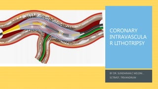
Coronary Intravascular Lithotripsy
- 2. SCOPE OF DISCUSSION • INTRODUCTION • CORONARY CALCIUM ASSESSMENT • CALCIUM MODIFICATION TECHNIQUES • CORONARY IVL SYSTEM • MECHANISM OF ACTION • DISRUPT CAD TRIALS • POTENTIAL USES OF IVL • CHALLENGING LESIONS • CONCLUSION
- 3. INTRODUCTION • Advancing age and the presence of comorbidities (e.g., diabetes and chronic kidney disease) predispose to coronary calcification. • Moderate or severe calcification is frequently present in patients undergoing percutaneous coronary interventions (PCI). • Lack of flexibility in calcified arteries makes it to difficult to advance stents down a tortuous anatomy. • Difficulty in stent delivery may lead to damage of the polymer/drug coating leading to impaired drug delivery. • Greater the arc, length or thickness of calcium, the greater the likelihood of stent under-expansion. • Moderate-to-severe calcification has been independently associated with increased major adverse cardiovascular events (MACE) and long-term rates of in-stent restenosis (ISR), stent thrombosis, target lesion revascularization (TLR), myocardial infarction (MI), and death.
- 7. CALCIUM SCORING BY OCT, IVUS
- 8. CALCIUM SCORING BY OCT, IVUS OCT: • The three-dimensional volume of calcific plaque is best quantified by OCT, since length, thickness, and circumferential arc of calcium can be visualized by the infrared spectrum of OCT. • Stent expansion was 99% in lesions with score 0, 85% with score 1, 86% with score 2, 80% with score 3, and 78% with score 4. • Therefore, OCT-based calcium score of ≥ 3 may indicate the need for calcium modification to induce calcium fracture, which has been associated with enhanced stent expansion IVUS: • Similar to OCT, an IVUS-based scoring system has been developed and validated. • Stent under-expansion (stent area at maximum calcification/average reference lumen area < 70%) by balloon and stenting was observed in lesions with scores ≥ 3, indicating the need for adjunctive calcium modification to achieve optimal stent expansion
- 9. CALCIUM MODIFICATION-AVAILABLE TECHNIQUES 1. NON-COMPLIANT BALLOON: • High and very high-pressure NC balloons allow more uniform balloon expansion and the application of higher forces in a focal segment of a coronary vessel, exerting excessive pressure at the edges and potentially causing coronary dissections or perforations. • The OPN NC balloon is a dedicated device for the treatment of in-stent restenosis, heavily calcified lesions or other lesions that cannot be dilated. 2. CUTTING BALLOON: • The cutting balloon has blades to make radially directed, longitudinal cuts to expand the vessel while limiting uncontrolled dissection. • Crossability and trackability are important limitations of cutting balloons. 3. SCORING BALLOON: • The scoring balloon has nitinol wires on the surface of the balloon in order to focus forces to induce plaque disruption and reduce balloon slippage.
- 10. 4. ROTATIONALATHERECTOMY: • Ablate calcified plaque using the “differential cutting” mechanism, i.e., selectively ablating rigid non-flexible calcium while minimizing interaction with soft, pliable non-calcified tissue. • The ROTABLATOR system utilizes the rotation of a front-cutting diamond-tipped burr (1.25 to 2.5 mm in diameter) that is mounted over an advancer drive shaft (RotaLink) connected to a motor that converts compressed gas into rotational energy. The RA burr is advanced over the dedicated 0.009-inch Rotawire. • The recommended burr size/artery ratio is 0.5–0.7 and the recommended range of burr revolution speed is 135,000– 180,000 rpm. • GUIDEWIRE BIAS: IVUS and OCT have demonstrated that the polishing effect of RA on the intimal surface of calcium produces a sharply delineated groove that follows the course of the guidewire.
- 11. 5. ORBITALATHERECTOMY: • The Diamondback 360 Coronary OA System is a newer atherectomy device that uses centrifugal force and differential “sanding” to modify calcified lesions. • The OA system uses a 1.25-mm eccentrically mounted diamond-coated crown connected to a drive shaft and to a controller powered by a pneumatic console that allows for bi-directional modification of calcium at 80,000 or 120,000 rpm. • Calcium modification by OA has a similar appearance to modification by RA (concave, polished lumen surface) that follows the guidewire course on IVUS or OCT.
- 12. 6. EXCIMER LASER CORONARY ATHERECTOMY (ELCA): • Excimer lasers are pulsed gas lasers that use a mixture of a rare gas and halogen as an active medium to generate pulses of short wavelength, high-energy ultraviolet (UV) light. • The depth of laser penetration is directly related to its wavelength, with UV laser (shorter wavelength) having less depth of penetration, heat production, and unwanted tissue damage. • Excimer laser coronary atherectomy (ELCA) is mediated through 3 distinct mechanisms: photochemical (breakdown of carbon–carbon bonds), photothermal (elevation of the temperature of intracellular water and generation of vapor bubbles at the catheter tip), and photomechanical (expansion and implosion of vapor bubbles disrupting the target lesion). • In severely calcified lesions through which a microcatheter cannot be delivered distally (e.g., in order to swap the workhorse guidewire for dedicated atherectomy wires to perform RA or OA), ELCA can be used over a standard 0.014-inch guide wire to modify the lesion and create a channel to allow for crossing of the microcatheter. • ELCA can also be used to ablate resistant lesions in CTO PCI where interventional equipment cannot cross the lesion or the proximal cap despite achieving distal wire position.
- 13. CORONARY INTRAVASCULAR LITHOTRIPSY • IVL uses acoustic shockwaves in a balloon-based system for treatment of concentric and/or eccentric vascular calcification, thereby improving vessel compliance and diameter. • The IVL balloon-catheter system includes miniaturized and arrayed emitting elements that are integrated within a semi-compliant balloon filled with a mixture of contrast and saline. • The coronary IVL system has 2 emitters integrated on a rapid-exchange balloon-based system and is available in diameters from 2.5 to 4.0 mm (in 0.5 mm increments) and is 12 mm in length. • The IVL balloon is selected in a 1:1 ratio to the reference coronary diameter, often guided by intravascular imaging, which is recommended for optimal lesion preparation. • The IVL catheter is connected via a connector cable to the generator that is preprogrammed to deliver 10 pulses in sequence at a frequency of 1 pulse/s for a maximum of 80 pulses per catheter.
- 14. • The emitting elements generate shockwaves that are similar in their waveform to the shockwaves generated by the lithotripters used in the extracorporeal lithotripsy of renal stones. • These shockwaves are characterized by short-duration of ~ 5 μs and generate a peak positive pressure of ≈ 50 atm. • The contrast-filled balloon is inflated at a subnominal pressure (4 atm) and is apposed to the vessel wall, providing an effective fluid–tissue interface with similar acoustic impedances to facilitate efficient coupling of the shockwave energy to the vessel wall.
- 16. MECHANISM OF ACTION Axial splitting by compressive circumferential forces generated by differential propagation of the acoustic shockwaves in the solid calcified plaque and the surrounding soft tissue Generation and violent collapse of cavitation bubbles by the shockwaves inside the saline-contrast-filled balloon that impact the surface of the calcific plaque. Fatigue mechanism involving progressive expansion of micro fractures into macro fractures by the cumulative impact of the repetitive multiple shockwave pulses. The surrounding vascular tissue is also subject to the shockwave energy; nevertheless, the non-calcified soft tissue transmits the energy as a pass-through due to its elasticity and is not generally impacted by the described forces that modify the calcific plaque.
- 20. DISRUPT CAD I • Single-arm, non-randomized, multicenter study. • IVL was feasible in all patients (n = 60) and facilitated the delivery of stents to all target lesions. • In an OCT substudy, circumferential calcium fracture was observed. • Multiple fractures in a single cross-section were detected in > 25% of lesions, leading to an average acute area gain of 2.1 mm2 with IVL alone. • IVL-induced fractures were independent of calcium depth, with multiple fractures per lesion occurring more frequently as the severity of the underlying calcification increased.
- 21. • IVL facilitated PCI resulted in stent apposition and expansion similar to expansions in drug-eluting stent (DES) implanted in non-calcified lesions. • Although plaque abrasion by RA and OA generate small microparticles that may embolize and impair microcirculatory perfusion, the calcium fragments that are generated by IVL remain subintimal with minimal disruption in intima; therefore, they do not tend to embolize distally.
- 30. DISRUPT CAD III TRIAL
- 36. POTENTIAL USES OF IVL 1. ACUTE CORONARY SYNDROMES: • Calcified lesions in culprit vessels are common in patients presenting with acute coronary syndromes (moderate calcification is found in 26.1% of these patients and severe calcification in 5.9%, and their presence is a strong predictor of definite stent thrombosis and target lesion revascularization. • The Disrupt CAD study included only patients with stable and unstable angina. Although there is not enough evidence to support the use of the IVL during primary PCI, early experience has shown favourable results. 2. UNPROTECTED LEFT MAIN CALCIFIED STENOSIS: • Calcification increases procedural complexity and therefore the risk of complications. • The Coronary IVL System, with controlled pulses delivered under low pressure, might potentially improve plaque modification with a lower risk of vessel closure, perforation or embolization.
- 37. 3. CHRONIC TOTAL OCCLUSION: • Moderate to severe calcification is frequently found in chronic total occlusions. • Debulking devices are usually avoided because the procedure is difficult and has a high risk of complications. • Coronary IVL System might be useful in facilitating lumen dilatation and communication with the subintimal space. 4. STENT UNDEREXPANSION DUE TO UNDERLYING CALCIFICATION: • Although the technique has been developed to treat calcified lesions in native coronary arteries before stenting, patients with severe stent under-expansion because of heavy calcification are at a higher risk of stent failure and future adverse events. • The circumferential sonic waves of the Coronary IVL System, conversely, have the advantage of extending beyond strut layers and fracture deeper calcium deposits.
- 38. ADVANTAGES OF IVL OVER ATHERECTOMY OR SPECIALITY BALLOONS • No specific training needed as the IVL device is delivered similar to standard catheter-based PCI. • IVL therapy is balloon based, and, therefore, the risk of atheromatous embolization may be lower than free debulking devices. • No guidewire bias: Sonic pressure waves are distributed uniformly across the inflated balloon, addressing calcium irrespective of its circumferential location leading to fracture. • Unlike traditional balloon technology, which is dependent on static barometric pressure, IVL delivers circumferential ultrashort pulses of high-intensity acoustic energy, which, by virtue of its compressive and decompressive components, results in effective circumferential modification of calcific atheroma. • IVL is typically performed at low atmospheric pressure balloon inflation lasting less than 2µs, minimizing mechanical vascular trauma. • Side-branch protection using a guidewire may be easily performed using IVL, without risk of wire entrapment or severing as may occur with rotational or orbital atherectomy.
- 39. CHALLENGING LESIONS • Severe tortuosity or angulation • Critical lumen reduction • Plaque indentation into the lumen and a very low vessel expansion compliance (small vessels and multiple stent layers present), could impact balloon deliverability and positioning. • Vessels with a diameter >4 mm (maximum shockwave balloon size) or plaque eccentricity preclude appropriate IVL balloon apposition to the vessel wall, and may reduce the efficacy of the therapy. • More data on the safety, efficacy, and clinical outcomes of IVL use for treatment of stent under-expansion are needed.
- 41. CONCLUSION • The Coronary IVL System is a promising new treatment modality to tackle moderate to severe calcified coronary lesions, with a high rate of success and a low risk of complications. • Larger studies and longer-term clinical data are needed to confirm the safety and efficacy of this technique with special attention to the effects on cardiac conduction and vessel healing response. • Randomised controlled clinical trials are required to evaluate its superiority against currently available calcium- modifying devices.
- 42. THANK YOU