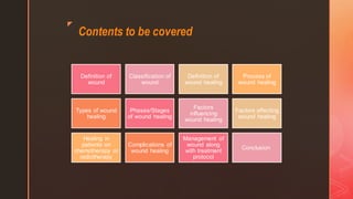
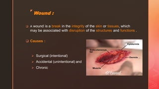
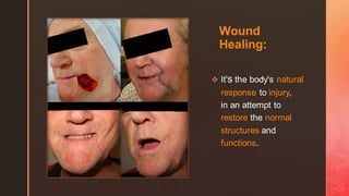
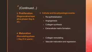
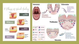
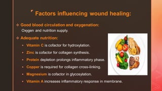
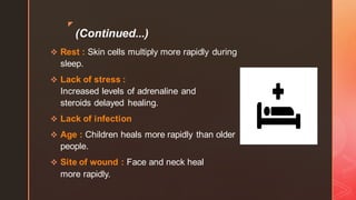
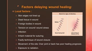
z
Wound
Healing
❖ Dr. Zannatul Tasnim (HMO),
❖ Dr. Tasneema Zannat Sohana (HMO)
Department of Oral & Maxillofacial
Surgery, ShSMC
"The wish for healing was always been half of health."
- Seneca
z
Contents to be covered
Definition of
wound
Classification of
wound
Definition of
wound healing
Process of
wound healing
Types of wound
healing
Phases/Stages
of wound healing
Factors
influencing
wound healing
Factors affecting
wound healing
Healing in
patients on
chemotherapy or
radiotherapy
Complications of
wound healing
Management of
wound along
with treatment
protocol
Conclusion
z
Wound :
❑ A wound is a break in the integrity of the skin or tissues, which
may be associated with disruption of the structures and functions .
❑ Causes :
➢ Surgical (intentional)
➢ Accidental (unintentional) and
➢ Chronic
z
Classifications of wound:
wounds can be classified as follows -
Rank and Wakefield
Classification: 2 types -
1. Tidy wounds :
Features:
I. Inflicted by sharp instruments.
II. Incised, clean-cut, usually be
single, healthy wounds with no
devitalized tissue.
III. Healing is occurred by primary
intention.
IV. e.g. Surgical incisions, glass-
cuts, knife wounds.
2. Untidy wounds:
Features:
I. Result from tearing, crushing,
avulsion, vascular injury or burn.
II. Often multiple and irregular,
containing devitalized tissue.
III. Healing is occurred by secondary
intention.
IV. e.g. Fractures - common and may
be multi-fragmentary.
z
Surgical wound classification (according
to the degree of contamination):
1. Class I (Clean wound):
Like surgical incisions, they're made
electively during surgical procedures in an
aseptic environment.
2. Class II (Clean-contaminated wound):
The kind of aseptic elective surgical
incision that's made in a contaminated
environment, like oral cavity, as it normally
contains huge number of oral flora carried
in saliva.
3. Class III (Contaminated wound):
Those aren't usually elective but traumatic in
origin; like fresh skin lacerations, open
fractures and penetrating wounds.
4. Class IV (Dirty contaminated wound):
Usually contains devitalized tissue or pre-
existing infection prior to operation.
z
According to the
depth of the wound:
1. Superficial wound:
Loss of epidermis only.
2. Partial thickness wound:
Involves the epidermis and
dermis.
3. Full thickness wound:
Involve the dermis, subcutaneous
fat and sometimes bone.
z
According to the origin of wound:
❖ 1. Open
wound:
➢ Abrasion
➢ Laceration
➢ Incision
➢ Puncture
➢ Penetrating
➢ Avulsion
➢ Amputation
❖ 2. Close
wound:
➢ Contusion
➢ Hematoma
➢ Crushing
injury
z
According to the duration of the
wound healing:
❖ 1. Acute wound:
-Usually heal in the anticipated time
frame
-Duration: Immediately to the few
weeks
-Example: Acquired as a result of
trauma or an operative procedure.
❖ 2. Chronic wound:
-Fails to heal in the anticipated
time frame and often re-occur
-Duration: 4 weeks to 3 months
-Example: Occurs as a result of
extended pressure on the tissue,
poor circulation or even poor
nutrition > pressure ulcers,
venous ulcers, diabetic foot
ulcers.
z
Wound
Healing:
❖ It's the body's natural
response to injury,
in an attempt to
restore the normal
structures and
functions.
z
Wound healing process:
It follows 2 distinct processes -
❖ Regeneration :
Healing by proliferation of parenchymal cells and usually results
in complete restoration of original tissue ( Structural and
functional).
❖ Repair :
Healing takes place by proliferation of connective tissue
elements resulting in fibrosis and scarring ( Granulation
tissue formation and contraction).
Both can occur at a time simultaneously.
z
Types of wound healing:
1. Healing by Primary Intention : Features -
➢ Incision edges of a clean surgical incision remain
close.
➢ Minimal tissue loss.
➢ Skin quickly regenerates.
➢ Faster healing with generally best cosmetic result.
➢ Treatment of choice for non-infected wound.
➢ Direct suture is done- if no tissue loss.
z
(Continued...)
2. Healing by Secondary Intention : Features -
➢ Open wound with jagged edges.
➢ There's gap between the edges.
➢ Tissue loss is greater than primary intention.
➢ Granulation tissue gradually fills the area of
defect with scar tissue (Contraction and
epithelialization).
➢ e.g. Ulcers and pressure sores.
z
(Continued...)
3. Healing by Tertiary Intention (Delayed Primary
Closure) :
Features -
➢ Utilized when there're high chances of wound infection.
➢ Wound left open for few days.
➢ Late suturing done to allow healing by primary intention.
➢ Usually a drain is left in situ.
z
Stages( and also phases) of wound
healing :
1. Hemostasis
(Immediate) :
2. Inflammation
(Reactive Phase>
Day 1-4) :
❖ Cellular and bio-physiologic events:
➢ Vascular constrictions
➢ Platelet aggregation, degranulation and
➢ Fibrin formation.
➢ Neutrophil infiltration
➢ Monocyte infiltration and differentiation to
Macrophage
➢ Lymphocyte infiltration.
z
(Continued...)
3. Proliferation
(Regenerative/repar
ative phase> Day 4-
21) :
4. Maturation
(Remodeling phase
> Day 21-2 years) :
❖ Cellular and bio-physiologic events:
➢ Re-epithelialization
➢ Angiogenesis
➢ Collagen synthesis
➢ Extracellular matrix formation.
➢ Collagen remodeling
➢ Vascular maturation and regression.
z
Factors influencing wound healing:
❖ Good blood circulation and oxygenation:
Oxygen and nutrition supply.
❖ Adequate nutrition:
▪ Vitamin C is cofactor for hydroxylation.
▪ Zinc is cofactor for collagen synthesis.
▪ Protein depletion prolongs inflammatory phase.
▪ Copper is required for collagen cross-linking.
▪ Magnesium is cofactor in glycosylation.
▪ Vitamin A increases inflammatory response in membrane.
z
(Continued...)
❖ Rest : Skin cells multiply more rapidly during
sleep.
❖ Lack of stress :
Increased levels of adrenaline and
steroids delayed healing.
❖ Lack of infection
❖ Age : Children heals more rapidly than older
people.
❖ Site of wound : Face and neck heal
more rapidly.
z
Factors delaying wound healing:
❖ Local factors :
➢ Skin edges not lined up
➢ Dead tissue in wound
➢ Foreign bodies in wound
➢ Tension on wound/ wound stress
➢ Infection
➢ Irritant material for suturing
➢ Faulty technique of wound closure
➢ Movement of the site: Over joint or back has poor healing prognosis
➢ Exposure to radiation.
z
(Continued...)
❖ General factors :
➢ Aging
➢ Malnutrition
➢ Anemia
➢ Cardiac insufficiency
➢ Pulmonary disease
➢ Diabetes Mellitus : Microangiopathy, arteriosclerosis, decrease
phagocytic activity.
➢ Smoking
➢ Immunosuppressive disease: e.g. HIV etc.
z (Continued...)
➢ Jaundice : Fibroblastic repair delayed
➢ Malignancies
➢ High Blood Urea
➢ Stress, lack of sleep
➢ Drug therapy:
• Steroids: inhibit macrophage function > less inflammatory response
(It's inhibitory effects is reversible by Vitamin A)
• Cytotoxic drugs (Antineoplastic agents > Cyclophosphamide, methotrexate, bisphosphonates) :
Decrease WBC, fibroblast proliferation, wound contraction and protein
synthesis.
• NSAIDS : Decrease collagen synthesis by 45% even at normal levels.
➢ Radiotherapy: Radiation dermatitis, radiation induced mucositis etc.
z
Healing in patients on Chemotherapy
or Radiotherapy:
❖ These patients have impaired wound healing
responses.
❖ Pulp may become necrotic during radiation
therapy.
❖ Symptomatic non-vital teeth should
be treated two-three weeks
before initiating radiation or chemotherapy,
whereas asymptomatic non-vital teeth may be
delayed.
z
Complications of wound healing:
❖ Dehiscence: Breakdown of suture
lines > Skin and tissue separation
❖ Evisceration: Protrusion of
visceral organs
❖ Hemorrhage: Massive blood loss
❖ Adhesions: Abnormal
attachments
❖ Infections: Purulent discharge
❖ Herniation
❖ Fistula formation
❖ Sinus formation
❖ Suture complications
❖ Hypertrophic scar
❖ Keloid
❖ Contracture
❖ Malignant change
z
Management of wound
along with treatment
protocol:
❖ Careful history taking:
➢ Evaluate the reason for
admission or referral
➢ Patient's expectations and
perceptions about wound
healing
➢ Evaluate psychosocial-cultural-
economic history
➢ Present medical comorbidities
z
(Continued...)
❖ Check current wound status and classify it : If vital area involve than
airway maintenance, bleeding control, fluid management, adequate
oxygen management.
❖ Administration of tetanus prophylaxis
❖ Administration of appropriate painkillers
❖ Cleaning should be done with normal saline
(Concentrated iodine, hydrogen peroxide and
organically based antibacterial shouldn't be used, as they impair healing,
due to injury to neutrophils and macrophages at wound site).
❖ Removal of foreign bodies with caution(sutures, other objects etc ).
z (Continued...)
❖ In case of massive hemorrhage, hemostasis should be maintained
> by pressure pad and start I.V. line.
In case of immediately after surgery
> Compression of surgical flap with sterile iced gauge is designed to
minimize the thickness of fibrin clot and thereby accelerate optimal wound
healing.
❖ If there's devitalized tissue : Debridement until bleeding occur.
❖ If hematoma present : Should be carefully evacuated and
bleeding sources should be controlled with ligature and/ cautery.
❖ Irregular wound edge should be debride for providing fresh edge
to reapproximate.
z
(Continued...)
✓ Surgeons can aid healing by ensuring adequate
opposition of wound edges (By surgical sutures,
glue or staples etc).
✓ Ensuring the correct tension of the sutures during
closure of wound is essential :
➢ If the suture is too loose and the wound edges
aren't properly opposed, it limits the primary
intention of healing and reduce wound strength.
➢ If it's too tight and the blood supply to the
region may become compromised and lead
to tissue necrosis and wound breakdown.
z (Continued...)
❖ Approximation of edges :
➢ Superficial layer : By non-absorbable sutures, staples, monofilament, octyl-
cyanoacrylate tissue glues.
➢ Deep layer : By absorbable suture.
❖ Mode of choice of wound closure :
➢ Primary suturing : For clean, incised wound.
➢ Delayed primary suturing : If the wound is lacerated or heavily infected ( e.g.
Major crush injury).
➢ Secondarysuturing : If we'd done primary suturing and any complications occur;
like > Gross oedema (Increases
tissue tension), hematoma, severe infection (Contamination of bacteria under the skin
increases pus) > It'd be reopened followed by frequent dressings and re-suturing (
After controlling infection).
➢ Skin graft : Big wound and impossible to reapproximate the wound edges, so
it's important to cover the denuded area to prevent bacterial infection and fluid loss.
z (Continued...)
❖ Drain may be placed in an area at risk of forming fluid collection (e.g. Craniotomy, intrathoracic
drain etc ).
❖ Antibiotics : Systemic antibiotic in case of obvious wound infection.
❖ Wound dressings:
I. Absorbent : Sterile cotton, saline soaked gauge.
II. Non-adherent dressing : Paraffin, petroleum jelly, Jelonet > Allow exudate to pass
through them.
III. Semipermeable films (Tegaderm) : Impermeable to bacteria, but permeable to air and
water.
IV. Hydrogel (Actiform cool, Sterigel) : Able to donate water in wound surface.
V. Hydrocolloid (Tegasorb) : Allow patient to bathe.
VI. Absorbent materials (Sorbsan, Kaltostat) : Biocompatible and non-occlusive.
VII. Medicated dressing : Used as drug delivery system ; agents are > Benzoyl Peroxide,
Zinc Oxide, Neomycin and Bacitracin-Zinc.
z
(Continued...)
❖Suture removal
time :
➢ Face : 3-4 days
➢ Scalp : 5 days
➢ Trunk : 7 days
➢ Arm/ Leg : 7-10
days
➢ Foot : 10-14 days
z
Conclusion :
✓ Understanding of wound healing is as
important as knowing the pathogenesis
of disease...
because satisfactory wound healing is the
ultimate goal of treatment.
✓ " It's no coincidence that four of the
six letters in 'Health' are 'Heal'. "-
Ed Northstrum.
THANK YOU !

WOUND HEALING.pdf

  • 1.
    z Wound Healing ❖ Dr. ZannatulTasnim (HMO), ❖ Dr. Tasneema Zannat Sohana (HMO) Department of Oral & Maxillofacial Surgery, ShSMC "The wish for healing was always been half of health." - Seneca
  • 2.
    z Contents to becovered Definition of wound Classification of wound Definition of wound healing Process of wound healing Types of wound healing Phases/Stages of wound healing Factors influencing wound healing Factors affecting wound healing Healing in patients on chemotherapy or radiotherapy Complications of wound healing Management of wound along with treatment protocol Conclusion
  • 3.
    z Wound : ❑ Awound is a break in the integrity of the skin or tissues, which may be associated with disruption of the structures and functions . ❑ Causes : ➢ Surgical (intentional) ➢ Accidental (unintentional) and ➢ Chronic
  • 4.
    z Classifications of wound: woundscan be classified as follows - Rank and Wakefield Classification: 2 types - 1. Tidy wounds : Features: I. Inflicted by sharp instruments. II. Incised, clean-cut, usually be single, healthy wounds with no devitalized tissue. III. Healing is occurred by primary intention. IV. e.g. Surgical incisions, glass- cuts, knife wounds. 2. Untidy wounds: Features: I. Result from tearing, crushing, avulsion, vascular injury or burn. II. Often multiple and irregular, containing devitalized tissue. III. Healing is occurred by secondary intention. IV. e.g. Fractures - common and may be multi-fragmentary.
  • 6.
    z Surgical wound classification(according to the degree of contamination): 1. Class I (Clean wound): Like surgical incisions, they're made electively during surgical procedures in an aseptic environment. 2. Class II (Clean-contaminated wound): The kind of aseptic elective surgical incision that's made in a contaminated environment, like oral cavity, as it normally contains huge number of oral flora carried in saliva. 3. Class III (Contaminated wound): Those aren't usually elective but traumatic in origin; like fresh skin lacerations, open fractures and penetrating wounds. 4. Class IV (Dirty contaminated wound): Usually contains devitalized tissue or pre- existing infection prior to operation.
  • 8.
    z According to the depthof the wound: 1. Superficial wound: Loss of epidermis only. 2. Partial thickness wound: Involves the epidermis and dermis. 3. Full thickness wound: Involve the dermis, subcutaneous fat and sometimes bone.
  • 9.
    z According to theorigin of wound: ❖ 1. Open wound: ➢ Abrasion ➢ Laceration ➢ Incision ➢ Puncture ➢ Penetrating ➢ Avulsion ➢ Amputation ❖ 2. Close wound: ➢ Contusion ➢ Hematoma ➢ Crushing injury
  • 10.
    z According to theduration of the wound healing: ❖ 1. Acute wound: -Usually heal in the anticipated time frame -Duration: Immediately to the few weeks -Example: Acquired as a result of trauma or an operative procedure. ❖ 2. Chronic wound: -Fails to heal in the anticipated time frame and often re-occur -Duration: 4 weeks to 3 months -Example: Occurs as a result of extended pressure on the tissue, poor circulation or even poor nutrition > pressure ulcers, venous ulcers, diabetic foot ulcers.
  • 12.
    z Wound Healing: ❖ It's thebody's natural response to injury, in an attempt to restore the normal structures and functions.
  • 13.
    z Wound healing process: Itfollows 2 distinct processes - ❖ Regeneration : Healing by proliferation of parenchymal cells and usually results in complete restoration of original tissue ( Structural and functional). ❖ Repair : Healing takes place by proliferation of connective tissue elements resulting in fibrosis and scarring ( Granulation tissue formation and contraction). Both can occur at a time simultaneously.
  • 15.
    z Types of woundhealing: 1. Healing by Primary Intention : Features - ➢ Incision edges of a clean surgical incision remain close. ➢ Minimal tissue loss. ➢ Skin quickly regenerates. ➢ Faster healing with generally best cosmetic result. ➢ Treatment of choice for non-infected wound. ➢ Direct suture is done- if no tissue loss.
  • 16.
    z (Continued...) 2. Healing bySecondary Intention : Features - ➢ Open wound with jagged edges. ➢ There's gap between the edges. ➢ Tissue loss is greater than primary intention. ➢ Granulation tissue gradually fills the area of defect with scar tissue (Contraction and epithelialization). ➢ e.g. Ulcers and pressure sores.
  • 17.
    z (Continued...) 3. Healing byTertiary Intention (Delayed Primary Closure) : Features - ➢ Utilized when there're high chances of wound infection. ➢ Wound left open for few days. ➢ Late suturing done to allow healing by primary intention. ➢ Usually a drain is left in situ.
  • 19.
    z Stages( and alsophases) of wound healing : 1. Hemostasis (Immediate) : 2. Inflammation (Reactive Phase> Day 1-4) : ❖ Cellular and bio-physiologic events: ➢ Vascular constrictions ➢ Platelet aggregation, degranulation and ➢ Fibrin formation. ➢ Neutrophil infiltration ➢ Monocyte infiltration and differentiation to Macrophage ➢ Lymphocyte infiltration.
  • 20.
    z (Continued...) 3. Proliferation (Regenerative/repar ative phase>Day 4- 21) : 4. Maturation (Remodeling phase > Day 21-2 years) : ❖ Cellular and bio-physiologic events: ➢ Re-epithelialization ➢ Angiogenesis ➢ Collagen synthesis ➢ Extracellular matrix formation. ➢ Collagen remodeling ➢ Vascular maturation and regression.
  • 22.
    z Factors influencing woundhealing: ❖ Good blood circulation and oxygenation: Oxygen and nutrition supply. ❖ Adequate nutrition: ▪ Vitamin C is cofactor for hydroxylation. ▪ Zinc is cofactor for collagen synthesis. ▪ Protein depletion prolongs inflammatory phase. ▪ Copper is required for collagen cross-linking. ▪ Magnesium is cofactor in glycosylation. ▪ Vitamin A increases inflammatory response in membrane.
  • 23.
    z (Continued...) ❖ Rest :Skin cells multiply more rapidly during sleep. ❖ Lack of stress : Increased levels of adrenaline and steroids delayed healing. ❖ Lack of infection ❖ Age : Children heals more rapidly than older people. ❖ Site of wound : Face and neck heal more rapidly.
  • 24.
    z Factors delaying woundhealing: ❖ Local factors : ➢ Skin edges not lined up ➢ Dead tissue in wound ➢ Foreign bodies in wound ➢ Tension on wound/ wound stress ➢ Infection ➢ Irritant material for suturing ➢ Faulty technique of wound closure ➢ Movement of the site: Over joint or back has poor healing prognosis ➢ Exposure to radiation.
  • 25.
    z (Continued...) ❖ General factors: ➢ Aging ➢ Malnutrition ➢ Anemia ➢ Cardiac insufficiency ➢ Pulmonary disease ➢ Diabetes Mellitus : Microangiopathy, arteriosclerosis, decrease phagocytic activity. ➢ Smoking ➢ Immunosuppressive disease: e.g. HIV etc.
  • 26.
    z (Continued...) ➢ Jaundice: Fibroblastic repair delayed ➢ Malignancies ➢ High Blood Urea ➢ Stress, lack of sleep ➢ Drug therapy: • Steroids: inhibit macrophage function > less inflammatory response (It's inhibitory effects is reversible by Vitamin A) • Cytotoxic drugs (Antineoplastic agents > Cyclophosphamide, methotrexate, bisphosphonates) : Decrease WBC, fibroblast proliferation, wound contraction and protein synthesis. • NSAIDS : Decrease collagen synthesis by 45% even at normal levels. ➢ Radiotherapy: Radiation dermatitis, radiation induced mucositis etc.
  • 27.
    z Healing in patientson Chemotherapy or Radiotherapy: ❖ These patients have impaired wound healing responses. ❖ Pulp may become necrotic during radiation therapy. ❖ Symptomatic non-vital teeth should be treated two-three weeks before initiating radiation or chemotherapy, whereas asymptomatic non-vital teeth may be delayed.
  • 28.
    z Complications of woundhealing: ❖ Dehiscence: Breakdown of suture lines > Skin and tissue separation ❖ Evisceration: Protrusion of visceral organs ❖ Hemorrhage: Massive blood loss ❖ Adhesions: Abnormal attachments ❖ Infections: Purulent discharge ❖ Herniation ❖ Fistula formation ❖ Sinus formation ❖ Suture complications ❖ Hypertrophic scar ❖ Keloid ❖ Contracture ❖ Malignant change
  • 30.
    z Management of wound alongwith treatment protocol: ❖ Careful history taking: ➢ Evaluate the reason for admission or referral ➢ Patient's expectations and perceptions about wound healing ➢ Evaluate psychosocial-cultural- economic history ➢ Present medical comorbidities
  • 31.
    z (Continued...) ❖ Check currentwound status and classify it : If vital area involve than airway maintenance, bleeding control, fluid management, adequate oxygen management. ❖ Administration of tetanus prophylaxis ❖ Administration of appropriate painkillers ❖ Cleaning should be done with normal saline (Concentrated iodine, hydrogen peroxide and organically based antibacterial shouldn't be used, as they impair healing, due to injury to neutrophils and macrophages at wound site). ❖ Removal of foreign bodies with caution(sutures, other objects etc ).
  • 32.
    z (Continued...) ❖ Incase of massive hemorrhage, hemostasis should be maintained > by pressure pad and start I.V. line. In case of immediately after surgery > Compression of surgical flap with sterile iced gauge is designed to minimize the thickness of fibrin clot and thereby accelerate optimal wound healing. ❖ If there's devitalized tissue : Debridement until bleeding occur. ❖ If hematoma present : Should be carefully evacuated and bleeding sources should be controlled with ligature and/ cautery. ❖ Irregular wound edge should be debride for providing fresh edge to reapproximate.
  • 33.
    z (Continued...) ✓ Surgeons canaid healing by ensuring adequate opposition of wound edges (By surgical sutures, glue or staples etc). ✓ Ensuring the correct tension of the sutures during closure of wound is essential : ➢ If the suture is too loose and the wound edges aren't properly opposed, it limits the primary intention of healing and reduce wound strength. ➢ If it's too tight and the blood supply to the region may become compromised and lead to tissue necrosis and wound breakdown.
  • 34.
    z (Continued...) ❖ Approximationof edges : ➢ Superficial layer : By non-absorbable sutures, staples, monofilament, octyl- cyanoacrylate tissue glues. ➢ Deep layer : By absorbable suture. ❖ Mode of choice of wound closure : ➢ Primary suturing : For clean, incised wound. ➢ Delayed primary suturing : If the wound is lacerated or heavily infected ( e.g. Major crush injury). ➢ Secondarysuturing : If we'd done primary suturing and any complications occur; like > Gross oedema (Increases tissue tension), hematoma, severe infection (Contamination of bacteria under the skin increases pus) > It'd be reopened followed by frequent dressings and re-suturing ( After controlling infection). ➢ Skin graft : Big wound and impossible to reapproximate the wound edges, so it's important to cover the denuded area to prevent bacterial infection and fluid loss.
  • 35.
    z (Continued...) ❖ Drainmay be placed in an area at risk of forming fluid collection (e.g. Craniotomy, intrathoracic drain etc ). ❖ Antibiotics : Systemic antibiotic in case of obvious wound infection. ❖ Wound dressings: I. Absorbent : Sterile cotton, saline soaked gauge. II. Non-adherent dressing : Paraffin, petroleum jelly, Jelonet > Allow exudate to pass through them. III. Semipermeable films (Tegaderm) : Impermeable to bacteria, but permeable to air and water. IV. Hydrogel (Actiform cool, Sterigel) : Able to donate water in wound surface. V. Hydrocolloid (Tegasorb) : Allow patient to bathe. VI. Absorbent materials (Sorbsan, Kaltostat) : Biocompatible and non-occlusive. VII. Medicated dressing : Used as drug delivery system ; agents are > Benzoyl Peroxide, Zinc Oxide, Neomycin and Bacitracin-Zinc.
  • 37.
    z (Continued...) ❖Suture removal time : ➢Face : 3-4 days ➢ Scalp : 5 days ➢ Trunk : 7 days ➢ Arm/ Leg : 7-10 days ➢ Foot : 10-14 days
  • 38.
    z Conclusion : ✓ Understandingof wound healing is as important as knowing the pathogenesis of disease... because satisfactory wound healing is the ultimate goal of treatment. ✓ " It's no coincidence that four of the six letters in 'Health' are 'Heal'. "- Ed Northstrum. THANK YOU !